. 2022 Feb 28;15(8):1542-1552.
doi: 10.1093/ckj/sfac061. eCollection 2022 Aug.

DLEU7-AS1 promotes renal cell cancer by silencing the miR-26a-5p/coronin-3 axis

Affiliations

DLEU7-AS1 promotes renal cell cancer by silencing the miR-26a-5p/coronin-3 axis

Xin-Jun Wang et al. Clin Kidney J. .

Abstract

Long non-coding RNAs (lncRNAs) have been implicated in the progression and development of many types of cancer by interacting with RNA, DNA and proteins, including DLEU7-AS1. However, the function of DLEU7-AS1 in renal cell cancer (RCC) remains unclear. In this study, two in silico prediction algorithms were used to discover the potential target of miR-26a-5p, which was determined to be a tumor suppressor gene, possibly DLEU7-AS1, through the downregulation of coronin-3 in RCC. Thus, we hypothesized that DLEU7-AS1 promotes RCC by silencing the miR-26a-5p/coronin-3 axis. To test our hypothesis, we confirmed that DLEU7-AS1 directly targets miR-26a-5p using the pmirGLO dual-luciferase reporter assay. Next, we observed that DLEU7-AS1 expression was markedly upregulated in RCC samples and inversely correlated with clinical prognosis and miR-26a-5p levels. Knockdown of DLEU7-AS1 significantly suppressed the growth and metastasis of RCC cells in vitro and attenuated tumor growth in vivo. Interestingly, exogenous expression of coronin-3 or miR-26a-5p inhibitor treatment almost completely rescued the DLEU7-AS1 knockdown-induced inhibitory effects on cell proliferation, migration and invasion. In conclusion, our data demonstrate that DLEU7-AS1 is an oncogene in RCC capable of regulating the growth and metastasis of RCC by silencing the miR-26a-5p/coronin-3 axis, suggesting that DLEU7-AS1 can be employed as a potential therapeutic target and prognostic biomarker for RCC.

Keywords: DLEU7-AS1; coronin-3; miR-26a-5p; renal cell cancer.


Figures

Figure 1:
Figure 1:
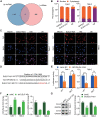
DLEU7-AS1 acts as a sponge for miR-26a-5p in RCC. (A) Venn diagram shows long non-coding RNA (lncRNA) targeting miR-26a-5p using lncRNA and LncBase. (B) The location of DLEU7-AS1 was confirmed in Caki-1 and 786-O cells using a subcellular fractionation assay. (C) RNA fluorescence in situ hybridization (FISH) assay confirmed the location of DLEU7-AS1. DLEU7-AS1 was largely located in the cytoplasm of Caki-1 and 786-O cells. Nuclei were stained with DAPI; DLEU7-AS1 was stained red. Reference genes 18S and U6 were stained red. Scale bar represents 25 μm. (D) The location of the miR-26a-5p target sites in DLEU7-AS1 and the mutation site of DLEU7-AS1. (E) Dual-luciferase reporter assay analyzed the effect of miR-26a-5p ectopic expression on the activity of DLEU7-AS1 WT and MUT in Caki-1 and 786-O cells. (F) The relative levels of miR-26a-5p in Caki-1 and 786-O cells, which were transfected using the negative control (shNC) and shDLEU7-AS1, were identified using real-time qRT-PCR (quantitative reverse transcription polymerase chain reaction). (G) Western blot was used to measure the protein levels of coronin-3 in Caki-1 and 786-O cells that were transfected with shNC and shDLEU7-AS1. Data represent mean ± SD of three biological replicates. Student's t-test was applied to obtain the P-value: ns: no significant difference, **P < 0.01, ***P < 0.001.
Figure 2:
Figure 2:
The expression level of DLEU7-AS1 in RCC. (A and B) The transcription level of DLEU7-AS1 in unpaired (A) and paired (B) tumor-normal RCC tissues was determined using data obtained from the Cancer Genome Atlas (TCGA) database. Data represent mean ± SD. (C and D) The correlation between the DLEU7-AS1 level and the expression of miR-26a-5p and coronin-3 was determined using the data obtained from the TCGA database. (E and F) Real-time quantitative reverse transcription polymerase chain reaction (qRT-PCR) analysis of DLEU7-AS1 expression in unpaired tumor-normal/paired tumor-normal RCC tissues. Data represent mean ± SD of three technical replicates. (G and H) Correlation between DLEU7-AS1 level and the levels of miR-26a-5p and coronin-3 (Coro3). Mann–Whitney test (A and E), Wilcoxon matched-pairs signed-rank test (B and F), ****P < 0.0001. Pearson correlation test (C, D, G and H).
Figure 3:
Figure 3:
Association between the level of DLEU7-AS1 and the clinicopathological characteristics of patients afflicted with RCC. Overview of DLEU7-AS1 expression (TCGA database) according to histologic grade (A), TNM stage (B), T status (C), N status (D) and M status (E). Data represent mean ± SD. Kruskal–Wallis one-way ANOVA followed by Dunn's multiple comparison test: ns, no significant difference; *P < 0.05, **P < 0.01. (F) Kaplan–Meier analysis of the OS of patients afflicted with RCC from the TCGA database by high or low expression of DLEU7-AS1. Log-rank test.
Figure 4:
Figure 4:
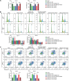
DLEU7-AS1 knockdown suppresses proliferation and promotes apoptosis in RCC cells via the miR-26a-5p/Coro3 axis. (A) DLEU7-AS1 deficiency inhibits cell viability via the miR-26a-5p/Coro3 axis. Data represent mean ± SD of four biological replicates. (B) DLEU7-AS1 knockdown induces cell-cycle arrest at the G0/G1 phase via the miR-26a-5p/Coro3 axis. Data represent mean ± SD of three biological replicates. (C) DLEU7-AS1 knockdown increases cell apoptosis via the miR-26a-5p/Coro3 axis. Data represent mean ± SD of three biological replicates. One-way ANOVA followed by Tukey's post-hoc test was applied for P-value: *P < 0.05, **P < 0.01, ***P < 0.001, ****P < 0.0001.
Figure 5:
Figure 5:
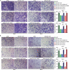
DLEU7-AS1 knockdown suppresses the ability of cell migration and invasion in RCC via the miR-26a-5p/Coro3 axis. (A) Transwell migration assays revealed that DLEU7-AS1 knockdown inhibits the migration ability of Caki-1 and 786-O cells. The effect of DLEU7-AS1 knockdown on the migration ability of Caki-1 and 786-O cell lines was rescued by using miR-26a-5p inhibitor and coronin-3. (B) Transwell Invasion assays demonstrated that DLEU7-AS1 knockdown inhibits the invasion of 786-O and Caki-1 cells. The effect of DLEU7-AS1 knockdown on the invasion ability of Caki-1 and 786-O cells was rescued by miR-26a-5p inhibitor and coronin-3. Data represent mean ± SD of three biological replicates, one-way ANOVA followed by Tukey's post-hoc test: **P < 0.01, ***P < 0.001, ****P < 0.0001.
Figure 6:
Figure 6:
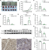
DLEU7-AS1 knockdown inhibits the tumor growth of RCC in vivo. (A) Images of isolated tumors. (B and C) Quantitation of tumor volumes (B) every 4 days and tumor weights (C) 34 days after injection with stably DLEU7-AS1 knockdown 786-O cells or negative control cells. Data represent mean ± SD of six mice. (D and F) Determining of DLEU7-AS1, miR-26-5p and Coro3 mRNA expression in tumors using RT-qPCR. Data represent mean ± standard error of mean of six mice. (G and H) Coro3 protein levels were detected using western blotting. Data represent mean ± SD of six mice. (I and J) Ki67-positive tumor cells were analyzed and quantified using IHC staining. Data represent mean ± SD of three mice. Student's t-test: *P < 0.05, **P < 0.01, ***P < 0.001, ****P < 0.0001.
Figure 7:
Figure 7:
Diagrammatic sketch of the regulation of DLEU7-AS1 on miR-26-5p/Coro3 axis.

References

    1. Hsieh JJ, Purdue MP, Signoretti Set al. . Renal cell carcinoma. Nat Rev Dis Primers 2017; 3: 17009. - PMC - PubMed
    1. Sung H, Ferlay Jet al. . Global Cancer Statistics 2020: GLOBOCAN Estimates of Incidence and Mortality Worldwide for 36 Cancers in 185 Countries. CA: A Cancer Journal for Clinicians2021; 71(3): 209–249 - PubMed
    1. Padala SA, Barsouk A, Thandra KCet al. . Epidemiology of renal cell carcinoma. World J Oncol 2020; 11: 79–87 - PMC - PubMed
    1. Barata PC, Rini BI. Treatment of renal cell carcinoma: current status and future directions. CA Cancer J Clin 2017; 67: 507–524 - PubMed
    1. Peng Y, Croce CM. The role of microRNAs in human cancer. Signal Transd Target Ther 2016; 1: 15004 - PMC - PubMed