. 2022 Oct;237(10):3845-3859.
doi: 10.1002/jcp.30838. Epub 2022 Jul 26.

Vasorin plays a critical role in vascular smooth muscle cells and arterial functions

Affiliations

Vasorin plays a critical role in vascular smooth muscle cells and arterial functions

Loïc Louvet et al. J Cell Physiol. 2022 Oct.

Abstract

Within the cardiovascular system, the protein vasorin (Vasn) is predominantly expressed by vascular smooth muscle cells (VSMCs) in the coronary arteries and the aorta. Vasn knockout (Vasn-/- ) mice die within 3 weeks of birth. In the present study, we investigated the role of vascular Vasn expression on vascular function. We used inducible Vasn knockout mice (VasnCRE-ERT KO and VasnSMMHC-CRE-ERT2 KO , in which respectively all cells or SMCs only are targeted) to analyze the consequences of total or selective Vasn loss on vascular function. Furthermore, in vivo effects were investigated in vitro using human VSMCs. The death of VasnCRE-ERT KO mice 21 days after tamoxifen injection was concomitant with decreases in blood pressure, angiotensin II levels, and vessel contractibility to phenylephrine. The VasnSMMHC-CRE-ERT2 KO mice displayed concomitant changes in vessel contractibility in response to phenylephrine and angiotensin II levels. In vitro, VASN deficiency was associated with a shift toward the SMC contractile phenotype, an increase in basal intracellular Ca2+ levels, and a decrease in the SMCs' ability to generate a calcium signal in response to carbachol or phenylephrine. Additionally, impaired endothelium-dependent relaxation (due to changes in nitric oxide signaling) was observed in all Vasn knockout mice models. Our present findings highlight the role played by Vasn SMC expression in the maintenance of vascular functions. The mechanistic experiments suggested that these effects are mediated by SMC phenotype switching and changes in intracellular calcium homeostasis, angiotensin II levels, and NO signaling.

Keywords: angiotensin II; artery; nitric oxide; smooth muscle cells; vascular function; vasorin.


Conflict of interest statement

The authors declare no conflicts of interest.

Figures
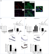
Figure 1
Figure 1
Vasn expression in ECs, SMCs, aorta, and organs of Vasn CRE‐ERT KO and Vasn SMMHC‐CRE‐ERT2 KO mice. (a) Sections of aorta from a Vasn Venus transgenic mouse. Immunofluorescence of ECs (CD31; red), aortic elastic fibers (green) and Venus+ cells (pink). Nuclei were counterstained with Hoechst reagent (blue). The red arrow indicates Venus‐positive ECs, and the yellow arrow indicates Venus‐positive SMCs. In Vasn CRE‐ERT KO mice (b) and Vasn SMMHC‐CRE‐ERT2 KO mice (c), total RNA from thoracic aortas, heart, kidney, and lung were measured and compared with control mice. Vasn protein expression in aortas was assessed in Western blots and compared with extracts from control mice. Representative immunoblots show the inhibition of Vasn protein synthesis. Quantification of Vasn expression in ECs and SMCs using Vasn immunostaining, scored from 0 (absence of immunostaining) to 4 (greatest possible immunostaining). In all qPCR experiments, Gapdh was used as the internal reference gene. For Vasn immunostaining, five thoracic aorta sections were averaged for each animal. EC, endothelial cell; SMC, smooth muscle cell. Negative control (Negative) corresponds to immunostaining without the primary anti‐Vasn antibody. Vasn protein expression was quantified and normalized against β‐ACTIN. Control mice n = 4–6, Vasn CRE‐ERT KO n = 5, Vasn SMMHC‐CRE‐ERT2 KO n = 4. *p < 0.05, **p < 0.01, ***p < 0.001 versus control, $p < 0.001 versus EC of Vasn SMMHC‐CRE‐ERT2 KO mice.
Figure 2
Figure 2
Effect of Vasn deletion on arterial blood pressure, heart rate, and Ang II levels in Vasn CRE‐ERT KO and Vasn SMMHC‐CRE‐ERT2 KO mice. Systolic, diastolic, and mean arterial blood pressures and heart rate 21 days after tamoxifen injection in (a) 8‐week‐old Vasn CRE‐ERT KO (b) 8‐week‐old Vasn SMMHC‐CRE‐ERT2 KO mice and (d) 1‐year‐old (Aged) Vasn CRE‐ERT KO mice. The results correspond to cumulative data from 13 to 16 control mice, 17 Vasn CRE‐ERT KO mice, 16 Vasn SMMHC‐CRE‐ERT2 KO mice, and 4 Aged Vasn CRE‐ERT KO mice. *p < 0.05, **p < 0.01, ***p < 0.0005, ****p < 0.0001 versus controls. (c) Ang II levels in control, Vasn CRE‐ERT KO and Vasn SMMHC‐CRE‐ERT2 KO mice after 14 days (D14) and 21 days (D21). Control mice n = 12, Vasn CRE‐ERT KO D14 n = 6, Vasn CRE‐ERT KO D21 n = 10, Vasn SMMHC‐CRE‐ERT2 KO D14 n = 6, Vasn SMMHC‐CRE‐ERT2 KO D21 n = 10. *p < 0.05, **p < 0.005 versus control mice. $$$$p < 0.0001 versus Vasn CRE‐ERT KO D14. £££p < 0.0005 versus Vasn SMMHC‐CRE‐ERT2 KO D21.
Figure 3
Figure 3
Impact of Vasn deletion on vascular function in Vasn CRE‐ERT KO and Vasn SMMHC‐CRE‐ERT2 KO mice, and correlation between Ang II levels and contraction in Vasn SMMHC‐CRE‐ERT2 KO mice. (a) Contraction obtained with phenylephrine and relaxation obtained with acetylcholine in Vasn CRE‐ERT KO mouse vessels 14 days (Vasn CRE‐ERT KO D14, n = 8) and 21 days (Vasn CRE‐ERT KO D21, n = 8) after Vasn KO. (b) Contraction obtained with phenylephrine and relaxation obtained with acetylcholine in Vasn SMMHC‐CRE‐ERT2 KO mouse vessels 14 days (Vasn SMMHC‐CRE‐ERT2 KO D14, n = 7) and 21 days (Vasn SMMHC‐CRE‐ERT2 KO D21, n = 4) after Vasn KO. Control mice n = 7. *p < 0.05, **p < 0.005, ***p < 0.0005 versus control mice; $p < 0.01, $$$p < 0.0001 versus D14 group. (c) Correlation between the contraction in response to phenylephrine and Ang II levels in Vasn SMMHC‐CRE‐ERT2 KO mice at 14 days and 21 days. Control mice n = 12, Vasn SMMHC‐CRE‐ERT2 KO D14 n = 6, Vasn SMMHC‐CRE‐ERT2 KO D21 n = 10. (d) p‐MYPT1 protein expression levels in aortas from Vasn CRE‐ERT KO and Vasn SMMHC‐CRE‐ERT2 KO mice were assessed using Western blot analysis and compared with extracts from control mice (n = 6, for each condition). *p < 0.05, versus control mice.
Figure 4
Figure 4
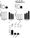
Influence of Vasn silencing on SMC phenotypic marker expression and STAT3 activation in aortic samples from Vasn CRE‐ERT KO and Vasn SMMHC‐CRE‐ERT2 KO mice. (a) mRNA expression in silenced (siVasn‐transfected) HVSMCs versus control cells treated with a scrambled siRNA (siNeg), representative Western blots of VASN protein expression, and quantification of VASN protein expression in siRNA‐transfected HVSMCs from six donors (n = 6); I, II and III are different donors; N and V correspond to the siNeg and siVasn conditions, respectively. (b) mRNA expression of collagen 8 (Coll8), collagen 1 (Coll1), and osteopontin (OPN) was analyzed using qPCR in siVasn HVSMCs and compared with control cells (siNeg). (c) mRNA expression of calponin (CNN1), α‐smooth muscle actin (α‐SMA), and smooth muscle myosin heavy chains (SMMHCs) was analyzed using qPCR in siVasn HVSMCs and compared with control cells (siNeg). For all mRNA expression analyses, the results correspond to cumulative data from two independent reverse transcriptions of total RNA from six donors (n = 12). The value in the siNeg condition was set to 1. *p < 0.05, **p < 0.01, ***p < 0.0001 versus siNeg. (d) STAT3 protein expression and activation in aorta samples from Vasn CRE‐ERT KO and Vasn SMMHC‐CRE‐ERT2 KO mice was assessed in Western blots and compared with extracts of aortas from control mice (n = 6, for each condition). The expression of the phosphorylated form of STAT3 (p‐STAT3) was quantified and normalized against total STAT3 (t‐STAT3). Representative immunoblots are shown above the quantification histograms. HVSMC, human vascular smooth muscle cell. *p < 0.05, **p < 0.01 versus control mice.
Figure 5
Figure 5
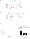
Effect of VASN silencing on calcium homeostasis in HVSMCs. (a) Typical frequency responses obtained to carbachol or phenylephrine stimulation in silenced (siVasn‐transfected cells) and control (scrambled siRNA, siNeg) cells (n = 45 cells). (b) Magnitude of Ca2+ responses and the basal Ca2+ concentration in siVasn‐transfected cells (n = 245) and siNeg control cells (n = 250). (c) Recordings of the extracellular application of Mn2+ (quenching the Fura‐2 fluorescent probe) and analysis of the corresponding slopes (siNeg, n = 32; siVasn, n = 24) in transfected HVSMCs. (d) Typical recordings of basal entry of Ca2+ and quantification in siNeg‐transfected HVSMCs (n = 19) and siVasn transfected HVSMCs (n = 21). HVSMC, human vascular smooth muscle cell. *p < 0.05, ***p < 0.001 versus siNeg.
Figure 6
Figure 6
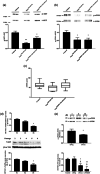
Western blot analysis of relaxation signaling pathways in aortas (a–c) of Vasn CRE‐ERT KOand Vasn SMMHC‐CRE‐ERT2 KO mice, and (d,e) HUVECs. Protein expression in aortas from Vasn CRE‐ERT KO and Vasn SMMHC‐CRE‐ERT2 KO mice was assessed using Western blots of selected markers and compared with extracts from control mice (n = 6 for each condition). The expression of the phosphorylated forms of AKT and eNOS proteins (p‐AKT and p‐eNOS) was quantified and normalized against the total protein forms (t‐AKT and t‐eNOS). Representative immunoblots are shown above each quantification histogram. (a) p‐AKT expression in Vasn CRE‐ERT KO and Vasn SMMHC‐CRE‐ERT2 KO mice. (b) p‐eNOS expression in Vasn CRE‐ERT KO and Vasn SMMHC‐CRE‐ERT2 KO KO mice. *p < 0.05, **p < 0.01 versus control mice. (c) ADMA levels in the different mice groups (n = 15–16). (d) VASN mRNA expression and VASN protein expression in HUVECs through passages (P). Results represent the accumulated data of independent qPCR experiments (n = 5) and the accumulated data of independent Western blot experiments (n = 3). The P2 condition was set to 1, *p < 0.05 versus P2. (e) The expression of the phosphorylated and total forms of eNOS proteins (p‐eNOS and t‐eNOS) was assessed by Western blot analysis. Activated p‐eNOS levels were quantified and normalized against those of t‐eNOS. I, II, and III indicate different independent HUVECs experiments; N and V correspond to the siNeg and siVasn conditions, respectively. Results represent the accumulated data of independent experiments (n = 4). The siNeg condition was set to 1, *p < 0.05 versus siNeg. NO production in VASN‐silenced HUVECs. NO production was assessed by measuring the fluorescence of DAF‐FM DA. Results represent the accumulated data of HUVECs independent experiments (n = 7). The Control condition was set to 1. HUVEC, human umbilical vein endothelial cell. *p < 0.05, **p < 0.01 versus Control, $p < 0.05 versus siNeg.

References

    1. Bonnet, A. L. , Chaussain, C. , Broutin, I. , Rochefort, G. Y. , Schrewe, H. , & Gaucher, C. (2018). From vascular smooth muscle cells to folliculogenesis: What about vasorin? Frontiers of Medicine, 5, 335. 10.3389/fmed.2018.00335 - DOI - PMC - PubMed
    1. Bravo‐Sagua, R. , Parra, V. , Muñoz‐Cordova, F. , Sanchez‐Aguilera, P. , Garrido, V. , Contreras‐Ferrat, A. , Chiong, M. , & Lavandero, S. (2020). Sarcoplasmic reticulum and calcium signaling in muscle cells: Homeostasis and disease. International Review of Cell and Molecular Biology, 350, 197–264. 10.1016/bs.ircmb.2019.12.007 - DOI - PubMed
    1. Campbell, D. J. (2013). Do intravenous and subcutaneous angiotensin II increase blood pressure by different mechanisms? Clinical and Experimental Pharmacology and Physiology, 40(8), 560–570. 10.1111/1440-1681.12085 - DOI - PubMed
    1. Choksi, S. , Lin, Y. , Pobezinskaya, Y. , Chen, L. , Park, C. , Morgan, M. , Li, T. , Jitkaew, S. , Cao, X. , Kim, Y. S. , Kim, H. S. , Levitt, P. , Shih, G. , Birre, M. , Deng, C. X. , & Liu, Z. G. (2011). A HIF‐1 target, ATIA, protects cells from apoptosis by modulating the mitochondrial thioredoxin, TRX2. Molecular Cell, 42(5), 597–609. 10.1016/j.molcel.2011.03.030 - DOI - PMC - PubMed
    1. Daya, H. A. , Kouba, S. , Ouled‐Haddou, H. , Benzerdjeb, N. , Telliez, M. S. , Dayen, C. , Sevestre, H. , Garçon, L. , Hague, F. , & Ouadid‐Ahidouch, H. (2021). Orai3‐mediates cisplatin‐resistance in non‐small cell lung cancer cells by enriching cancer stem cell population through PI3K/AKT pathway. Cancers, 13(10), 2314. 10.3390/cancers13102314 - DOI - PMC - PubMed

Publication types