Review
. 2022 Jul;12(7):220174.
doi: 10.1098/rsob.220174. Epub 2022 Jul 27.

Endocrine cybernetics: neuropeptides as molecular switches in behavioural decisions

Affiliations
Review

Endocrine cybernetics: neuropeptides as molecular switches in behavioural decisions

Dick R Nässel et al. Open Biol. 2022 Jul.

Abstract

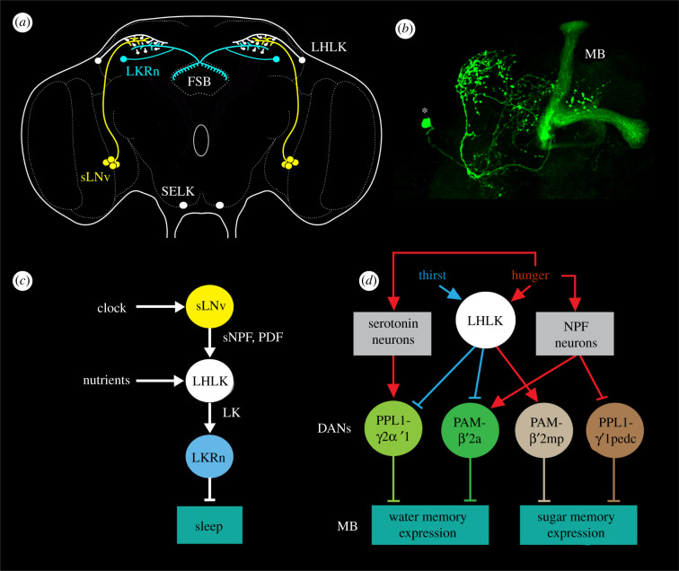
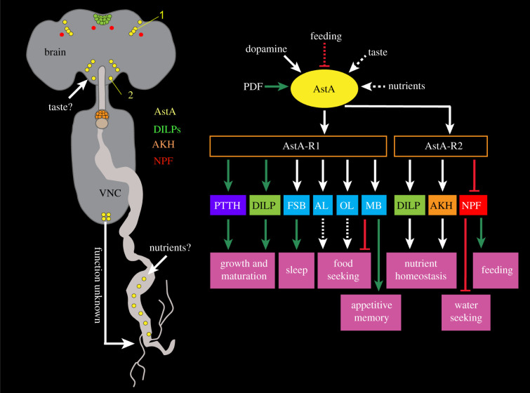
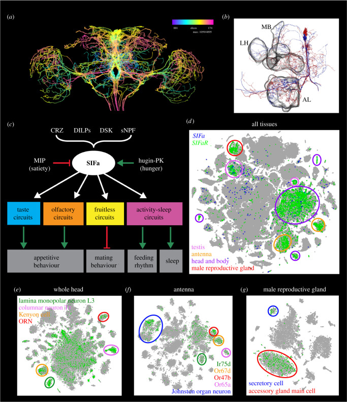
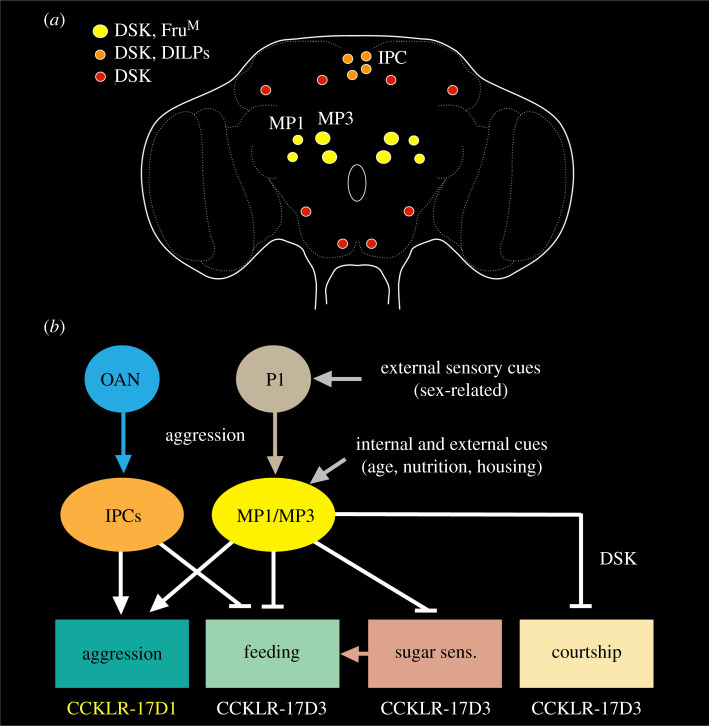
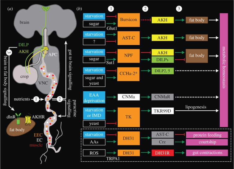
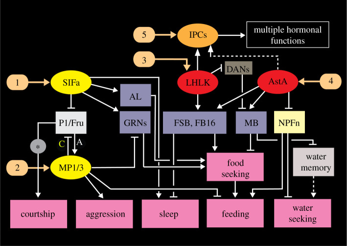
Plasticity in animal behaviour relies on the ability to integrate external and internal cues from the changing environment and hence modulate activity in synaptic circuits of the brain. This context-dependent neuromodulation is largely based on non-synaptic signalling with neuropeptides. Here, we describe select peptidergic systems in the Drosophila brain that act at different levels of a hierarchy to modulate behaviour and associated physiology. These systems modulate circuits in brain regions, such as the central complex and the mushroom bodies, which supervise specific behaviours. At the top level of the hierarchy there are small numbers of large peptidergic neurons that arborize widely in multiple areas of the brain to orchestrate or modulate global activity in a state and context-dependent manner. At the bottom level local peptidergic neurons provide executive neuromodulation of sensory gain and intrinsically in restricted parts of specific neuronal circuits. The orchestrating neurons receive interoceptive signals that mediate energy and sleep homeostasis, metabolic state and circadian timing, as well as external cues that affect food search, aggression or mating. Some of these cues can be triggers of conflicting behaviours such as mating versus aggression, or sleep versus feeding, and peptidergic neurons participate in circuits, enabling behaviour choices and switches.

Keywords: Drosophila melanogaster; brain circuits; interneurons; neuromodulation; peptide hormones.


Conflict of interest statement

We declare we have no competing interests.

Figures

Figure 1.
Figure 1.
The Drosophila brain and hierarchical organization of peptidergic signalling. (a) The brain of adult Drosophila with some of the neuropils indicated in colour. The central complex (CX) is highlighted in blue and red. Pars intercerebralis (PI), optic lobe (OL), mushroom bodies (MB), antennal lobes (AL), lateral horn (LH) and suboesophageal zone (SEZ) are also highlighted. (b) Three levels of modulatory peptide signalling. (1) Widely arborizing orchestrating neurons innervate many neuropils and receive multiple inputs of different kinds. (2) Intermediate size peptidergic neurons (often in one hemisphere only) interconnect several regions. They may for instance receive inputs from clock neurons and others and supply outputs to the fan-shaped body to mediate nutrition-dependent sleep activity. (3) Local neurons mediate executive (intrinsic) neuromodulation, here exemplified by a local neuron in the antennal lobe. Such neurons can serve as an interface between different odour channels and also receive higher-level modulatory inputs. At this level also some of the odorant-sensitive neurons are known to signal locally with neuropeptides to modulate sensory gain. (c–f) Confocal images of GFP labelled neurons of the types indicated in (b). (c) SIFamide-expressing neurons widely arborizing throughout the brain (level 1 in (b)). (d) Leucokinin-expressing neurons. One of the LHLK neurons is encircled (level 2 in (b)). (e) A small number of local neurons (LNs) innervating each of the antennal lobes (level 3 in (b)). Several such neurons express tachykinin. (f) A single drosulfakinin (DSK)-expressing neurons (MP1a type) with wide branches (level 1 in (b)). (g) Interorgan signalling. Neurosecretory cells in the brain (1) and endocrine cells in corpora cardiaca (2) release hormones into the circulation that act on peripheral targets such as the intestine (3). The intestine and the fat body (4) produce peptide hormones that signal to the brain. Brains in (a) and (b) were made in RStudio using the Natverse package and (g) was generated in BioRender.
Figure 2.
Figure 2.
Different levels of peptidergic neuromodulation exemplified in odour signal processing. This figure illustrates two main levels of neuromodulation, orchestrating (extrinsic) and executive (local). (a) Orchestrating neurons expressing the neuropeptide SIFamide have widely arborizing processes in most regions of the Drosophila brain including the antennal lobe (AL). Four neuronal cell bodies (at arrow) give rise to the processes. The inset shows branches in the antennal lobe. (b) Localized, executive peptide signalling occurs in select glomeruli in the antennal lobe by short neuropeptide F (sNPF) produced in olfactory sensory neurons (OSNs) innervating the glomeruli. Three glomeruli innervated by sNPF-producing OSNs are seen here (DL3, DA1 and Va1v). The local sNPF-mediated modulation is described in the text. (c) Neurotransmitters and neuropeptides involved in intrinsic and extrinsic neuromodulation in the antennal lobe. Two glomeruli are shown here with OSNs and projection neurons (PNs). The substances used for intrinsic modulation are produced by local neurons (LNs) and some OSNs (light blue). Substances used for extrinsic modulation are produced by various types of large neurons that originate outside the olfactory system. Two LNs are highlighted in red and yellow. In addition, insulin-like peptides (DILPs) act presynaptically on OSNs via the circulation. All the OSNs use acetylcholine, but some subpopulations of the OSNs additionally use sNPF or myoinhibitory peptide (not shown). The PNs are also cholinergic and some of these co-express sNPF or TK. (d) This figure shows different levels of modulation of olfactory signals related to food odour attraction and aversion. Three channels responding to food odours from receptor (Or) to antennal lobe glomerulus (DM) are shown (red, blue and green). Two are modulated by nutrient-dependent hormonal DILP signals (1 and 2), and a third by orchestrating SIFamide (SIFa) neurons (3). These represent orchestrating signals. At the executive level, the Or42b receptor expressing OSNs also express sNPF and the sNPF receptor (sNPFR). In hungry flies, low levels of DILPs upregulate sNPF expression (1) and thus the signal strength to higher order olfactory neurons increases and food search increases (attraction). TK released from local neurons inactivates synaptic activity in Or85a neurons, and low levels of DILPs (in hungry flies) inhibit TK action (2) leading to aversion for high concentrations of food odour (vinegar). SIFa neurons modulate food attraction via Or47a-DM3 at the level of projection neurons (3) and are under regulation of myoinhibitory peptide (MIP) and Hugin-pyrokinin (Hugin-PK). (a) and (b) are altered from Carlsson et al. [85], (c) is redrawn from Nässel [36] which was based on an idea from Lizbinski & Dacks [86] and (d) is altered from Nässel et al. [35] which in turn was based on Sayin et al. [10].
Figure 3.
Figure 3.
Executive peptide signalling: multiple distributed local functions illustrated by the numerous neurons producing tachykinin (TK). (a) Schematic of neuronal TK distribution in the adult Drosophila brain (frontal view). Neuronal cell bodies are shown in different colours (see legend in figure). Some have been studied functionally in some detail (several colours); others remain unexplored (white). The light red neurons (SMP, MPP, LPP1a) innervate different layers of the fan-shaped body of the central complex (CX) [97] and modulate explorative walking [98]. The green ones (DC1, DC2) are local neurons of the antennal lobe that are part of circuitry that modulates odour sensitivity in olfactory sensory neurons (OSNs) [99]. In male flies the light blue neurons (LPP1b) express FruM and probably acetylcholine (Ach) and regulate levels of aggression [100]. The pink ones (ITPn) are lateral neurosecretory cells that co-express TK, ion transport peptide (ITP) and short neuropeptide F (sNPF) [101] and regulate aspects of metabolic and water homeostasis [101,102]. The arrow indicates axons destined for peripheral neurohemal sites. The DN neurons are involved in pheromone sensing [103] and the SEZ neurons in regulation of larval insulin-producing cells (IPCs) [104]. The terminology (except ITPn) is from Winther et al. [105] and specifications of neurons are compiled from papers cited above. This figure was updated from Nässel et al. [106]. (b–e) Executive (intrinsic) peptidergic neuromodulation in the Drosophila antennal lobe, exemplified by TK signalling in modulation of food odour sensing. (b) TK peptides are expressed in local neurons (LN) of the antennal lobe and innervate most glomeruli. Two glomeruli are shown here (DM1 and DM5). Of these, DM1 mediates food odour attraction (Or42b) and DM5 food odour aversion (Or85b). (c) Image of TK immunoreactive LNs (green) in the clusters DC1 and DC2 innervating the antennal lobe (AL). (d,e) Role of TK signalling in the DM5 glomerulus, which relays aversive odour signals from olfactory sensory neurons (OSNs) that express odorant receptors Or85b to DM5 projection neurons (PN), which in turn signal to higher order neurons that control food search. (d) In fed flies the circulating level of insulin-like peptides (DILPs) is high, which suppresses expression of the TK receptor DTKR. When DTKR signalling is low there is no suppression of Ca2+ channel activity. Hence there is an increased release of acetylcholine (ACh) when the OSN is activated and as a consequence the DM5 PN relays strong aversive signals and food search is reduced. (e) In the hungry fly the DILP level is low, DTKR expression is high and therefore TK signalling activates DTKR and the OSN releases less Ach. This suppresses activation of the aversive DM5 PN and results in increased food search. The (b), (d) and (e) were redrawn from figures in Ko et al. [107].
Figure 4.
Figure 4.
Context-specific signalling, exemplified by neurons signalling with leucokinin (LK). (a) Schematic depiction of LK neurons in relation to some neurosecretory cells in the adult Drosophila brain. The LHLKs act on (white arrows) insulin (DILP) producing cells (IPCs), dopaminergic neurons (PPL1 and PAM subtypes) and at least two types of LK receptor (LKR)-expressing neurons (LKRn; FSB 1 and 2; see (b)) innervating the FSB. SELKs may act (grey arrow) on the ALK/ITPn that express the LKR. The Hugin neurons of the suboesophageal zone are shown since they form a link between gustatory sensory cells and feeding circuits, including IPCs. The numbered boxes (1–4) indicate sites of interaction between neurons. Data derived from [9,93,94,172,173]. (b) Schematic diagram of functional connections between LK neurons (yellow boxes) and other neurons, circuits and peripheral targets. Arrows indicate various actions, dashed arrows (and ?) suggest actions yet to be confirmed, and stop bars indicate inhibitory action. The LK neurons in the brain are shown as yellow boxes and the IPCs as a green box. Targets of LK signalling are shown as dark blue boxes. LHLKs signal to two types of LKR expressing neurons of the fan-shaped body (FSB 1, 2; these are LKR neurons and FBl6 neurons), and via dopaminergic neurons (DAN) to mushroom body-associated neurons (MB). The LKR-expressing FSB neurons inhibit sleep [172] and the FBl6 neurons regulate food choice [9], whereas the MB neurons, via dopamine (DA) inputs, mediate water (and sugar) memory [34]. LHLKs respond to decreased glucose and receive inputs from neuronal circuits of the circadian clock and systems sensing thirst and hunger (red ellipses). LHLKs signal with LK to IPCs, which regulates sleep–metabolism interactions [93,94]. IPCs are nutrient-sensing and use DILPs to regulate multiple functions, including carbohydrate and lipid metabolism, feeding, stress responses and fecundity; IPCs also express drosulfakinin, DSK (see [174]). IPCs are likely to act on the ALK/ITPn with DILP2 (dashed arrow) (see [175]). SELK neurons may receive gustatory inputs [171], but their actions are not functionally confirmed (dashed arrows and ?). The ALK/ITPn are neurosecretory cells that use ion transport peptide (ITP) to systemically regulate water homeostasis via the intestine and hindgut and also to regulate feeding and drinking [102]. These cells also use tachykinin (TK) and short neuropeptide F (sNPF) to regulate responses to starvation and desiccation [101], probably by paracrine signalling (asterisk), but the neuronal circuitry is not yet known. The magenta box represents neurons expressing receptors for LK, TK and sNPF (and ITP; not shown) in the brain that are yet to be identified. The role of LK in ALK/ITPn cells is not yet known. (b) is updated and modified from Nässel [176].
Figure 5.
Figure 5.
Context-specific LK signalling in regulation of sleep–metabolism interactions and water memory in Drosophila. (a) Schematic of brain neurons connecting clock, nutrient-sensing and sleep regulation. There is one pair of LHLK neurons in the brain. Clock neurons (sLNv) have outputs on each LHLK that in turn inhibit LK receptor expressing neurons (LKRn) that innervate the fan-shaped body (FSB) and thereby inhibit sleep in a nutrient-dependent fashion. Figure compiled from data in [93,172,178,179]. (b) Image of the LHLK neuron in the left hemisphere of the brain, with cell body at *. MB, mushroom body. From Zandawala et al. [94]. (c) A schematic of the connections in (a). PDF, pigment-dispersing factor; sNPF, short neuropeptide F. (d) LHLK neurons and a circuit regulating water–sugar based memory. The LHLK neurons receive hunger and thirst signals and act on dopaminergic neurons (DANs) of PPL1 and PAM subtypes to regulate expression of water and sugar memory in mushroom body (MB) circuits. Some of these neurons also receive inputs from serotonin and neuropeptide F (NPF) producing neurons. Redrawn from Senapati et al. [34].
Figure 6.
Figure 6.
Context-specific signalling with Allatostatin-A (AstA). AstA neuropeptide from the brain and gut regulates diverse feeding associated behaviours and physiology. (a) A schematic showing the locations of select AstA-expressing cells in the nervous system and midgut, as well as some of its downstream neuronal targets. The morphology and location of AstA cells suggest that the suboesophageal zone AstA neurons may receive taste inputs from the proboscis, and AstA-expressing enteroendocrine cells in the gut may be nutrient-sensitive. A PLP neuron is indicated by 1 and a Janu-AstA by a 2; these are discussed in the text. (b) Inputs and behavioural outputs of AstA cells. AstA-expressing neurons receive inputs from the pigment-dispersing factor (PDF) expressing clock neurons [91] and dopaminergic inputs via the Dop1R1 receptor [9]. In addition, feeding inhibits AstA neurons and they may also receive gustatory inputs via the proboscis and nutrient information via the gut. AstA, in turn, mediates its effects via its two receptors, AstA-R1 and AstA-R2 expressed in various peptidergic cells/neurons. These include cells expressing prothoracicotropic hormone (PTTH), insulin-like peptides (DILPs), adipokinetic hormone (AKH) and neuropeptide F (NPF). AstA-R1 is also expressed in neurons in the fan-shaped body (FSB) and mushroom body (MB). Moreover, axonal projections of AstA neurons to the antennal lobe (AL) and optic lobe (OL), coupled with single-cell transcriptome data from the Fly Cell Atlas suggest expression of AstA-R1 in the AL and OL. AstA modulation of peptidergic neurons and other neuronal targets influences various feeding-related behaviours including feeding, appetitive memory and water seeking. Green arrows represent stimulation, red bars represent inhibition, white arrows represent unclear valence and dashed arrows represent postulated actions. (b) is based on [,,,,–183].
Figure 7.
Figure 7.
Orchestrating neuromodulation and behavioural switches illustrated by SIFamide (SIFa) neurons. Expression of SIFa and SIFaR correlates with the functions of this signalling system. (a) SIFa expression in four neurons with extensive arborizations throughout the brain, including the central complex, mushroom bodies antennal and optic lobes. Image obtained from https://neuronbridge.janelia.org [188,189]. (b) Partial reconstructions of two SIFa neurons from serial electron microscopic sections. MB, mushroom body, LH, lateral horn, AL, antennal lobe. Data obtained from neuPRINT (https://neuprint.janelia.org) [188,190]. (c) Peptidergic inputs and behavioural outputs of SIFa neurons. SIFa influences appetitive behaviour via modulation of taste (neurons not identified) and olfactory circuits (neurons in DM3 glomerulus; see also (f)), mating via actions on fruitless-expressing neurons and sleep via modulation of activity-sleep circuits. CRZ, corazonin; DILPs, insulin-like peptides; DSK, drosulfakinin; sNPF, short neuropeptide F; MIP myoinhibitory peptide, Hugin-PK, Hugin pyrokinin. Based on [87,90,186,191,192]. (d) t-SNE visualization of single-cell transcriptomes derived from all the tissues of the adult fly. SIFa and SIFaR are expressed in the head and body, and testis clusters whereas SIFaR is additionally expressed in the antenna and male reproductive gland. (e–g) t-SNE visualization of single-cell transcriptomes showing the expression of SIFaR in different cell populations of the (e) whole head, (f) antenna and (g) male reproductive gland. Within the whole head, SIFaR is expressed in the lamina monopolar neurons L3, columnar neurons T1, Kenyon cells of the mushroom body and olfactory receptor neurons (ORNs). SIFaR is broadly expressed in the antenna with prominent expression in the Johnston organ and olfactory neurons expressing Ir75d, Or67d, Or47b and Or65a odorant receptors. Lastly, SIFaR is predominantly expressed in the male accessory gland main cells and secretory cells of the male reproductive gland. Data for (d–g) were mined using Scope (http://scope.aertslab.org) [193].
Figure 8.
Figure 8.
Orchestrating neuromodulation and behavioural switches illustrated by drosulfakinin (DSK)-producing neurons. (a) Distribution of DSK-expressing neurons. The neurons whose functions have been investigated are the MP1 and MP3 neurons that in male flies co-express fruitless (FruM) and a subpopulation of the 14 insulin-producing cells (IPCs). (b) Regulatory roles of the DSK neurons in male flies. DSK signalling is shown with white arrows (activation) and stop bars (inhibition). Note that the MP1/MP3 neurons stimulate aggression (via the DSK receptor CCKLR-17D1) and inhibit courtship as well as sugar sensing gustatory receptors (Gr64f) and feeding (via CCKLR-17D3). DSK from IPCs stimulate aggression and inhibits feeding (but DSK receptor type was not investigated). The MP1/3 neurons are modulated by internal states and external cues as well as by a population of P1 neurons that mediate male-specific behaviours and receive sensory inputs from conspecific male and female flies (sex pheromones, visual, etc.). MP1/MP3 neurons also feed back onto P1 neurons to suppress courtship (not shown here, but see electronic supplementary material, figure S3). The IPC mediated aggression (and courtship; not shown) is regulated by OAN. For further details see electronic supplementary material, figure S3. This figure is compiled from [,,–202].
Figure 9.
Figure 9.
Interorgan peptide signalling: brain, intestine and fat body. Peptides from the gut modulate metabolic homeostasis, behaviours and gut physiology via state-dependent (mainly nutritional) gut-to-brain and paracrine signalling. See electronic supplementary material, figure S5 for distribution of gut peptides. (a) A schematic showing the signalling pathways mediating the effects of peptides produced by gut enteroendocrine cells (EECs; orange) and enterocytes (ECs; white). (1) These cells predominantly sense nutrients such as carbohydrates, yeast and amino acids, but they can also sense ROS, and receive inputs from the innate IMD pathway. (2) Once these cells are activated, they release peptides into the circulation for local effects (paracrine signalling) on gut enterocytes or muscles (red). In addition, the peptides target the nervous system, including the adipokinetic hormone (AKH)-producing cells (APCs) and Drosophila insulin-like peptide (DILP)-producing cells (IPCs). (3) AKH and DILPs in turn target their receptors (AKHR and dInR, respectively) on the fat body to influence metabolic homeostasis. (b) Various intestinal peptidergic pathways influence metabolic homeostasis in a feeding-state-dependent manner. Sugar activates Bursicon EECs via the Glut1 glucose transporter and Bursicon indirectly inhibits APCs to regulate lipid homeostasis [241]. Starvation triggers release of Allatostatin-C (AstC), which activates APCs to promote energy mobilization [242]. Neuropeptide F (NPF)-producing EECs respond to sugar via the Sut1 transporter, and released NPF inhibits APCs and activates IPCs to regulate lipid metabolism [243]. Sugars and yeast activate, while starvation inhibits release of CCHa-2 from cells in the gut and fat body. CCHa-2 stimulates release of DILP2 and 5 to influence organismal growth [244]. Deprivation of EAAs activates CNMa-producing ECs, to regulate metabolic homeostasis via actions on CNMaR-expressing neurons [245]. Both starvation and IMD pathway activate, whereas yeast inhibits tachykinin (TK)-producing EECs [161,162]. TK acts locally on enterocytes via its receptor TKR99D to affect lipogenesis. Amino acids activate diuretic hormone 31 (DH31) producing EECs [45]. DH31 regulates the balance between feeding and courtship via actions on AstC- and corazonin (CRZ)-producing neurons, respectively [39]. Moreover, DH31 EECs can also sense reactive oxygen species (ROS) via the TRPA1 receptor to regulate gut contractions [246]. Boxes in (b) are colour-coded to match the cell types in (a). Dashed arrows indicate indirect actions. The two receptors in the fat body were generated in BioRender.
Figure 10.
Figure 10.
Summary diagram of peptide signalling that regulates competing behaviours. SIFamide (SIFa) and drosulfakinin (DSK)-expressing (MP1/3) neurons interact with several brain circuits to regulate behaviours in an orchestrating fashion, some of which are competing. One hub that regulates courtship versus aggression under peptidergic influence is a set of FruM-expressing P1 neurons. The asterisk indicates additional neuronal circuitry (see electronic supplementary material, figure S3). SIFa and DSK also target antennal lobe (AL) neurons and gustatory receptor neurons (GRNs) to regulate food search and feeding. LHLKs are leucokinin-expressing neurons that target different circuits in the fan-shaped body (FSB in general, or layer 16, FBl6, specifically) or mushroom bodies (MB) via dopaminergic neurons (DANs) to regulate sleep and food choice. LHLKs also regulate insulin-producing cells (IPCs) to affect sleep/metabolism interactions. Allatostatin-A neurons (AstA) target FSB and MB to regulate food choice and sleep, and neuropeptide F-expressing neurons (NPFn) to modulate feeding and water seeking. The IPCs release insulin-like peptides (DILPs) to regulate feeding, metabolism, sleep and other functions. The numbers (1–5) indicate inputs to the different neurons systems as follows, 1 (SIFa): myoinhibitory peptide, hugin-PK, clock inputs, and probably corazonin (CRZ), DILPs, DSK and short neuropeptide F (sNPF); 2 (MP1/MP3): nutritional state, P1 neurons, age, housing conditions; 3 (LHLK): nutritional state, thirst, clock; 4 (AstA): dopamine, pigment-dispersing factor, nutritional state; 5 (IPCs) nutritional state, clock, GABA, serotonin, octopamine, dopamine, DILPs, allatostatin-C (AstC), neuropeptide F (NPF), short NPF (sNPF), CCHamide2, tachykinin (TK), adipokinetic hormone (AKH), and CRZ. For references, see text.

References

    1. Wei D, Talwar V, Lin D. 2021. Neural circuits of social behaviors: innate yet flexible. Neuron 109, 1600-1620. (10.1016/j.neuron.2021.02.012) - DOI - PMC - PubMed
    1. Yapici N, Zimmer M, Domingos AI. 2014. Cellular and molecular basis of decision-making. EMBO Rep. 15, 1023-1035. (10.15252/embr.201438993) - DOI - PMC - PubMed
    1. Miroschnikow A, Schlegel P, Pankratz MJ. 2020. Making feeding decisions in the Drosophila nervous system. Curr. Biol. 30, R831-R840. (10.1016/j.cub.2020.06.036) - DOI - PubMed
    1. Ishimoto H, Kamikouchi A. 2021. Molecular and neural mechanisms regulating sexual motivation of virgin female Drosophila. Cell. Mol. Life Sci. 78, 4805-4819. (10.1007/s00018-021-03820-y) - DOI - PMC - PubMed
    1. Lee SS, Wu MN. 2020. Neural circuit mechanisms encoding motivational states in Drosophila. Curr. Opin. Neurobiol. 64, 135-142. (10.1016/j.conb.2020.05.002) - DOI - PMC - PubMed

Publication types

LinkOut - more resources