. 2022 Jul;19(192):20220268.
doi: 10.1098/rsif.2022.0268. Epub 2022 Jul 27.

Structural mechanics of filamentous cyanobacteria

Affiliations

Structural mechanics of filamentous cyanobacteria

Mixon K Faluweki et al. J R Soc Interface. 2022 Jul.

Abstract

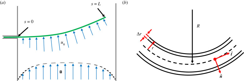
Filamentous cyanobacteria, forming long strands of connected cells, are one of the earliest and most successful forms of life on Earth. They exhibit self-organized behaviour, forming large-scale patterns in structures like biomats and stromatolites. The mechanical properties of these rigid structures have contributed to their biological success and are important to applications like algae-based biofuel production. For active polymers like these cyanobacteria, one of the most important mechanical properties is the bending modulus, or flexural rigidity. Here, we quantify the bending stiffness of three species of filamentous cyanobacteria, of order Oscillatoriales, using a microfluidic flow device where single filaments are deflected by fluid flow. This is complemented by measurements of Young's modulus of the cell wall, via nanoindentation, and the cell wall thickness. We find that the stiffness of the cyanobacteria is well-captured by a simple model of a flexible rod, with most stress carried by a rigid outer wall. Finally, we connect these results to the curved shapes that these cyanobacteria naturally take while gliding, and quantify the forces generated internally to maintain this shape. The measurements can be used to model interactions between cyanobacteria, or with their environment, and how their collective behaviour emerges from such interactions.

Keywords: Young’s modulus; bending stiffness; biomechanics; cyanobacteria; gliding motility; microfluidics.


Figures

Figure 1.
Figure 1.
Filamentous cyanobacteria under confocal fluorescence imaging. (a) Under ideal conditions active gliding specimens of Oscillatoria lutea appear as long thin curved filaments. (b) When rendered inactive, for example by being briefly cooled, the same filaments adopt a more random shape. (c) Under higher magnification O. lutea is seen to be composed of one-cell-wide strands of connected cells.
Figure 2.
Figure 2.
An example of filamentous cyanobacteria structure (O. lutea) showing a reticulate pattern.
Figure 3.
Figure 3.
Modelling cyanobacteria bending and structure. (a) The filament of bacteria is treated as a slender rod of length L, which bends under a drag force arising from the normal component of the channel flow, un. (b) Close up the filament is approximated as a hollow cylinder of radius r and wall thickness Δr. The radius of curvature (R = 1/κ), tangent (t^) and normal (n^) vectors are defined with reference to the neutral axis of the cylinder, which follows path s.
Figure 4.
Figure 4.
Microfluidic flow cell. The sketch to the left gives a schematic of the flow cell, showing how the components were connected. The close-up view to the right highlights the interactions between a cyanobacteria filament and the flow in the channel. The filament is introduced through a narrow inlet, with tight corners to pin it in place and anchor it at the point along the wall where it enters into the main channel. The view here of the chip in use is a superposition of two white-light microscopy images of the rest and deflected configurations of a single filament, as it is pushed by a laminar flow u travelling along the y-axis of the channel.
Figure 5.
Figure 5.
Measurements of the bending stiffness of filamentous cyanobacteria. (a) Each flow test consisted of a sequence of different flow speeds, separated by stopped flow conditions, while we tracked the deflection of a single filament of cyanobacteria in the channel. As shown here, the total deflection was proportional to the applied flow. (b) Each deflection profile was fit to the expected elastic deformation, with the magnitude of the elastic response (effectively, β) and a solid-body rotation, α, as fitting parameters; shown here are the fits for one example filament. (c) After accounting for any rotation through a global correction, a bending stiffness, β(s), was then extracted at every point along the filament. (d) We show the distribution of the values of β observed for the three species of filamentous cyanobacteria studied. In each case, results are shown before (yellow, left) and after (blue, right) accounting for rotational effects. On the boxes, whiskers indicate extreme points, a line gives the median and the bottom and top edges of the box indicate the 25th and 75th percentile, respectively.
Figure 6.
Figure 6.
Cyanobacteria elasticity was measured by nanoindentation. (a) A summary here shows Young’s modulus measurements of the three species of filamentous cyanobacteria, probed to 50 nm depth. On each box, the whiskers indicate extreme points while the red line is the median and the bottom and top edges of the box indicate the 25th and 75th percentile, respectively. The inset gives an example map of the reduced modulus E* for O. lutea. (b) Repeating the tests for different indentation depths showed no significant effect on the measured E, over the range of depths used. Error bars represent standard deviations of measurements.
Figure 7.
Figure 7.
Cross-sectional images of filamentous cyanobacteria, taken using transmission electron microscopy. (a) K. animale, (b) L. lagerheimii, (c) O. lutea. Arrows indicate: PG, peptidoglycan layer; CM, cytoplasmic membrane; OM, outer membrane and EL, external layer.
Figure 8.
Figure 8.
Freely gliding filamentous cyanobacteria adopt a curved shape. (a) A plot of the orientation angle versus position along a filament shows that the filament shape has a constant rate of change of angle, i.e. a constant curvature. Shown are four typical cases of O. lutea, corresponding to the profiles shown in the upper left. There is a variation of curvature between filaments. (b) Examples of active, gliding filamentous cyanobacteria (black) showing curved configurations are given along with best-fit circles.
Figure 9.
Figure 9.
Curvature distributions for filamentous cyanobacteria, as measured by fitting the filament shape to a circular arc, for (a) active gliding filaments at 20C and (b) inactive filaments at 2C.
Figure 10.
Figure 10.
Measured persistence length in inactive filaments of K. animale, L. lagerheimii and O. lutea, evaluated by analysing 164, 162 and 79 filaments for each species, respectively. Fits for the persistence length use equation (4.2). The shaded region represents extreme points of the distributions (using shadedErrorBar function [68]).

References

    1. Schopf JW, Packer BM. 1987. Early Archean (3.3-billion to 3.5-billion-year-old) microfossils from Warrawoona Group, Australia. Science 237, 70-73. (10.1126/science.11539686) - DOI - PubMed
    1. Allwood AC, Walter MR, Kamber BS, Marshall CP, Burch IW. 2006. Stromatolite reef from the Early Archaean era of Australia. Nature 441, 714-718. (10.1038/nature04764) - DOI - PubMed
    1. Rasmussen B, Fletcher IR, Brocks JJ, Kilburn MR. 2008. Reassessing the first appearance of eukaryotes and cyanobacteria. Nature 455, 1101-1104. (10.1038/nature07381) - DOI - PubMed
    1. Walter MR, Bauld J, Brock TD. 1976. Microbiology and morphogenesis of columnar stromatolites (Conophyton, Vacerrilla) from hot springs in Yellowstone National Park. In Stromatolites (ed. MR Walter), vol. 20 of Developments in Sedimentology, pp. 273–310. Amsterdam, The Netherlands: Elsevier.
    1. Jones B, Renaut RW, Rosen MR, Ansdell KM. 2002. Coniform stromatolites from geothermal systems, North Island, New Zealand. Palaios 17, 84-103. (10.1669/0883-1351(2002)017<0084:CSFGSN>2.0.CO;2) - DOI

Publication types