. 2022 Jul 22;12(8):1012.
doi: 10.3390/biom12081012.

Coaxial Synthesis of PEI-Based Nanocarriers of Encapsulated RNA-Therapeutics to Specifically Target Muscle Cells

Affiliations

Coaxial Synthesis of PEI-Based Nanocarriers of Encapsulated RNA-Therapeutics to Specifically Target Muscle Cells

Raquel de la Hoz et al. Biomolecules. .

Abstract

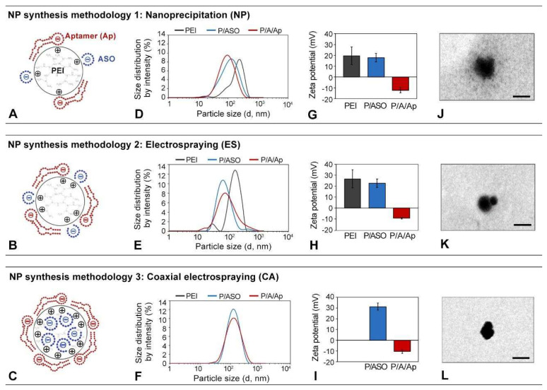
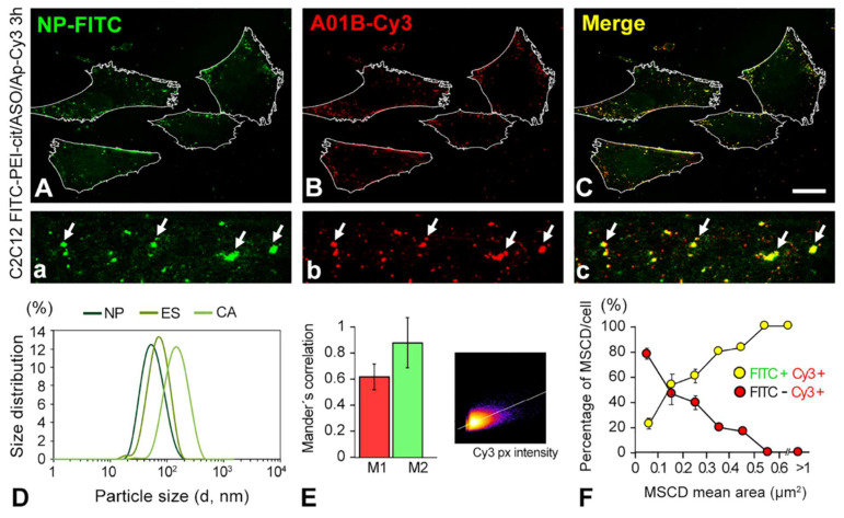
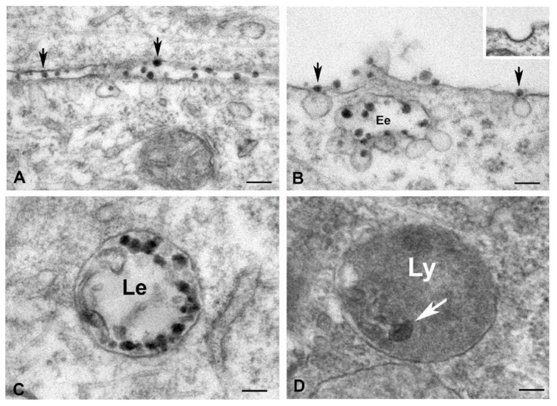
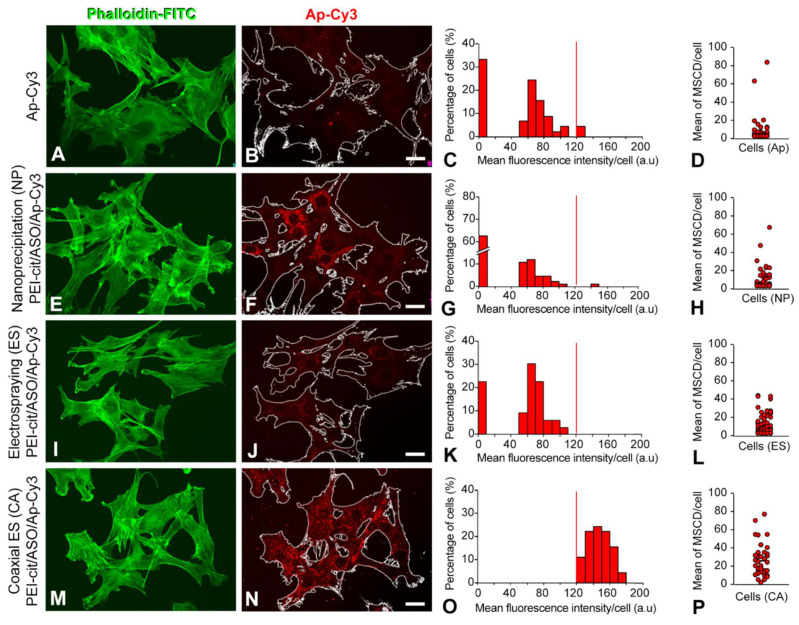
In this work, we performed a methodological comparative analysis to synthesize polyethyleneimine (PEI) nanoparticles using (i) conventional nanoprecipitation (NP), (ii) electrospraying (ES), and (iii) coaxial electrospraying (CA). The nanoparticles transported antisense oligonucleotides (ASOs), either encapsulated (CA nanocomplexes) or electrostatically bound externally (NP and ES nanocomplexes). After synthesis, the PEI/ASO nanoconjugates were functionalized with a muscle-specific RNA aptamer. Using this combinatorial formulation methodology, we obtained nanocomplexes that were further used as nanocarriers for the delivery of RNA therapeutics (ASO), specifically into muscle cells. In particular, we performed a detailed confocal microscopy-based comparative study to analyze the overall transfection efficiency, the cell-to-cell homogeneity, and the mean fluorescence intensity per cell of micron-sized domains enriched with the nanocomplexes. Furthermore, using high-magnification electron microscopy, we were able to describe, in detail, the ultrastructural basis of the cellular uptake and intracellular trafficking of nanocomplexes by the clathrin-independent endocytic pathway. Our results are a clear demonstration that coaxial electrospraying is a promising methodology for the synthesis of therapeutic nanoparticle-based carriers. Some of the principal features that the nanoparticles synthesized by coaxial electrospraying exhibit are efficient RNA-based drug encapsulation, increased nanoparticle surface availability for aptamer functionalization, a high transfection efficiency, and hyperactivation of the endocytosis and early/late endosome route as the main intracellular uptake mechanism.

Keywords: Nusinersen; RNA-therapeutics; aptamers; coaxial electrospraying; muscle-specific therapy; nanoparticle; polyethylenimine; spinal muscular atrophy.


Conflict of interest statement

The authors declare no conflict of interest.

Figures

Figure 1
Figure 1
(AC) Schematic depiction of the electrostatic structure of the PEI-cit/ASO/Ap nanocomplex attained with the three synthesis methodologies: nanoprecipitation (NP, (A)), electrospraying (ES, (B)), and coaxial electrospraying (CA, (C)). (DF) Particle size distribution of the PEI-cit nanocomplexes at different levels of functionalization (PEI-cit, PEI-cit/ASO, and PEI-cit/ASO/Ap) synthesized by the three methodologies: NP (D), ES (E), and CA (F). (GI) Z-potential of the surface of the PEI-cit nanocomplexes at different levels of functionalization (PEI-cit, PEI-cit/ASO, and PEI-cit/ASO/Ap) synthesized by NP (G), ES (H), and CA (I). Note the decrement of the Z-potential due to the addition of the electronegative oligonucleotides (ASO, Ap) to the positively charged nanocore. (JL) TEM images of PEI-cit/ASO nanocomplex morphology by: NP (J), ES (K), and CA (L). Scale bars: 180 nm.
Figure 2
Figure 2
(AC) Confocal microscopy images of C2C12 cells transfected with the FITC-PEI-cit/ASO/Ap-Cy3 nanocomplexes. Images show the colocalization from the PEI green (FITC) signal (A) and the aptamer (Ap) red signal (Cy3) (B), as well as (C) the overlap of both channels within the cells. Note that in the higher magnification details (ac), only the micron-sized structures of both channels, denoted by arrows, colocalized (yellow). (D) Particle size distribution curves of the fluorescent nanocomplexes (FITC-conjugated PEI) synthetized by the three studied synthesis methodologies (NP, ES and CA). (E) Colocalization analysis of the endocytic nanocomplexes transfected in C2C12 cells by Mander’s colocalization coefficients (M1 and M2) showing the amount of fluorescence of the pixels in cooccurrence in both color channels (green channel, FITC-PEI; red channel, Ap-Cy3), and a representative colocalization intensity histogram of the confocal microscopy images. (F) Percentage of the nanoparticle-loaded vesicles (MSCD) per cell vs. their mean area. Note that the percentage of the colocalizing MSCD per cell increased as the MSCD size became larger. Scale bar: 15 µm (AC).
Figure 3
Figure 3
(AD) Electron microscopy illustrating the endocytic pathway of the PEI-cit/ASO/Ap nanocomplexes in cultured myoblasts. (A) Interactions of nanocomplexes (electron dense particles) with the plasma membrane (arrows). (B) Incorporation of nanocomplexes in non-coated endocytic vesicles (arrows). Note the fusion of internalized vesicles containing nanocomplexes with an early endosome (Ee). Inset: Detail of a clathrin coated vesicle. (C) Late endosome (Le) loaded with numerous electron dense particles of nanocomplexes. (D) Electron dense body of lysosomal nature containing residual nanocomplexes. Scale bars: 100 nm (AC) and 50 nm (D).
Figure 4
Figure 4
(A,E,I,M) Confocal images showing the actin filaments (FITC-conjugated phalloidin) of the cytoskeleton from C2C12 cells. (B,F,J,N) Analogous confocal images showing the same C2C12 cells outlined transfected with either free Cy3-conjugated aptamer (B) or with the PEI-cit/ASO/Ap-Cy3 nanocomplexes synthetized by nanoprecipitation ((E), NP), electrospraying ((I), ES), and coaxial electrospraying ((N), ES). (C,G,K,O) Mean fluorescence intensity histogram of the Cy3 aptamer signal only from nanocomplexes located at MSCD. Vertical red lines correspond to the threshold limit that discriminates the MFI corresponding to MSCD of areas above and below 0.5 µm2. (D,H,L,P) Dispersion diagram of the mean number of MSCD of mean area ≥ 0.5 µm2 per cell. Note that most cells transfected with free aptamer (D), do not exhibit MSCD. Conversely, all cells transfected with CA nanocomplexes enclosed at least 1 MSCD in their cytoplasm. Scale bar: 20 µm.
Figure 5
Figure 5
Confocal images of C2C12 cells showing the cellular uptake of a FAM-conjugated non-targeting GapmeR (FAM, green) achieved using either Dharmafect 1 (DF1) as RNA delivery liposomal-based methodology (DF1) (AD) or by a muscle receptor-mediated mechanism with CA synthesized nanocomplex functionalized with the Cy3-conjugated specific A01B aptamer (Cy3, red) (EH). Cells were fixed and counter stained with DAPI (blue) to visualize cell nuclei. (I,J) Higher magnification (100×) confocal images showing the cytoplasmic FAM-GapmeR accumulation, surrounding the cell nuclei, using both DF1 (I) and CA nanocomplexes (J). Note the strong colocalization of FAM-GapmeR with the Cy3-aptamer in yellow foci, corresponding to MSCD. (K) Fluorescence intensity analysis using 3D interactive surface plot from the Fiji software from the cell indicated in ((J), white box). The graphical representation allows a clear visualization of intracellular FAM-conjugated Gapmers (green) and Cy3-conjugated Aptamer (red). Each yellow peak represents Gapmer/Aptamer (FAM/Cy3) colocalization in MSCD. Scale bars: 100 µm (AH) and 15 µm (IJ).

References

    1. Burnett J.C., Rossi J.J. RNA-Based Therapeutics: Current Progress and Future Prospects. Chem. Biol. 2012;19:60–71. doi: 10.1016/j.chembiol.2011.12.008. - DOI - PMC - PubMed
    1. Atri C., Guerfali F.Z., Laouini D. MicroRNAs in Diagnosis and Therapeutics. Elsevier Inc.; Amsterdam, The Netherlands: 2019.
    1. Rinaldi C., Wood M.J.A. Antisense Oligonucleotides: The next Frontier for Treatment of Neurological Disorders. Nat. Rev. Neurol. 2018;14:9–22. doi: 10.1038/nrneurol.2017.148. - DOI - PubMed
    1. Bajan S., Hutvagner G. RNA-Based Therapeutics: From Antisense Oligonucleotides to MiRNAs. Cells. 2020;9:137. doi: 10.3390/cells9010137. - DOI - PMC - PubMed
    1. Patel S., Kim J., Herrera M., Mukherjee A., Kabanov A.V., Sahay G. Brief Update on Endocytosis of Nanomedicines. Adv. Drug Deliv. Rev. 2019;144:90–111. doi: 10.1016/j.addr.2019.08.004. - DOI - PMC - PubMed

Publication types

LinkOut - more resources