Review
. 2022 Jul 26;12(8):1030.
doi: 10.3390/biom12081030.

Molecular, Subcellular, and Arrhythmogenic Mechanisms in Genetic RyR2 Disease

Affiliations
Review

Molecular, Subcellular, and Arrhythmogenic Mechanisms in Genetic RyR2 Disease

Ewan Douglas Fowler et al. Biomolecules. .

Abstract

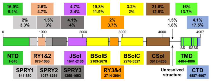
The ryanodine receptor (RyR2) has a critical role in controlling Ca2+ release from the sarcoplasmic reticulum (SR) throughout the cardiac cycle. RyR2 protein has multiple functional domains with specific roles, and four of these RyR2 protomers are required to form the quaternary structure that comprises the functional channel. Numerous mutations in the gene encoding RyR2 protein have been identified and many are linked to a wide spectrum of arrhythmic heart disease. Gain of function mutations (GoF) result in a hyperactive channel that causes excessive spontaneous SR Ca2+ release. This is the predominant cause of the inherited syndrome catecholaminergic polymorphic ventricular tachycardia (CPVT). Recently, rare hypoactive loss of function (LoF) mutations have been identified that produce atypical effects on cardiac Ca2+ handling that has been termed calcium release deficiency syndrome (CRDS). Aberrant Ca2+ release resulting from both GoF and LoF mutations can result in arrhythmias through the Na+/Ca2+ exchange mechanism. This mini-review discusses recent findings regarding the role of RyR2 domains and endogenous regulators that influence RyR2 gating normally and with GoF/LoF mutations. The arrhythmogenic consequences of GoF/LoF mutations will then be discussed at the macromolecular and cellular level.

Keywords: arrhythmias; calcium release deficiency syndrome; calcium sparks; catecholaminergic polymorphic ventricular tachycardia; delayed afterdepolarizations; early afterdepolarizations; long QT syndrome; ryanodine receptor.


Conflict of interest statement

The authors declare no conflict of interest.

Figures

Figure 1
Figure 1
A schematic illustration of human RyR2 domain organization and boundaries. Color-coded distribution of mutations associated with disease (top value) and their frequency within individual structural domains (bottom value). The six transmembrane segments (S) are represented by a vertical line. See Figure 2 below for a corresponding cartoon representation showing the arrangement of domains in 3D (using same color coding).
Figure 2
Figure 2
A schematic representation of the RyR2 3D architecture depicting 4 regulatory layers from the cytoplasmic side. The pore-forming region, comprised of the TMD and CTD, is modulated by the CSol (Layer 1), NTD, JSol and BSol (Layer 2), SPRY1/2/3 and RY1/2/3/4 (Layer 3), and accessory proteins (Layer 4), e.g., FKBP. The colors of regions in the RyR2 3D structure correspond to the colors in the RyR2 sequence in Figure 1.
Figure 3
Figure 3
Simplified schematic to illustrate the proposed pathways linking RyR2 loss-of-function (LoF, red) and gain-of-function (GoF, blue) abnormalities to pro-arrhythmic behavior. In GoF, excess Ca2+ leak leads to diastolic Ca2+ waves and DADs which tend to decrease the Ca2+ load. In LoF, evoked Ca2+ release is impaired resulting in Ca2+ loading and less Ca2+-dependent inactivation (CDI) of ICa, ultimately resulting in prolonged systolic Ca2+ release and EADs. Increasing Ca2+ load worsens Ca2+ regulation and hastens arrhythmia development.

References

    1. Bers D. Cardiac excitation-contraction coupling. Nature. 2002;415:198–205. doi: 10.1038/415198a. - DOI - PubMed
    1. Tester D.J., Ackerman M.J. Cardiomyopathic and channelopathic causes of sudden unexplained death in infants and children. Annu. Rev. Med. 2009;60:69–84. doi: 10.1146/annurev.med.60.052907.103838. - DOI - PubMed
    1. Eisner D.A., Caldwell J.L., Kistamas K., Trafford A.W. Calcium and Excitation-Contraction Coupling in the Heart. Circ. Res. 2017;121:181–195. doi: 10.1161/CIRCRESAHA.117.310230. - DOI - PMC - PubMed
    1. Cheng H., Lederer W.J., Cannell M.B. Calcium sparks: Elementary events underlying excitation-contraction coupling in heart muscle. Science. 1993;262:740–744. doi: 10.1126/science.8235594. - DOI - PubMed
    1. Marx S., Reiken S., Hisamatsu Y., Jayaraman T., Burkhoff D., Rosemblit N., Marks A. PKA phosphorylation dissociates FKBP12.6 from the calcium release channel (ryanodine receptor): Defective regulation in failing hearts. Cell. 2000;101:365–376. doi: 10.1016/S0092-8674(00)80847-8. - DOI - PubMed

Publication types

MeSH terms

Substances