Review
. 2022 Jul 1;7(3):87.
doi: 10.3390/biomimetics7030087.

The Role of the Extracellular Matrix (ECM) in Wound Healing: A Review

Affiliations
Review

The Role of the Extracellular Matrix (ECM) in Wound Healing: A Review

Robert B Diller et al. Biomimetics (Basel). .

Abstract

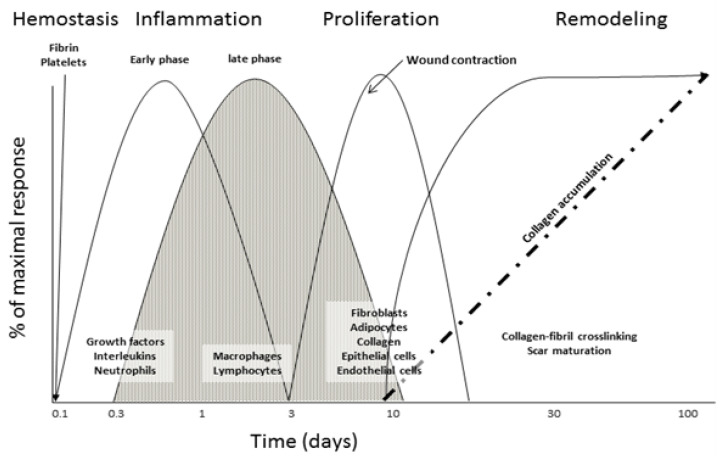
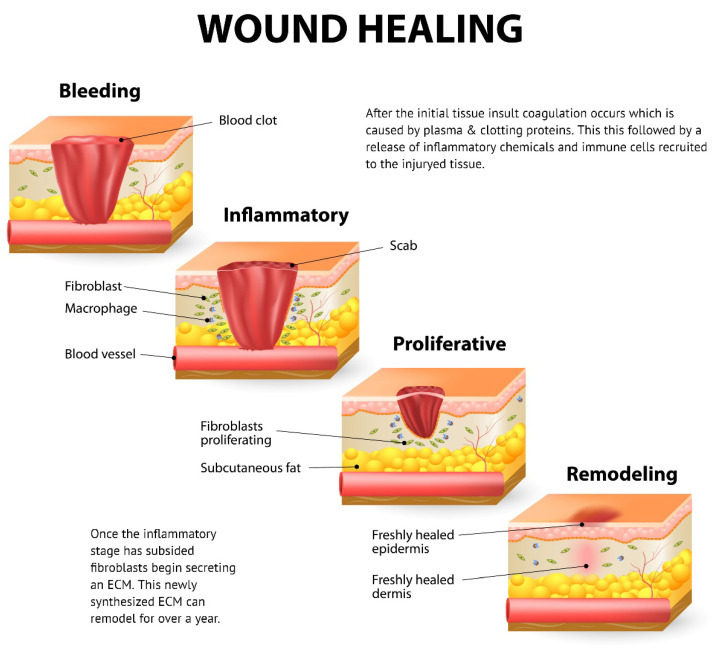
The extracellular matrix (ECM) is a 3-dimensional structure and an essential component in all human tissues. It is comprised of varying proteins, including collagens, elastin, and smaller quantities of structural proteins. Studies have demonstrated the ECM aids in cellular adherence, tissue anchoring, cellular signaling, and recruitment of cells. During times of integumentary injury or damage, either acute or chronic, the ECM is damaged. Through a series of overlapping events called the wound healing phases-hemostasis, inflammation, proliferation, and remodeling-the ECM is synthesized and ideally returned to its native state. This article synthesizes current and historical literature to demonstrate the involvement of the ECM in the varying phases of the wound healing cascade.

Keywords: collagen; dermal mimics; extracellular matrix (ECM); fibroblast(s); first-intention healing; full-thickness wound; granulation; hemostasis; inflammation; proliferation; second-intention healing; tissue remodeling; wound healing.


Conflict of interest statement

The authors have the following relevant disclosures: Robert B. Diller is employed by Amnio Technology (Phoenix, AZ, USA) and Aaron J. Tabor is employed by Axolotl Biologix (Flagstaff, AZ, USA). Both companies offer membrane based wound care products.

Figures

Figure 1
Figure 1
Schematic of the classical wound healing cascade with important stages of cellular infiltration and protein deposition.
Figure 2
Figure 2
(A) Clinical image of chronic diabetic injury with granulation tissue presence. The wound is on the medial aspect of the foot. Notice the white arrow pointing to red tissue indicating cellular infiltrate and neovascularization. The black arrow is pointing to the area in the middle of the wound which is indicative of hyper-granulation. (Patient consent provided). (B) Heamatoxalin and Eosin-stained murine skin the black frame is a gross image (scale bar = 2 mm) of the skin sample, and the red framed image (scale bar = 100 µm) is a magnified image of the area in the small red box. The white arrows point to fibroblasts in the granulation tissue. The orange arrows point to immature collagen deposition. The green arrows point to neovasculature. (C) CD-163 (Inflammatory cell marker) reacted immunohistochemistry-stained murine skin the black frame is a gross image (scale bar = 2 mm) of the skin sample and the red framed image (scale bar = 100 µm) is a magnified image of the area in the small red box. The black arrows point to fibroblasts in the granulation tissue. The purple arrows point to immature collagen deposition. The blue arrows point to inflammatory cells: macrophages, and monocytes. (D) CD-31 (endothelial cell marker) reacted immunohistochemistry-stained murine skin, the black frame is a gross image (scale bar = 2 mm) of the skin sample, and the red framed image (scale bar = 100 µm) is a magnified image of the area in the small red box. The black arrows point to neovasculature in the granulation tissue. The purple arrows point to immature collagen deposition.
Figure 3
Figure 3
Phases of the wound healing response including hemostasis (coagulation), inflammation, proliferation, and tissue remodeling (Designua/Shutterstock.com).

References

    1. Childs D.R., Murthy A.S. Overview of wound healing and management. Surg. Clin. 2017;97:189–207. doi: 10.1016/j.suc.2016.08.013. - DOI - PubMed
    1. Guo S.A., DiPietro L.A. Factors affecting wound healing. J. Dent. Res. 2010;89:219–229. doi: 10.1177/0022034509359125. - DOI - PMC - PubMed
    1. Sanford J.A., Gallo R.L. Seminars in Immunology. Volume 25. Academic Press; Cambridge, MA, USA: 2013. Functions of the skin microbiota in health and disease; pp. 370–377. - PMC - PubMed
    1. Robson M.C., Steed D.L., Franz M.G. Wound healing: Biologic features and approaches to maximize healing trajectories. Curr. Probl. Surg. 2001;38:72–140. doi: 10.1067/msg.2001.111167. - DOI - PubMed
    1. Anderson J.M. Biological responses to materials. Annu. Rev. Mater. Res. 2001;31:81–110. doi: 10.1146/annurev.matsci.31.1.81. - DOI

LinkOut - more resources