Review
. 2022 Jul 22;11(8):1421.
doi: 10.3390/antiox11081421.

Oxidative Stress in Tauopathies: From Cause to Therapy

Affiliations
Review

Oxidative Stress in Tauopathies: From Cause to Therapy

Fernando Bartolome et al. Antioxidants (Basel). .

Abstract

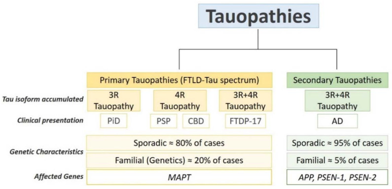
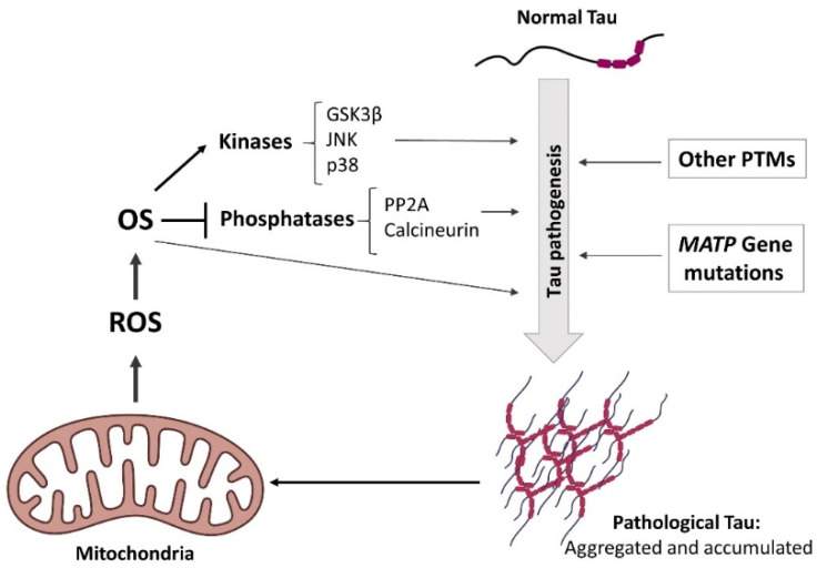
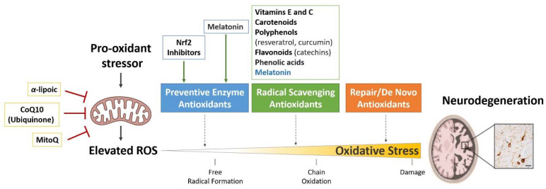
Oxidative stress (OS) is the result of an imbalance between the production of reactive oxygen species (ROS) and the antioxidant capacity of cells. Due to its high oxygen demand, the human brain is highly susceptible to OS and, thus, it is not a surprise that OS has emerged as an essential component of the pathophysiology of several neurodegenerative diseases, including tauopathies. Tauopathies are a heterogeneous group of age-related neurodegenerative disorders characterized by the deposition of abnormal tau protein in the affected neurons. With the worldwide population aging, the prevalence of tauopathies is increasing, but effective therapies have not yet been developed. Since OS seems to play a key role in tauopathies, it has been proposed that the use of antioxidants might be beneficial for tau-related neurodegenerative diseases. Although antioxidant therapies looked promising in preclinical studies performed in cellular and animal models, the antioxidant clinical trials performed in tauopathy patients have been disappointing. To develop effective antioxidant therapies, the molecular mechanisms underlying OS in tauopathies should be completely understood. Here, we review the link between OS and tauopathies, emphasizing the causes of OS in these diseases and the role of OS in tau pathogenesis. We also summarize the antioxidant therapies proposed as a potential treatment for tauopathies and discuss why they have not been completely translated to clinical trials. This review aims to provide an integrated perspective of the role of OS and antioxidant therapies in tauopathies. In doing so, we hope to enable a more comprehensive understanding of OS in tauopathies that will positively impact future studies.

Keywords: antioxidants; oxidative stress; tau; tauopathies.


Conflict of interest statement

The authors declare no conflict of interest.

Figures

Figure 1
Figure 1
Pathologic and genetic characteristics of tauopathies. Tauopathies are complex diseases regarding pathology, clinical presentation and genetics. Pathologically, tauopathies are classified in primary and secondary tauopathies. Primary tauopathies fall under the umbrella of frontotemporal lobar degeneration with tau inclusions (FTLD-Tau) and include Pick’s disease (PiD), progressive supranuclear palsy (PSP), corticobasal degeneration (CBD), and frontotemporal dementia with Parkinsonism (FTDP-17). Alzheimer’s disease in a secondary tauopathy characterized by the presence of extracellular inclusions containing Amyloid-β (Aβ) protein. Depending on tau isoform composing the intracellular inclusions, tauopathies are classified in 3R-tauopathies (PiD), 4R tauopathies (PSP and CBD) and 3R:4R tauopathies (FTDP-17 and AD). The majority of tauopathy cases are sporadic. Mutations in MAPT account of around 20% of the FTLD-tau cases. Mutations in amyloid-beta precursor protein (APP) Presenilin-1 and Presenilin-2 (PSEN-1,2) genes account for around 5% of AD cases.
Figure 2
Figure 2
Schematic representation of the positive loop between elevated OS and tau pathogenesis. Mitochondria is the major ROS resource in neurons. Increased ROS leads to elevated OS, which induces tau pathogenesis by the regulation of the activity of tau kinases/phosphatases. On the other hand, the accumulation of pathological forms of tau protein might induce mitochondrial damage and thus, ROS production. We speculate that there is a positive loop where the OS generated in the early stages of the disease induces tau pathology and, in consequence, pathological tau promotes mitochondrial impairment and more OS, leading to neuronal death.
Figure 3
Figure 3
Summary of the antioxidants therapies proposed as good candidates to treat tauopathies. OS is a lineal process that start in the mitochondria by the production of ROS and, if is not prevented, ends with cellular damage and/or cell death. Antioxidants prevent OS by counteracting the damaging action of ROS. Antioxidant enzymes act in the first line of defense suppressing the generation of free radicals. Free radical-scavengers act in the second line of defense interacting with free radicals and neutralizing them to prevent cell damage. The third line of defense consists of restoring the impairment affected by free radicals and they work repairing the damage and reconstituting membranes. Tauopathies antioxidant therapies research has been focused on targeting the first two lines of OS defense by activating antioxidant enzymes and treating with free-radical scavengers. Because mitochondria is the first resource of ROS in the cells, mitochondria-targeting therapies have also been developed to reduce OS.

References

    1. Das K., Roychoudhury A. Reactive oxygen species (ROS) and response of antioxidants as ROS-scavengers during environmental stress in plants. Front. Environ. Sci. 2014;2:53. doi: 10.3389/fenvs.2014.00053. - DOI
    1. Brand M.D., Affourtit C., Esteves T.C., Green K., Lambert A.J., Miwa S., Pakay J.L., Parker N. Mitochondrial superoxide: Production, biological effects, and activation of uncoupling proteins. Free Radic. Biol. Med. 2004;37:755–767. doi: 10.1016/j.freeradbiomed.2004.05.034. - DOI - PubMed
    1. Finkel T. Signal transduction by reactive oxygen species. J. Cell Biol. 2011;194:7–15. doi: 10.1083/jcb.201102095. - DOI - PMC - PubMed
    1. Dröge W. Free Radicals in the Physiological Control of Cell Function. Physiol. Rev. 2002;82:47–95. doi: 10.1152/physrev.00018.2001. - DOI - PubMed
    1. Auten R.L., Davis J.M. Oxygen Toxicity and Reactive Oxygen Species: The Devil Is in the Details. Pediatr. Res. 2009;66:121–127. doi: 10.1203/PDR.0b013e3181a9eafb. - DOI - PubMed

LinkOut - more resources