. 2022 Jul 26;10(8):1795.
doi: 10.3390/biomedicines10081795.

Sanguinarine Induces H2O2-Dependent Apoptosis and Ferroptosis in Human Cervical Cancer

Affiliations

Sanguinarine Induces H2O2-Dependent Apoptosis and Ferroptosis in Human Cervical Cancer

Ameer Alakkal et al. Biomedicines. .

Abstract

Sanguinarine (SNG) is a benzophenanthridine alkaloid isolated mainly from Sanguinaria canadensis, Chelidonium majus, and Macleaya cordata. SNG is considered an antineoplastic agent based on its cytotoxic activity against various tumors. However, the exact molecular mechanism through which SNG mediates this activity has not been elucidated. Here, we report that SNG induces death in human cervical cancer (HeLa) cells through activation of two interdependent cell death pathways-apoptosis and ferroptosis. SNG-induced apoptosis was characterized by caspase activation and PARP cleavage, while ferroptosis involved solute carrier family 7 member 11 (SLC7A11) down-regulation, glutathione (GSH) depletion, iron accumulation, and lipid peroxidation (LPO). Interestingly, incubation with caspase inhibitor z-VAD-fmk not only inhibited the features of apoptosis, but also negated markers of SNG-induced ferroptosis. Similarly, pretreatment with ferroptosis inhibitor ferrostatin-1 (Fer-1), apart from rescuing cells from SNG-induced ferroptosis, also curbed the features of SNG-induced apoptosis. Our study implies that, together, apoptosis and ferroptosis act as partners in the context of SNG mediated tumor suppression in HeLa cells. Importantly, SNG increased the generation of reactive oxygen species (ROS), and ROS inhibition blocks the induction of both apoptosis and ferroptosis. These findings highlight the value of continued investigation into the potential use of SNG as an antineoplastic agent.

Keywords: LPO; ROS; apoptosis; ferroptosis; labile iron; sanguinarine.


Conflict of interest statement

The authors have no conflict of interest to declare.

Figures

Figure 1
Figure 1
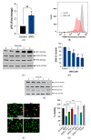
SNG-induced cell death is associated with features of apoptosis. HeLa cells were treated with (A) indicated concentrations of SNG for 6 h and (B) 3 μM SNG for the indicated time period. Cell viability was measured using an MTT assay. Data shown are means ± SD (n = 3) (* p < 0.05, ** p < 0.01, and *** p < 0.001) vs. the respective control. Cells were treated with indicated concentrations of SNG for 6 h. Following treatment, (C) morphological changes were assessed by microscopy, (D) live-dead assay was performed, and a (E) Western blot analysis of indicated proteins was performed. Actin was used as the loading control, and (F) apoptosis analysis by flow cytometry was done using Annexin V-FITC/PI staining. HeLa cells pretreated with z-VAD-fmk were treated with SNG (3 μM) for 6 h. Following treatment, (G) Western blot analysis of indicated proteins was performed. Actin was used as a loading control, (H) apoptosis analysis by flow cytometry using the Annexin V-FITC/PI staining, and (I) cell viability was assessed by the MTT assay. Data shown are mean ± SD (n = 3). ** p < 0.01 and *** p < 0.001. (J) HeLa cells were pretreated with Baf A1 or NSA were treated with SNG (3 μM) and cell viability was measured by the MTT assay. Data shown are mean ± SD (n = 3). *** p < 0.001. ns, no significance.
Figure 2
Figure 2
SNG induces the features of ferroptosis in HeLa cells. HeLa cells were treated with SNG. Following treatment, (A) LPO was determined by flow cytometry using C11-BODIPY 581/591 prob. Data shown are mean ± SD (n = 3). Significant differences, ** p < 0.01, (B) intracellular labile iron was determined by FACS using the fluorescent indicator PGSK, (C,E) and Western blot analysis of the indicated proteins was performed. Actin was used as a loading control, and the (D) GSH levels were measured. Data shown are means ± SD (n = 4) (* p < 0.05, ** p < 0.01, and *** p < 0.001 vs. control). HeLa cells pretreated with Fer-1, DFO, or Trolox were treated with SNG (3 μM). Following treatment, (F) a live/dead assay was performed and (G) cell viability was assessed using an MTT assay. Data shown are means ± SD (n = 3) (* p < 0.05 and *** p < 0.001).
Figure 3
Figure 3
H2O2 is the ROS molecule responsible for SNG-induced apoptosis and ferroptosis in HeLa cells. Cells were treated with the indicated concentration of SNG for 1 h. Following treatment, (A) the cells were stained with DCFH-DA and analyzed using fluorometry. H2O2 (1 mM) was used as a positive control. Data shown are mean ± SD (n = 3), (** p < 0.01). The cells were pretreated with NAC for 1 h, followed by SNG treatment. Following treatment, (B) cells were stained with DCFH-DA and were analyzed by fluorometry. Data shown are mean ± SD (n = 3), ** p < 0.01 and *** p < 0.001, (C) Western blot analysis of indicated proteins were performed. Actin was used as a loading control, (D) LPO was determined by flow cytometry using the C11-BODIPY 581/591 prob. Data shown are mean ± SD (n = 3). Significant differences, *** p < 0.001, and (E) cell viability were assessed using an MTT assay. Data shown are means ± SD (n = 4) (** p < 0.01, and *** p < 0.001). (F) Cells were treated with MitoPY1 were stained with Hoechst 33,342 (1.5 µL; 10 mg/mL stock solution). After treatment with SNG in the presence or absence of Sod-Py, the fluorescence of MitoPY1 (green) and Hoechst (blue) was detected using fluorescent microscopy. Cells pretreated with Sod-Py were treated with SNG. After treatment, (G) the Western blot analysis of the indicated proteins were performed. Actin was used as a loading control, (H) LPO was determined by flow cytometry using the C11-BODIPY 581/591 prob. Data shown are mean ± SD (n = 3). Significant differences, *** p < 0.001, and (I) cell viability were assessed using an MTT assay. Data shown are means ± SD (n = 3) (*** p < 0.001). The cells pretreated with Cat or SOD were treated with SNG. Following the treatment, (J) Western blot analysis of the indicated proteins were performed. Actin was used as a loading control, and the (K) GSH levels were measured. Data shown are means ± SD (n = 3) (** p < 0.01 and *** p < 0.001), (L) LPO was determined by flow cytometry using the C11-BODIPY 581/591 prob. Data shown are mean ± SD (n = 3) (*** p < 0.001), and (M) cell viability was assessed using an MTT assay. Data shown are means ± SD (n = 3) (*** p < 0.001).
Figure 3
Figure 3
H2O2 is the ROS molecule responsible for SNG-induced apoptosis and ferroptosis in HeLa cells. Cells were treated with the indicated concentration of SNG for 1 h. Following treatment, (A) the cells were stained with DCFH-DA and analyzed using fluorometry. H2O2 (1 mM) was used as a positive control. Data shown are mean ± SD (n = 3), (** p < 0.01). The cells were pretreated with NAC for 1 h, followed by SNG treatment. Following treatment, (B) cells were stained with DCFH-DA and were analyzed by fluorometry. Data shown are mean ± SD (n = 3), ** p < 0.01 and *** p < 0.001, (C) Western blot analysis of indicated proteins were performed. Actin was used as a loading control, (D) LPO was determined by flow cytometry using the C11-BODIPY 581/591 prob. Data shown are mean ± SD (n = 3). Significant differences, *** p < 0.001, and (E) cell viability were assessed using an MTT assay. Data shown are means ± SD (n = 4) (** p < 0.01, and *** p < 0.001). (F) Cells were treated with MitoPY1 were stained with Hoechst 33,342 (1.5 µL; 10 mg/mL stock solution). After treatment with SNG in the presence or absence of Sod-Py, the fluorescence of MitoPY1 (green) and Hoechst (blue) was detected using fluorescent microscopy. Cells pretreated with Sod-Py were treated with SNG. After treatment, (G) the Western blot analysis of the indicated proteins were performed. Actin was used as a loading control, (H) LPO was determined by flow cytometry using the C11-BODIPY 581/591 prob. Data shown are mean ± SD (n = 3). Significant differences, *** p < 0.001, and (I) cell viability were assessed using an MTT assay. Data shown are means ± SD (n = 3) (*** p < 0.001). The cells pretreated with Cat or SOD were treated with SNG. Following the treatment, (J) Western blot analysis of the indicated proteins were performed. Actin was used as a loading control, and the (K) GSH levels were measured. Data shown are means ± SD (n = 3) (** p < 0.01 and *** p < 0.001), (L) LPO was determined by flow cytometry using the C11-BODIPY 581/591 prob. Data shown are mean ± SD (n = 3) (*** p < 0.001), and (M) cell viability was assessed using an MTT assay. Data shown are means ± SD (n = 3) (*** p < 0.001).
Figure 4
Figure 4
Crosstalk between apoptosis and ferroptosis in SNG-induced cell death. Cells were treated with SNG (3 μM) in the presence or absence of z-VAD-fmk for 6 h. Following treatment, (A) Western blot analysis of the indicated proteins were performed. Actin was used as a loading control and the (B) GSH levels were measured. Data shown are means ± SD (n = 3) (*** p < 0.001). Cells were treated with SNG (3 μM) in the presence or absence of Fer-1 for 6 h. Following treatment, (C) Western blot analysis of the indicated proteins was performed and actin was used as the loading control, (D) apoptosis was analyzed by Annexin V-FITC/PI staining. (E) The cells were treated with SNG (3 μM) in the presence or absence of a combination of z-VAD-fmk and Fer-1 for 6 h. Following treatment, cell viability was assessed using an MTT assay. Data shown are means ± SD (n = 3) (* p < 0.05, ** p < 0.01, and *** p < 0.001).
Figure 5
Figure 5
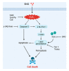
Schematic representation of SNG-mediated cell death. SNG induced the accumulation of intracellular ROS, promoting caspase activation and apoptosis. In addition, ROS further triggered down-regulation of SLC7A11 and GSH depletion and LPO, resulting in the activation of ferroptosis.

References

    1. Galadari S., Rahman A., Pallichankandy S., Thayyullathil F. Molecular targets and anticancer potential of sanguinarine-a benzophenanthridine alkaloid. Phytomedicine. 2017;34:143–153. doi: 10.1016/j.phymed.2017.08.006. - DOI - PubMed
    1. Pallichankandy S., Rahman A., Thayyullathil F., Galadari S. ROS-dependent activation of autophagy is a critical mechanism for the induction of anti-glioma effect of sanguinarine. Free Radic. Biol. Med. 2015;89:708–720. doi: 10.1016/j.freeradbiomed.2015.10.404. - DOI - PubMed
    1. Rahman A., Pallichankandy S., Thayyullathil F., Galadari S. Critical role of H2O2 in mediating sanguinarine-induced apoptosis in prostate cancer cells via facilitating ceramide generation, ERK1/2 phosphorylation, and Par-4 cleavage. Free Radic. Biol. Med. 2019;134:527–544. doi: 10.1016/j.freeradbiomed.2019.01.039. - DOI - PubMed
    1. Gu S., Yang X.C., Xiang X.Y., Wu Y., Zhang Y., Yan X.Y., Xue Y.N., Sun L.K., Shao G.G. Sanguinarine-induced apoptosis in lung adenocarcinoma cells is dependent on reactive oxygen species production and endoplasmic reticulum stress. Oncol. Rep. 2015;34:913–919. doi: 10.3892/or.2015.4054. - DOI - PubMed
    1. Han M.H., Kim G.Y., Yoo Y.H., Choi Y.H. Sanguinarine induces apoptosis in human colorectal cancer HCT-116 cells through ROS-mediated Egr-1 activation and mitochondrial dysfunction. Toxicol. Lett. 2013;220:157–166. doi: 10.1016/j.toxlet.2013.04.020. - DOI - PubMed

LinkOut - more resources