Review
. 2022 Jul 22;14(15):3583.
doi: 10.3390/cancers14153583.

Endoscopic Management of Esophageal Cancer

Affiliations
Review

Endoscopic Management of Esophageal Cancer

Christopher Paiji et al. Cancers (Basel). .

Abstract

Advances in technology and improved understanding of the pathobiology of esophageal cancer have allowed endoscopy to serve a growing role in the management of this disease. Precursor lesions can be detected using enhanced diagnostic modalities and eradicated with ablation therapy. Furthermore, evolution in endoscopic resection has provided larger specimens for improved diagnostic accuracy and offer potential for cure of early esophageal cancer. In patients with advanced esophageal cancer, endoluminal therapy can improve symptom burden and provide therapeutic options for complications such as leaks, perforations, and fistulas. The purpose of this review article is to highlight the role of endoscopy in the diagnosis, treatment, and palliation of esophageal cancer.

Keywords: diagnosis; endoscopic surgery; esophageal cancer; oncologic outcomes; staging.


Conflict of interest statement

The authors declare no conflict of interest.

Figures

Figure 1
Figure 1
Ligation-assisted endoscopic mucosal resection (EMR). (a) Nodular mucosa representing high grade dysplasia in Barrett’s esophagus (b) Band deployed to create pseudopolyp (c) EMR performed using hot snare (d) Site of resection after ligation-assisted EMR.
Figure 2
Figure 2
Endoscopic Submucosal Dissection (ESD). (a) Nodular mass lesion representing focally invasive adenocarcinoma (b) Distal mucosa incision performed after submucosal injection (c) Tunnel creation by ESD (d) Defect after completion of en bloc resection.
Figure 3
Figure 3
Radiofrequency ablation (RFA). (a) Evidence of Barrett’s esophagus and stenosis associated with prior endoscopic mucosal resection (b) RFA performed using BarrxTM Halo Ultra Long Catheter (c) Segment of Barrett’s esophagus after RFA.
Figure 4
Figure 4
Hybrid argon plasma coagulation (Hybrid-APC). (a) Focal islands of Barrett’s esophagus (b) Submucosal lift created using a hybrid-APC catheter (c) Focal islands after treatment with hybrid-APC (d) Neosquamous epithelium identified on narrow band imaging at follow-up endoscopy.
Figure 5
Figure 5
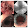
Esophageal stent placement. (a) Moderate luminal narrowing from squamous cell carcinoma (b) Placement of partially-covered metal stent over a wire under fluoroscopic guidance (c) Proximal end of partially-covered metal stent (d) Distal end of partially-covered metal stent.
Figure 6
Figure 6
Palliative endoscopic cryotherapy. (a) Ulcerated and friable mass lesion representing squamous cell carcinoma (b) Application of liquid nitrogen using a spray catheter (c) Treatment of another area of nodularity (d) Site of cancer after cryotherapy.

References

    1. GBD 2017 Oesophageal Cancer Collaborators The global, regional, and national burden of oesophageal cancer and its attributable risk factors in 195 countries and territories, 1990-2017: A systematic analysis for the Global Burden of Disease Study 2017. Lancet Gastroenterol. Hepatol. 2020;5:582–597. doi: 10.1016/S2468-1253(20)30007-8. - DOI - PMC - PubMed
    1. Edgren G., Adami H.O., Weiderpass E., Nyrén O. A global assessment of the oesophageal adenocarcinoma epidemic. Gut. 2013;62:1406–1414. doi: 10.1136/gutjnl-2012-302412. - DOI - PubMed
    1. Then E.O., Lopez M., Saleem S., Gayam V., Sunkara T., Culliford A., Gaduputi V. Esophageal Cancer: An Updated Surveillance Epidemiology and End Results Database Analysis. World J. Oncol. 2020;11:55–64. doi: 10.14740/wjon1254. - DOI - PMC - PubMed
    1. Uhlenhopp D.J., Then E.O., Sunkara T., Gaduputi V. Epidemiology of esophageal cancer: Update in global trends, etiology and risk factors. Clin. J. Gastroenterol. 2020;13:1010–1021. doi: 10.1007/s12328-020-01237-x. - DOI - PubMed
    1. Jain S., Dhingra S. Pathology of esophageal cancer and Barrett’s esophagus. Ann. Cardiothorac. Surg. 2017;6:99–109. doi: 10.21037/acs.2017.03.06. - DOI - PMC - PubMed

LinkOut - more resources