

Epidemiology and Management of Cerebral Venous Thrombosis during the COVID-19 Pandemic
- PMID: 35892907
- PMCID: PMC9332165
- DOI: 10.3390/life12081105
Epidemiology and Management of Cerebral Venous Thrombosis during the COVID-19 Pandemic
Abstract
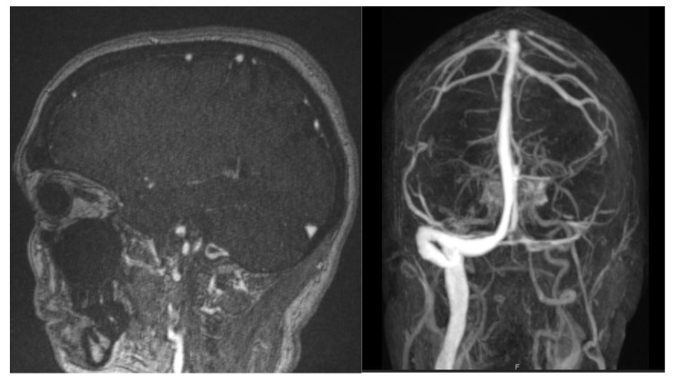
Cerebral venous thrombosis (CVT) is a rare type of stroke that may cause an intracranial hypertension syndrome as well as focal neurological deficits due to venous infarcts. MRI with venography is the method of choice for diagnosis, and treatment with anticoagulants should be promptly started. CVT incidence has increased in COVID-19-infected patients due to a hypercoagulability state and endothelial inflammation. CVT following COVID-19 vaccination could be related to vaccine-induced immune thrombotic thrombocytopenia (VITT), a rare but severe complication that should be promptly identified because of its high mortality rate. Platelet count, D-dimer and PF4 antibodies should be dosed. Treatment with non-heparin anticoagulants and immunoglobulin could improve recuperation. Development of headache associated with seizures, impaired consciousness or focal signs should raise immediate suspicion of CVT. In patients who received a COVID-19 adenovirus-vector vaccine presenting thromboembolic events, VITT should be suspected and rapidly treated. Nevertheless, vaccination benefits clearly outweigh risks and should be continued.
Keywords: COVID-19; adenovirus vaccine; cerebral venous thrombosis; vaccine-induced immune thrombotic thrombocytopenia (VITT).
Conflict of interest statement
The authors declare no conflict of interests.
Figures


References
Publication types
LinkOut - more resources
Full Text Sources
Miscellaneous
