. 2022 Jul 26;12(8):1118.
doi: 10.3390/life12081118.

MGRN1 as a Phenotypic Determinant of Human Melanoma Cells and a Potential Biomarker

Affiliations

MGRN1 as a Phenotypic Determinant of Human Melanoma Cells and a Potential Biomarker

Marta Abrisqueta et al. Life (Basel). .

Abstract

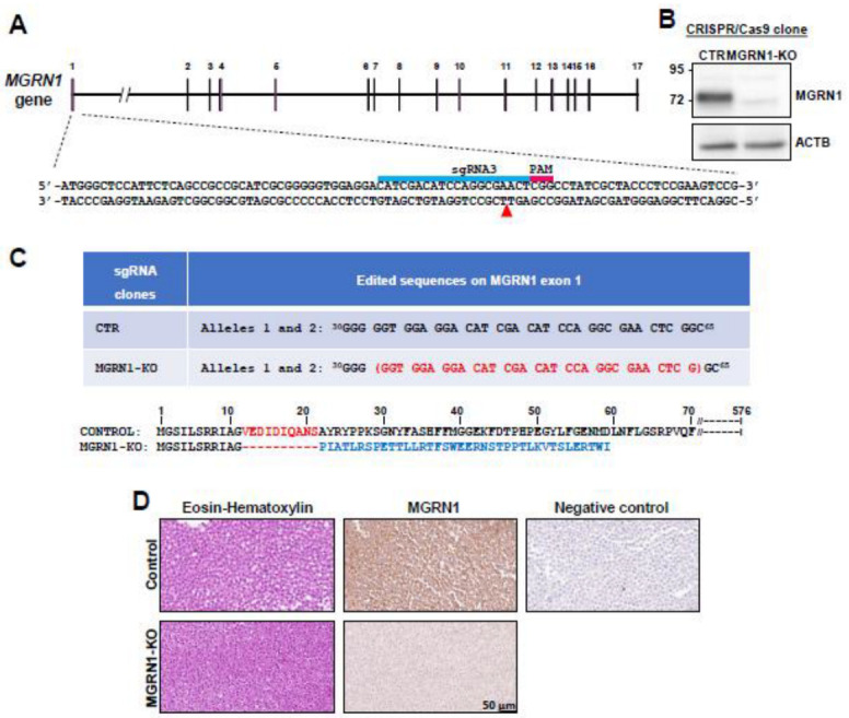
Mahogunin Ring Finger 1 (MGRN1), a ubiquitin ligase expressed in melanocytes, interacts with the α melanocyte-stimulating hormone receptor, a well-known melanoma susceptibility gene. Previous studies showed that MGRN1 modulates the phenotype of mouse melanocytes and melanoma cells, with effects on pigmentation, shape, and motility. Moreover, MGRN1 knockdown augmented the burden of DNA breaks in mouse cells, indicating that loss of MGRN1 promoted genomic instability. However, data concerning the roles of MGRN1 in human melanoma cells remain scarce. We analyzed MGRN1 knockdown in human melanoma cells. Transient MGRN1 depletion with siRNA or permanent knockdown in human melanoma cells by CRISPR/Cas9 caused an apparently MITF-independent switch to a more dendritic phenotype. Lack of MGRN1 also increased the fraction of human cells in the S phase of the cell cycle and the burden of DNA breaks but did not significantly impair proliferation. Moreover, in silico analysis of publicly available melanoma datasets and estimation of MGRN1 in a cohort of clinical specimens provided preliminary evidence that MGRN1 expression is higher in human melanomas than in normal skin or nevi and pointed to an inverse correlation of MGRN1 expression in human melanoma with patient survival, thus suggesting potential use of MGRN1 as a melanoma biomarker.

Keywords: DNA damage; Mahogunin Ring Finger 1 (MGRN1); biomarker; melanocytes; melanoma.


Conflict of interest statement

The authors declare no conflict of interest.

Figures

Figure 1
Figure 1
CRISPR/Cas9-mediated knockdown of MGRN1 in HBL human melanoma cells. (A) Schematic representation of the MGRN1 gene highlighting the sequence targeted by sgRNA3 in exon 1 (blue bar). This 20 mer oligonucleotide was designed to bind the DNA target directly upstream of a 5′-NGG adjacent motif (PAM, red bar). (B) Representative immunoblot of MGRN1 in control and MGRN1-KO 3.7 clonal cells. Cell-free detergent-solubilized extracts were electrophoresed in 10% SDS-PAGE gels and immunoblotted for MGRN1 and actin B (ACTB) as a control for comparable loading. Note the different electrophoretic mobility of the MGRN1 band in the control lane and the faint non-specific band in the extract of MGRN1-KO cells. (C) Top: edited sequence in exon 1 of MGRN1 in MGRN1-KO-3.7 cells, compared with the consensus sequence in control (CTR) HBL cells (nucleotides 30 to 65). Deleted nucleotides are shown in red, between brackets. Bottom: amino acid sequence of control and the resulting truncated MGRN1 protein in MGRN1-KO-3.7 cells. (D) Homogeneity of clonal MGRN1-KO cell cultures. Control and MGRN1-KO cells were eosin–hematoxylin-stained or immunostained for MGRN1, as indicated. Note the lack of significant staining for MGRN1-KO cells, indicating the homogeneity of the clonal culture. Negative control stands for samples of control MGRN1-expressing cells treated with a secondary antibody in the absence of anti-MGRN1.
Figure 2
Figure 2
Normal growth of MGRN1-null human melanoma cells. (A) Phase-contrast images of control (HBL-CTR) and MGRN1-KO cells (HBL MGRN1-KO) in 2D cultures. The smaller images on the top right corners show a magnification of a representative region of the micrograph. Scale bar, 50 μm. (B) Changes in the morphology of HBL cells depleted of MGRN1 by siRNA transfection and stimulated with NDP-MSH, as specified in the upper scheme depicting the experimental design. Representative phase-contrast micrographs are shown below. (C) Efficient repression of MGRN1 expression by the siRNA transfection employed above. The upper graph represents the levels of MGRN1 mRNA as estimated by real-time RT-PCR, normalized to the expression in cells treated with control, non-targeting siRNA. ACTB mRNA was used as a loading control. Further details concerning primer sequences are provided under Material and Methods. A representative Western blot of detergent-solubilized cell extracts, stained for MGRN1 and GAPDH as loading control, is shown below, along with the quantification of 3 independent blots. (D) Western blot analysis of MITF expression in cells treated as in panel B, immunostained for MITF and GAPDH (as loading control). A representative blot out of 3 independent experiments and the quantification of the fold change of the normalized MITF signal relative to control unstimulated cells is shown below. (E) Comparable expression of MITF in control HBL cells and MGRN1-KO clones obtained by permanent knockdown of MGRN1. Detergent-solubilized, cell-free extracts were analyzed for MGRN1 by Western blot. A representative blot out of 3 independent experiments and the corresponding quantification are shown. The numbers on top of the MGRN1-KO lanes refer to the individual clone analyzed. GAPDH was used as a loading control. The numbers below these lanes correspond to the normalized MITF signal relative to the control lane (mean of 2 independent experiments). (F) Cell cycle progression in human melanoma cells lacking MGRN1. Control HBL cells and MGRN1-KO cells (clone 3.7) were stained with propidium iodide and analyzed in an FACScanto cytometer. The graph shows the percentage of cells in the G0/G1, S, and G2/M phases (mean ± sem, n = 5 for control and n = 2 for MGRN1-KO cells). (G) Comparable proliferation rates of control and MGRN1-KO cells. The proliferation of control and MGRN1-KO cells was compared with 3 independent methods: metabolic activity measured with MTT (left), metabolic labeling of DNA with BrdU (right), and counting viable cells over 72 h (graph on the bottom). In this last case, doubling times of roughly 23 h were obtained upon nonlinear regression, without statistically significant differences.
Figure 3
Figure 3
Increased DNA damage in MGRN1-null human melanoma cells. (A) Representative images of control (CTR) and MGRN1-KO cells immunostained for γ-H2AX (left) and quantification of γ-H2AX staining intensity (right). (B) Alkaline comet assay was performed to detect DNA breaks in control (CTR) and MGRN1-KO cells. Representative images of comets are shown. The graph shows the median of relative tail moments measured using CASPLAB software.
Figure 4
Figure 4
Expression of MGRN1 in melanomas, nevi, and normal skin. (A) Analysis of MGRN1 expression across different tumor types (TCGA dataset) and normal tissues (GTEx dataset), available in the GEPIA database. Median expression is represented by the height of the bar. Tumor abbreviations in X axis highlighted with red boxes indicate a statistically significant higher expression of MGRN1 in tumors than in paired normal samples. Green boxes represent lower expression of MGRN1 in tumor. SKCM stands for skin cutaneous melanoma. ANOVA was used for statistical analysis. (B) Representation of the normalized MGRN1 expression levels in normal skin and melanoma samples, available in the GEPIA database. (C) Comparison of the normalized MGRN1 expression levels in normal skin, nevi, and melanoma samples, using six independent datasets available at the GEO database. (D) Relative expression of MGRN1 in nevi and melanoma samples obtained at the BBHRI. MGRN1 mRNA levels were quantified by dPCR and GAPDH mRNA was used for normalization. About 40 samples per group were analyzed. Graphs show mean ± sem, and one-way ANOVA was used for statistical analysis. * p < 0.05. In panels (BD), MM stands for melanoma.
Figure 5
Figure 5
Correlation of lower MGRN1 expression in melanomas with longer patient survival. (A) Kaplan–Meier curves for the survival of melanoma patients according to data from the TCGA dataset. Patients were stratified as a function of the highest and lowest 10% levels of MGRN1 in primary tumor specimens. (B,C) Kaplan–Meier curves show metastasis-free (B) and disease-free (C) survival for patients of the BBHRI upon resection of primary melanomas. Patients were stratified as a function of the 50% high (blue curves, n = 6 for left graph and n = 8 for right graph) and 50% low (red curves, n = 6 for left graph and n = 8 for right graph) MGRN1 mRNA levels in primary tumor specimens. p values are indicated in the graphs. (D) Comparison of the Breslow thickness at diagnosis in melanoma samples. Data correspond to the TCGA dataset (left) with the 25% higher and lower expression of MGRN1, and to the BBHRI cohort (right) with the 50% higher and lower MGRN1 expression. Graphs show mean ± sem (right) or median (left), and a t-test was used for statistical analysis.

References

    1. Parker E.R. The Influence of Climate Change on Skin Cancer Incidence—A Review of the Evidence. Int. J. Women’s Dermatol. 2021;7:17–27. doi: 10.1016/j.ijwd.2020.07.003. - DOI - PMC - PubMed
    1. Lopes F.C.P.S., Sleiman M.G., Sebastian K., Bogucka R., Jacobs E.A., Adamson A.S. UV Exposure and the Risk of Cutaneous Melanoma in Skin of Color. JAMA Dermatol. 2021;157:213. doi: 10.1001/jamadermatol.2020.4616. - DOI - PubMed
    1. Raimondi S., Suppa M., Gandini S. Melanoma Epidemiology and Sun Exposure. Acta Derm. Venereol. 2020;100:adv00136. doi: 10.2340/00015555-3491. - DOI - PMC - PubMed
    1. O’Leary M.A., Wang S.J. Epidemiology and Prevention of Cutaneous Cancer. Otolaryngol. Clin. N. Am. 2021;54:247–257. doi: 10.1016/j.otc.2020.11.001. - DOI - PubMed
    1. García-Borrón J.C., Abdel-Malek Z., Jiménez-Cervantes C. MC1R, the CAMP Pathway, and the Response to Solar UV: Extending the Horizon beyond Pigmentation. Pigment Cell Melanoma Res. 2014;27:699–720. doi: 10.1111/pcmr.12257. - DOI - PMC - PubMed

LinkOut - more resources