Review
. 2022 Jul 25;12(8):682.
doi: 10.3390/metabo12080682.

Infective Endocarditis in High-Income Countries

Affiliations
Review

Infective Endocarditis in High-Income Countries

Francesco Nappi et al. Metabolites. .

Abstract

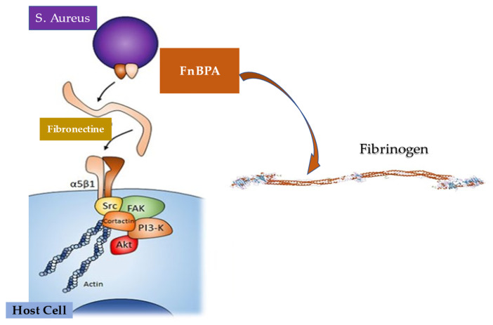
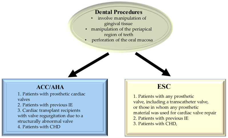
Infective endocarditis remains an illness that carries a significant burden to healthcare resources. In recent times, there has been a shift from Streptococcus sp. to Staphylococcus sp. as the primary organism of interest. This has significant consequences, given the virulence of Staphylococcus and its propensity to form a biofilm, rendering non-surgical therapy ineffective. In addition, antibiotic resistance has affected treatment of this organism. The cohorts at most risk for Staphylococcal endocarditis are elderly patients with multiple comorbidities. The innovation of transcatheter technologies alongside other cardiac interventions such as implantable devices has contributed to the increased risk attributable to this cohort. We examined the pathophysiology of infective endocarditis carefully. Inter alia, the determinants of Staphylococcus aureus virulence, interaction with host immunity, as well as the discovery and emergence of a potential vaccine, were investigated. Furthermore, the potential role of prophylactic antibiotics during dental procedures was also evaluated. As rates of transcatheter device implantation increase, endocarditis is expected to increase, especially in this high-risk group. A high level of suspicion is needed alongside early initiation of therapy and referral to the heart team to improve outcomes.

Keywords: Staphylococcus aureus; biofilm; fibronectin; immune response; infective endocarditis.


Conflict of interest statement

The authors declare no conflict of interest.

Figures

Figure 1
Figure 1
Depiction of Duke criteria for the diagnosis of infective endocarditis. HACEK indicates haemophilus species, Aggregatibacter (formerly Actinobacillus) actinomycetemcomitans, Cardiobacterium hominis, Eikenella corrodens, and Kingella kingae. For patients who have prosthetic valves and possible infective endocarditis according to clinical criteria, transoesophageal echocardiography is recommended. For patients with native valve endocarditis, transthoracic echocardiography is recommended.
Figure 2
Figure 2
Depiction of the incidence of infective endocarditis according to age and sex. The incidence peak was reported as 194 cases per million in a population of men with IE aged 75–79 years. Blue box, female; dark yellow box, male.
Figure 3
Figure 3
Depiction of the incidence of infective endocarditis according to the previous cardiac history in a French population study reporting data from 497 adults. Red dark box, no known cardiac disease; yellow box, prosthetic valve with or without intracardiac device; blue box, intracardiac device; green box, other cardiac device including individuals who received LVAD. Abbreviation; LVAD, left ventricular assist device.
Figure 4
Figure 4
Major incidence of IE is revealed in elderly history of CIEDs and in younger population with history of PWID. Minor incidence in patients with central venous catheters, HIV, CHD, and immunosuppression. 26.6% of cases of IE are due to Staphylococcus aureus and CoNS are involved in 9.7% of cases * Low numbers of Coxiella Burnetii, Bartonella quintana, Pseudomonas aeruginosa, Tropheryma whipplei, Enterobacteriaceae, Acinetobacter ursingii, Listeria monocytogenes, Propionibacterium acnes, Lactobacillus spp., Corynebacterium spp, Francisella tularensis, Erysipelothrix rhusiopathiae, Gordonia bronchialis, Bacillus spp., Catabacter hongkongensi, Moraxella catarrhalis, Campylobacter fetus, Neisseria elongata and Veillonella spp. collected Abbreviations: CIED, cardiac implantable electronic devices; CHD, congenital heart disease; CoNS, coagulase negative; HIV; immunodeficiency virus; IE, infective endocarditis; PWID; persons who inject drugs.
Figure 5
Figure 5
The main constituents that form EPS are molecules of polysaccharides, proteins, nucleic acids, and lipids. Abbreviation: EPS, extracellular polymeric substances.
Figure 6
Figure 6
Depiction of virulent factors of S. aureus. MSCRAMMs have a substantial key role in driving the initiation of endovascular, bone and joint, and prosthetic device infections. These structures can bind to molecules such as collagen (mostly via Cna), fibronectin (via FnbAB), and fibrinogen (with ClfAB and Fib), and thus evade the immune system. The development of infection is induced by Coa and von Willebrand factor-binding protein that led to critical virulence. Coa binds preferentially to soluble fibrinogen, while vWbp does not disclose any preference between the two forms of fibrinogen. Abbreviations: Clf, cell-bound clumping factor; Coa, coagulase; Fnb, fibronectin binding protein; MSCRAMM, microbial surface components recognizing adhesive matrix molecules; vWbp, von Willebrand factor-binding protein.
Figure 7
Figure 7
Bacterial adhesion induces the pathophysiological process of infective endocarditis. The first step led to inflammatory response with the involvement of inflammatory cells (PMN, monocyte, and macrophage). The inflammation is mediated by the production of cytokines (TNF, α, Il 1,6 and 8), integrins, tissue factor, and adhesion molecules (ICAM, VCAM), which in turn attract monocytes and platelets with associated production of fibronectin, due to the effect induced by chemokines. S. Aureus releases Cytoxins that trigger the immunity response both innate and mediate (T-cell and B-cell). Abbreviations: ICAM, Inter Cellular Adhesion Molecule; S. Aureus, Staphylococcus aureus; IL; interleukine; PMN, polymorphonuclear; TNF, tumor necrosis factor; VCAM, vascular cell adhesion molecule.
Figure 8
Figure 8
Staphylocytotoxins interfere (great blue arrow) with the cells of the innate (blue box) and adaptive (green box) immune response. Cytoxins (TSS-1, Staphylococcal endotoxin, and alpha toxin) are capable of lysing immune cells, including PMN, monocytes, and macrophages involved in the clearance of S. aureus (red arrow). Cytotoxins can also impair the function of adaptive immune cells (green arrows) represented by T and B lymphocytes. Finally, cytotoxins can impair the interaction between innate and adaptive immune cells (blue arrows). Abbreviation: TSS-1, Toxic Shock Syndrome-1.
Figure 9
Figure 9
The substantial pathogenic action of Staphylococcus Aureus is depicted during infection of the heart and endothelial cells. The process involves three crucial stages: an increase in the inflammatory response (blue box), adhesion and internalization of the pathogen (green box), and the development of an active immune response (brown box). The external envelope of S. Aureus, consisting of the cell wall and peptidoglycans, expresses the different molecules involved in the three physopathological processes. Abbreviations: Atl, autolysin; Clfa, clumping factor A: FnBP, fibronectin binding protein; Hsc70, Heat shock cognate; Hsp60, Heat shock protein; SdrD, Serine Aspartate repeat containing protein D; TLR2, Toll-like receptor 2; TNFR1, tumor necrosis factor receptor 1.
Figure 10
Figure 10
Experimental endocarditis induced by S. aureus marked the crucial function of sequential fibrinogen binding responsible for valve colonization and the paramount action of fibronectin binding leading to endothelial invasion. FnBPA responsible for fibrinogen and fibronectin binding may promote cell invasion in vivo endocarditis.
Figure 11
Figure 11
2020 ACC/AHA and 2021 ESC Guidelines on Use of Antibiotic Prophylaxis for the Prevention of Infection of Heart Structure. Abbreviations: ACC; American College of Cardiology; AHA, American Heart Association; ESC, European Society of Cardiology.

Similar articles

Cited by

References

    1. Murdoch D.R., Corey G.R., Hoen B., Miró J.M., Fowler V.G., Bayer A.S., Karchmer A.W., Olaison L., Pappas P.A., Moreillon P., et al. Clinical presentation, etiology, and outcome of infective endocarditis in the 21st century: The International Collaboration on Endocarditis-Prospective Cohort Study. Arch. Intern. Med. 2009;169:463–473. doi: 10.1001/archinternmed.2008.603. - DOI - PMC - PubMed
    1. Liaqat W., Palaiodimos L., Li W., Karamanis D., Tahir A., Tzoumas A., Nagraj S., Tiwari N., Grushko M., Kokkinidis D., et al. Epidemiologic and clinical characteristics of infective endocarditis: A single-center retrospective study in the Bronx, New York. Infection. 2022:1–13. doi: 10.1007/s15010-022-01846-3. - DOI - PubMed
    1. Paul G., Ochs L., Hohmann C., Baldus S., Michels G., Meyer-Schwickerath C., Fätkenheuer G., Mader N., Wahlers T., Weber C., et al. Surgical Procedure Time and Mortality in Patients with Infective Endocarditis Caused by Staphylococcus aureus or Streptococcus Species. J. Clin. Med. 2022;11:2538. doi: 10.3390/jcm11092538. - DOI - PMC - PubMed
    1. Selton-Suty C., Célard M., Le Moing V., Doco-Lecompte T., Chirouze C., Iung B., Strady C., Revest M., Vandenesch F., Bouvet A. Preeminence of Staphylococcus aureus in infective endocarditis: A 1-year population-based survey. Clin. Infect. Dis. 2012;54:1230–1239. doi: 10.1093/cid/cis199. - DOI - PubMed
    1. Chen H., Zhan Y., Zhang K., Gao Y., Chen L., Zhan J., Chen Z., Zeng Z. The Global, Regional, and National Burden and Trends of Infective Endocarditis From 1990 to 2019: Results from the Global Burden of Disease Study 2019. Front. Med. 2022;9:774224. doi: 10.3389/fmed.2022.774224. - DOI - PMC - PubMed