. 2022 Jul 23;12(8):1202.
doi: 10.3390/jpm12081202.

Protective Effect of Resveratrol against Hypoxia-Induced Neural Oxidative Stress

Affiliations

Protective Effect of Resveratrol against Hypoxia-Induced Neural Oxidative Stress

Amogh Auti et al. J Pers Med. .

Abstract

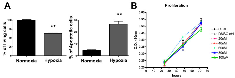
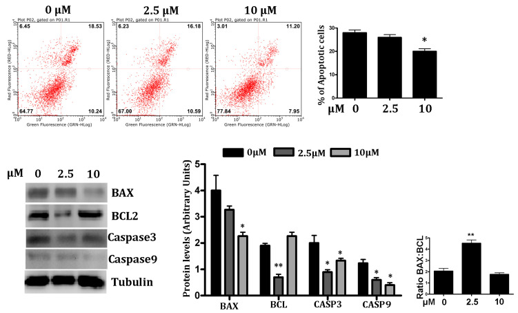
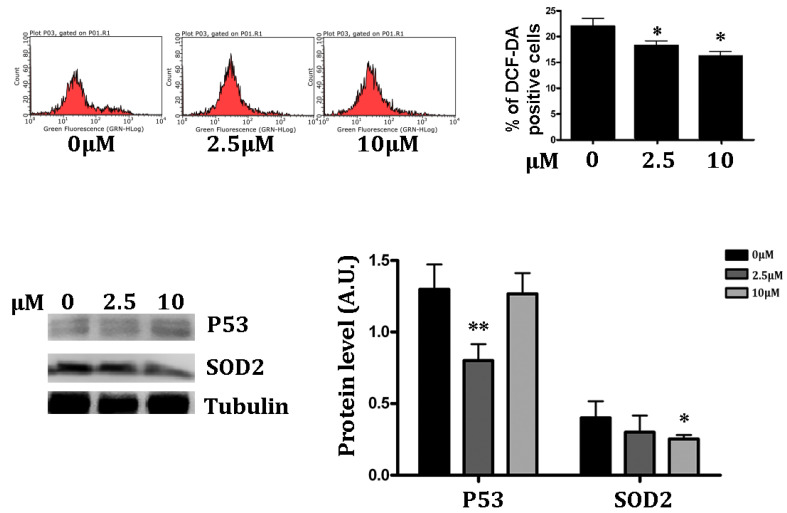
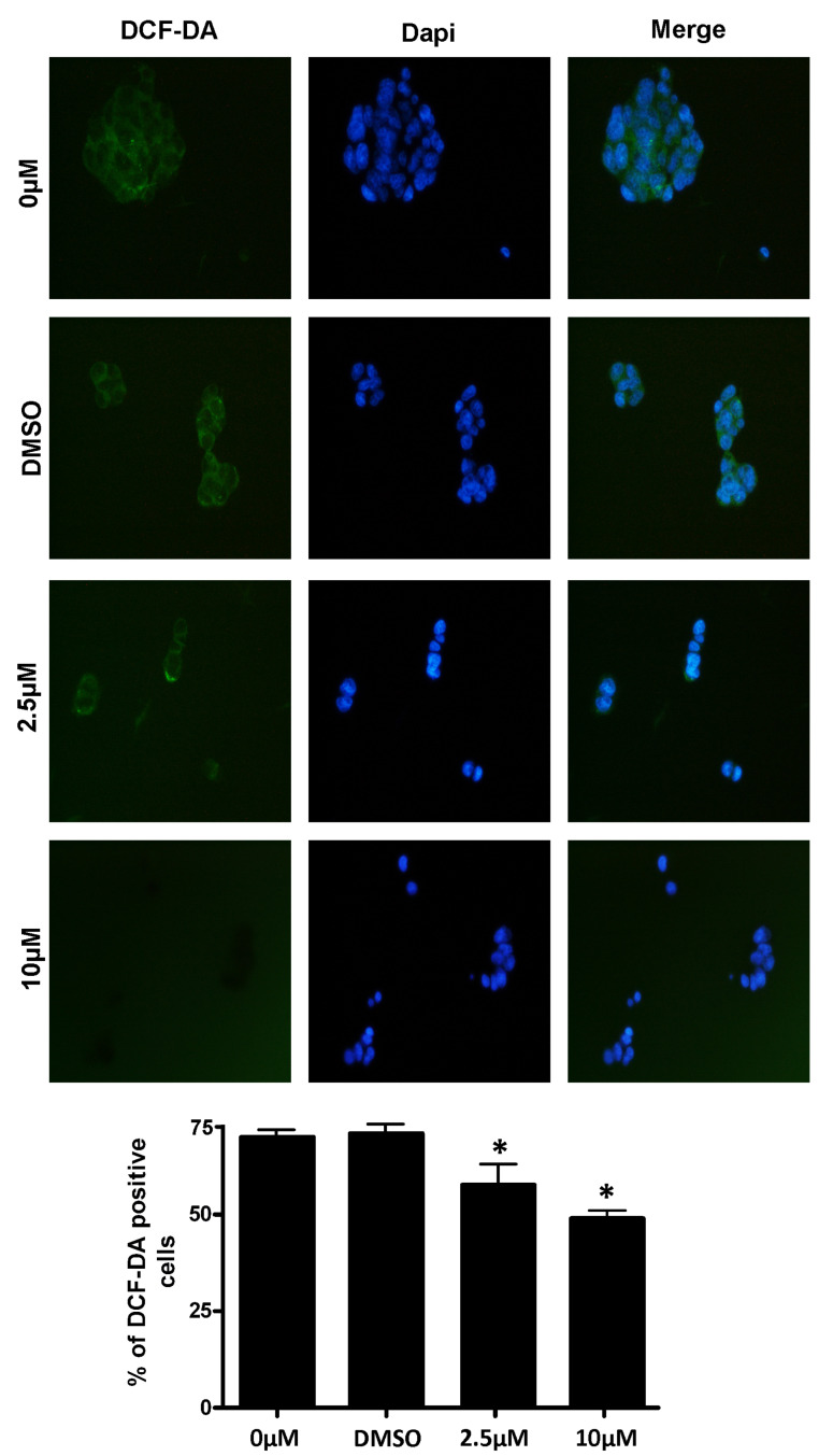
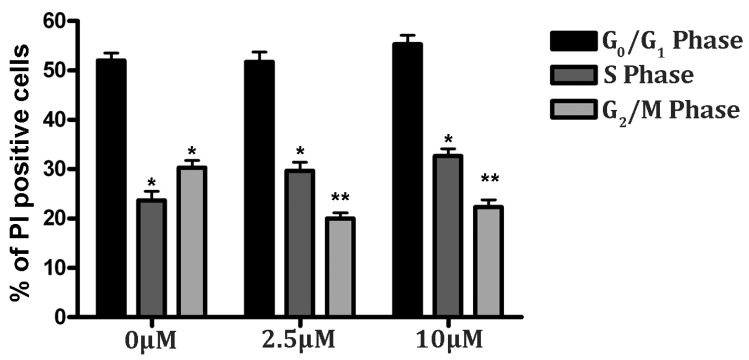
Oxidative stress plays an important role in brain aging and in neurodegenerative diseases. New therapeutic agents are necessary to cross the blood-brain barrier and target disease pathogenesis without causing disagreeable side effects. Resveratrol (RSV) may act as a neuroprotective compound, but little is known about its potential in improving the cognitive and metabolic aspects that are associated with neurodegenerative diseases. The objective of this study was to investigate the protective effects and the underlying mechanisms of RSV against hypoxia-induced oxidative stress in neuronal PC12 cells. For the induction of the hypoxia model, the cells were exposed to oxygen-deprived gas in a hypoxic chamber. Cell cycle and apoptosis were analyzed by a fluorescence activated cell sorting (FACS) analysis. The intracellular reactive oxygen species (ROS) level was analyzed by using dichlorodihydrofluorescein diacetate (DCFDA) and 5-(and-6)-chloromethyl-2',7'-dichlorodihydrofluorescein diacetate, acetyl ester (CM-H2DCFDA) tests. The expression of activated caspase-3, -9, Bcl-2, Bax, p53, and SOD was investigated by a Western blot analysis. We found that hypoxia reduced PC12 viability by inducing apoptosis, while RSV treatment attenuated the ROS-induced damage by reducing caspase-3, -9, and the Bax/Bcl-2 ratio. The RSV treated groups were found to improve cellular health, with a 7.41% increase in the S phase population in the 10 µM group, compared to the control. Hence, RSV has a protective effect in neuronal cells and may halt the cell cycle in the G1/S phase to repair the intracellular damage. Therefore, RSV could be a good candidate to act as an antioxidant and promising preventive therapeutic agent in neurodegenerative diseases for personalized medicine.

Keywords: PC12 cells; hypoxia; ischemia; oxidative stress; personalized medicine; resveratrol; translational research.


Conflict of interest statement

The authors declare no conflict of interest.

Figures

Figure 1
Figure 1
Hypoxic treatment induces cell death in PC12 cells. PC12 cells were seeded in two 6 well plates and subjected to hypoxia in an airtight chamber supplied with hypoxic gases for 6 h at 37 °C. The other plate was maintained at standard culture conditions (normoxia). Post-hypoxic treatment: both the culture plates were maintained in fresh medium at standard culture conditions and harvested after for 24 h. (A) Apoptotic cells were detected using a fluorescein conjugated with Annexin V kit. The bar graph represents quantification in percentage values of live cells and the apoptotic cell death comparison between normoxic vs. hypoxia-treated cells. (B) The cytotoxic effect of RSV was analyzed by CCK-8 assay. Data expressed as mean ± SD (** p < 0.01) of triplicate experiments.
Figure 2
Figure 2
Effect of RSV on hypoxia-induced apoptosis in PC12 cells. Upper Panel—Each quadrant of FACS analysis shows proportion of cells (values in %). Lower left quadrant (absence of both markers) indicates viable cells; upper left quadrant (PI positive) indicates cellular necrosis; upper right quadrant (Annexin V positive and PI positive) indicates late-stage apoptosis; lower right quadrant (Annexin V positive) indicates early-stage apoptosis. The bar graph represents the mean % of late apoptotic cells in RSV treated under hypoxic conditions, (n = 3), (* p < 0.05, ** p < 0.01). Lower Panel—Western blot analysis of apoptotic regulator proteins. Quantification of Bcl-2, Bax, both cleaved caspase-3 and caspase 9 expression were presented in bar graphs as the fold-increase, respectively. All protein expression values were neutralized by tubulin expression, which was used as internal control. Ratio of Bax/Bcl-2 is represented in bar graph (Extreme right). Data are represented as means ± SD (n = 3), (* p < 0.05, ** p < 0.01) of triplicate experiments.
Figure 3
Figure 3
RSV treatment increases intracellular antioxidant potential. Upper Panel—RSV-treated and control PC12 cells were harvested after 24 h post hypoxia and then stained with DCF-DA fluorescent dye. The FACS analysis results are represented in % positive cells in the bar graph. Data were represented as means ± SD (n = 3), (* p <0.05, ** p <0.01). Lower Panel—The antioxidant potential of the cell was assessed by a protein expression analysis of SOD2 and p53 by Western blot. The blotted protein bands were quantified and represented in the bar chart, according to RSV treatment. Data were represented as means ± SD, (n = 3), (* p < 0.05, ** p < 0.001).
Figure 4
Figure 4
Measurement of intracellular ROS levels in RSV-treated PC12 cells under hypoxic conditions. Post-hypoxia and RSV treatment: the harvested PC12 cells were stained by CM-H2DCFDA to stain the intracellular ROS. DAPI was used to stain the nucleus. The images were obtained by a fluorescence microscope under low light conditions. The qualitative data of intracellular ROS in all concentration treatments of PC12 cells were captured through two different filters and a merged image was created. The lower panel contains the represented quantitative data, shown in the bar graph. Data were represented as means ± SD (* p < 0.05), of n = 3.
Figure 5
Figure 5
RSV improves cell cycle progression in PC12 cells under hypoxic stress. Cells were treated with RSV for 24 h prior to hypoxic treatment. Cells were cultured in fresh media and harvested post-24 h. Next, cells were fixed in ethanol and stained with PI for a cell cycle analysis by flow cytometry. Results of the cell cycle analysis were represented in percentage values (* p < 0.05, ** p < 0.01). The representative results of three independent experiments are shown in the bar graph. Data are represented as means ± SD of triplicate experiments.

References

    1. Birben E., Sahiner U.M., Sackesen C., Erzurum S., Kalayci O. Oxidative Stress and Antioxidant Defense. World Allergy Organ. J. 2012;5:9–19. doi: 10.1097/WOX.0b013e3182439613. - DOI - PMC - PubMed
    1. Di Domenico M., Pinto F., Quagliuolo L., Contaldo M., Settembre G., Romano A., Coppola M., Ferati K., Bexheti-Ferati A., Sciarra A., et al. The Role of Oxidative Stress and Hormones in Controlling Obesity. Front. Endocrinol. 2019;10:540. doi: 10.3389/fendo.2019.00540. - DOI - PMC - PubMed
    1. Vanacore D., Messina G., Lama S., Bitti G., Ambrosio P., Tenore G., Messina A., Monda V., Zappavigna S., Boccellino M., et al. Effect of restriction vegan diet’s on muscle mass, oxidative status, and myocytes differentiation: A pilot study. J. Cell. Physiol. 2018;233:9345–9353. doi: 10.1002/jcp.26427. - DOI - PubMed
    1. Giudice A., Montella M., Boccellino M., Crispo A., D’Arena G., Bimonte S., Facchini G., Ciliberto G., Botti G., Quagliuolo L., et al. Epigenetic Changes Induced by Green Tea Catechins are Associated with Prostate Cancer. Curr. Mol. Med. 2017;17:405–420. doi: 10.2174/1566524018666171219101937. - DOI - PubMed
    1. Saberi-Karimian M., Katsiki N., Caraglia M., Boccellino M., Majeed M., Sahebkar A. Vascular endothelial growth factor: An important molecular target of curcumin. Crit. Rev. Food Sci. Nutr. 2017;59:299–312. doi: 10.1080/10408398.2017.1366892. - DOI - PubMed