

Bio-Guided Isolation of SARS-CoV-2 Main Protease Inhibitors from Medicinal Plants: In Vitro Assay and Molecular Dynamics
- PMID: 35893619
- PMCID: PMC9332707
- DOI: 10.3390/plants11151914
Bio-Guided Isolation of SARS-CoV-2 Main Protease Inhibitors from Medicinal Plants: In Vitro Assay and Molecular Dynamics
Abstract
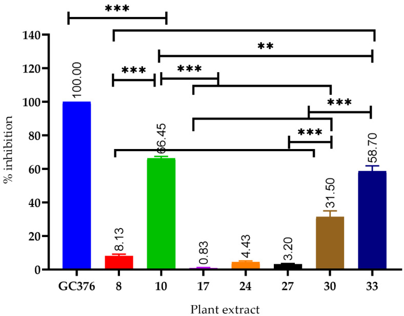
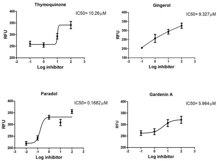
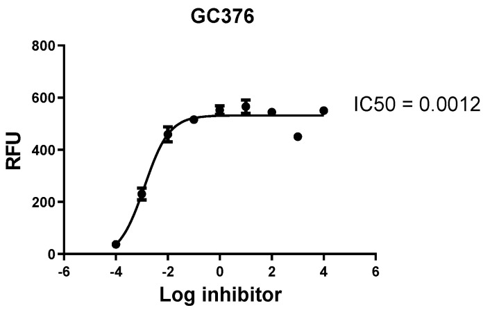
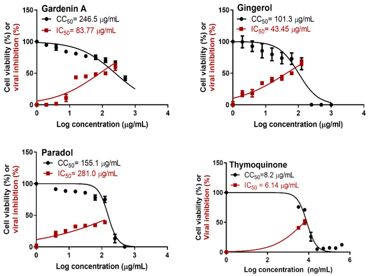
Since the emergence of the pandemic of the coronavirus disease (COVID-19) caused by severe acute respiratory syndrome coronavirus 2 (SARS-CoV-2), the discovery of antiviral phytoconstituents from medicinal plants against SARS-CoV-2 has been comprehensively researched. In this study, thirty-three plants belonging to seventeen different families used traditionally in Saudi Arabia were tested in vitro for their ability to inhibit the SARS-CoV-2 main protease (MPRO). Major constituents of the bio-active extracts were isolated and tested for their inhibition potential against this enzyme; in addition, their antiviral activity against the SARS-CoV-2 Egyptian strain was assessed. Further, the thermodynamic stability of the best active compounds was studied through focused comparative insights for the active metabolites regarding ligand-target binding characteristics at the molecular level. Additionally, the obtained computational findings provided useful directions for future drug optimization and development. The results revealed that Psiadia punctulata, Aframomum melegueta, and Nigella sativa extracts showed a high percentage of inhibition of 66.4, 58.7, and 31.5%, against SARS-CoV-2 MPRO, respectively. The major isolated constituents of these plants were identified as gardenins A and B (from P. punctulata), 6-gingerol and 6-paradol (from A. melegueta), and thymoquinone (from N. sativa). These compounds are the first to be tested invitro against SARS-CoV-2 MPRO. Among the isolated compounds, only thymoquinone (THY), gardenin A (GDA), 6-gingerol (GNG), and 6-paradol (PAD) inhibited the SARS-CoV-2 MPRO enzyme with inhibition percentages of 63.21, 73.80, 65.2, and 71.8%, respectively. In vitro assessment of SARS-CoV-2 (hCoV-19/Egypt/NRC-03/2020 (accession number on GSAID: EPI_ISL_430820) revealed a strong-to-low antiviral activity of the isolated compounds. THY showed relatively high cytotoxicity and was anti-SARS-CoV-2, while PAD demonstrated a cytotoxic effect on the tested VERO cells with a selectivity index of CC50/IC50 = 1.33 and CC50/IC50 = 0.6, respectively. Moreover, GNG had moderate activity at non-cytotoxic concentrations in vitro with a selectivity index of CC50/IC50 = 101.3/43.45 = 2.3. Meanwhile, GDA showed weak activity with a selectivity index of CC50/IC50 = 246.5/83.77 = 2.9. The thermodynamic stability of top-active compounds revealed preferential stability and SARS-CoV-2 MPRO binding affinity for PAD through molecular-docking-coupled molecular dynamics simulation. The obtained results suggest the treating potential of these plants and/or their active metabolites for COVID-19. However, further in-vivo and clinical investigations are required to establish the potential preventive and treatment effectiveness of these plants and/or their bio-active compounds in COVID-19.
Keywords: 6-gingerol; 6-paradol; SARS-CoV-2 Egyptian strain; SARS-CoV-2 main protease; coronavirus; gardenin A; thymoquinone.
Conflict of interest statement
The authors declare no conflict of interest.
Figures
Similar articles
-
Exploring the therapeutic potential of Thai medicinal plants: in vitro screening and in silico docking of phytoconstituents for novel anti-SARS-CoV-2 agents.BMC Complement Med Ther. 2024 Jul 19;24(1):274. doi: 10.1186/s12906-024-04586-z. BMC Complement Med Ther. 2024. PMID: 39030504 Free PMC article.
-
Targeting COVID-19 (SARS-CoV-2) main protease through active phytochemicals of ayurvedic medicinal plants - Withania somnifera (Ashwagandha), Tinospora cordifolia (Giloy) and Ocimum sanctum (Tulsi) - a molecular docking study.J Biomol Struct Dyn. 2022 Jan;40(1):190-203. doi: 10.1080/07391102.2020.1810778. Epub 2020 Aug 27. J Biomol Struct Dyn. 2022. PMID: 32851919 Free PMC article.
-
In Silico and In Vitro Identification of Pan-Coronaviral Main Protease Inhibitors from a Large Natural Product Library.Pharmaceuticals (Basel). 2022 Mar 3;15(3):308. doi: 10.3390/ph15030308. Pharmaceuticals (Basel). 2022. PMID: 35337106 Free PMC article.
-
Mechanistic Aspects of Medicinal Plants and Secondary Metabolites against Severe Acute Respiratory Syndrome Coronavirus 2 (SARS-CoV-2).Curr Pharm Des. 2021;27(38):3996-4007. doi: 10.2174/1381612827666210705160130. Curr Pharm Des. 2021. PMID: 34225607 Review.
-
Nigella sativa and its chemical constituents: pre-clinical and clinical evidence for their potential anti-SARS-CoV-2 effects.Inflammopharmacology. 2024 Feb;32(1):273-285. doi: 10.1007/s10787-023-01385-9. Epub 2023 Nov 15. Inflammopharmacology. 2024. PMID: 37966624 Review.
Cited by
-
Assessing the antibacterial potential of 6-gingerol: Combined experimental and computational approaches.Saudi Pharm J. 2024 May;32(5):102041. doi: 10.1016/j.jsps.2024.102041. Epub 2024 Mar 18. Saudi Pharm J. 2024. PMID: 38558886 Free PMC article.
-
Potential Therapeutic Target and Vaccines for SARS-CoV-2.Pathogens. 2023 Jul 10;12(7):926. doi: 10.3390/pathogens12070926. Pathogens. 2023. PMID: 37513773 Free PMC article.
-
Food Plant Secondary Metabolites Antiviral Activity and Their Possible Roles in SARS-CoV-2 Treatment: An Overview.Molecules. 2023 Mar 8;28(6):2470. doi: 10.3390/molecules28062470. Molecules. 2023. PMID: 36985442 Free PMC article. Review.
-
The effect of Ni gella sativa and vitamin D3 supplementation on the clinical outcome in COVID-19 patients: A randomized controlled clinical trial.Front Pharmacol. 2022 Nov 8;13:1011522. doi: 10.3389/fphar.2022.1011522. eCollection 2022. Front Pharmacol. 2022. PMID: 36425571 Free PMC article.
-
Predicting the Anti-SARS-CoV-2 Potential of Isoquinoline Alkaloids from Brazilian Siparunaceae Species Using Chemometric Tools.Int J Mol Sci. 2025 Jan 13;26(2):633. doi: 10.3390/ijms26020633. Int J Mol Sci. 2025. PMID: 39859347 Free PMC article.
References
Grants and funding
LinkOut - more resources
Full Text Sources
Miscellaneous
