. 2022 Jul 23;14(8):1610.
doi: 10.3390/v14081610.

TRIM7 Restricts Coxsackievirus and Norovirus Infection by Detecting the C-Terminal Glutamine Generated by 3C Protease Processing

Affiliations

TRIM7 Restricts Coxsackievirus and Norovirus Infection by Detecting the C-Terminal Glutamine Generated by 3C Protease Processing

Jakub Luptak et al. Viruses. .

Abstract

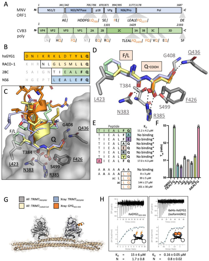
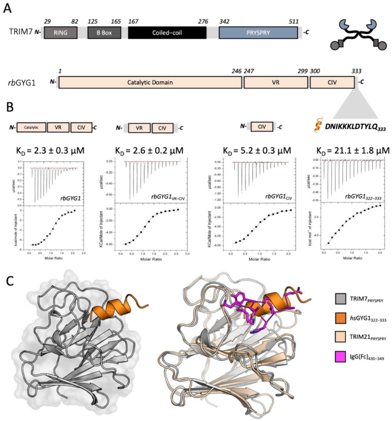
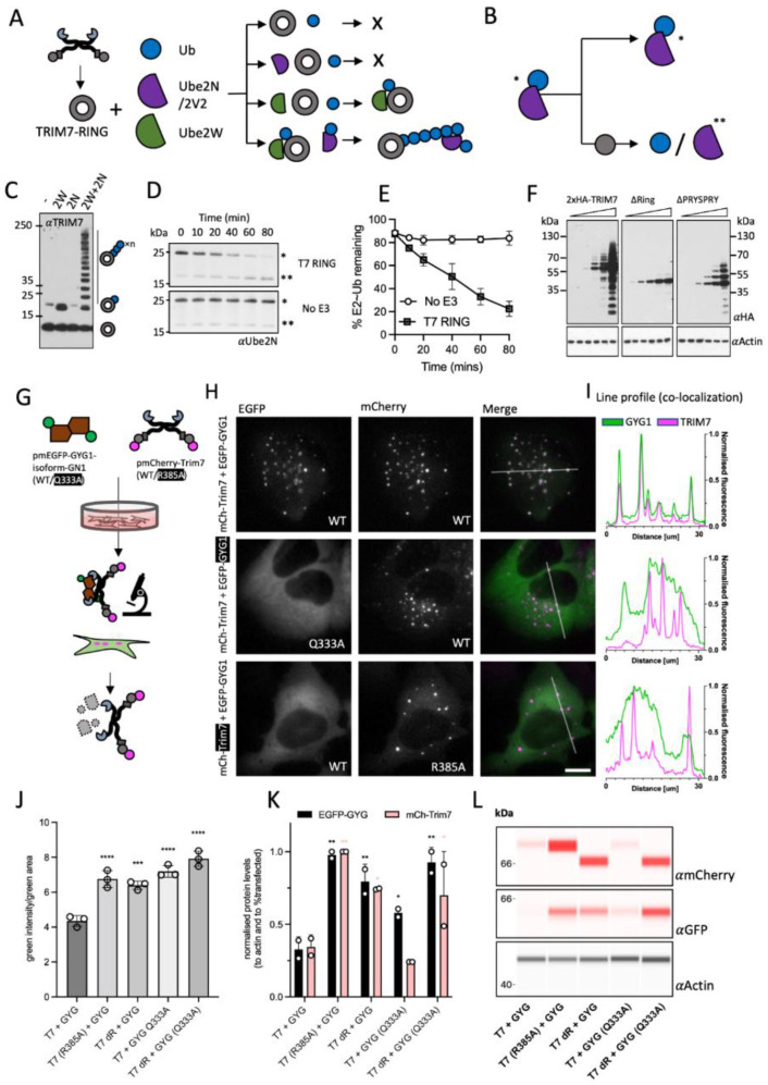
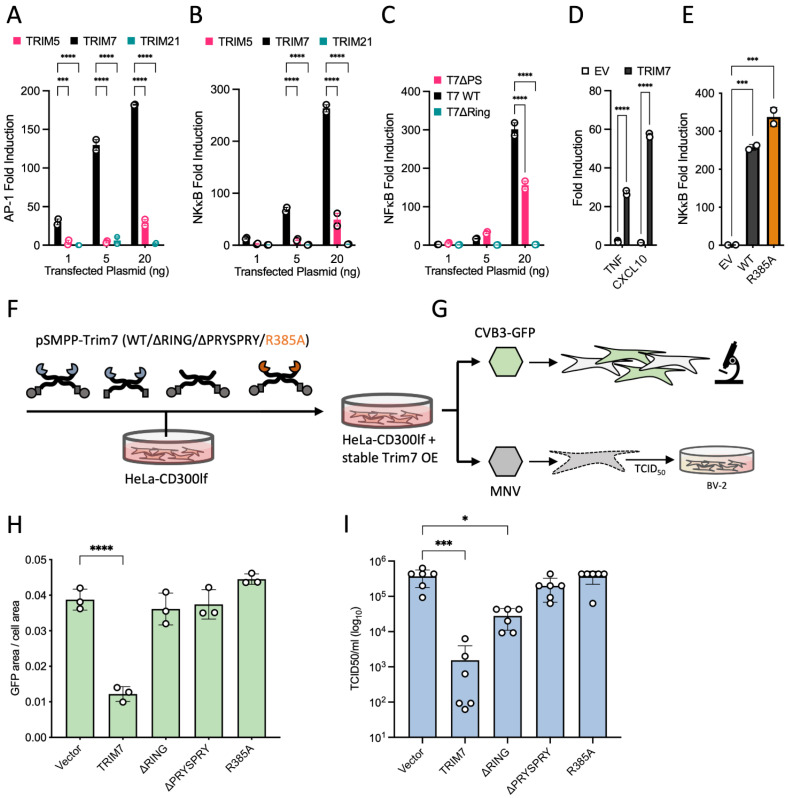
TRIM7 catalyzes the ubiquitination of multiple substrates with unrelated biological functions. This cross-reactivity is at odds with the specificity usually displayed by enzymes, including ubiquitin ligases. Here we show that TRIM7's extreme substrate promiscuity is due to a highly unusual binding mechanism, in which the PRYSPRY domain captures any ligand with a C-terminal helix that terminates in a hydrophobic residue followed by a glutamine. Many of the non-structural proteins found in RNA viruses contain C-terminal glutamines as a result of polyprotein cleavage by 3C protease. This viral processing strategy generates novel substrates for TRIM7 and explains its ability to inhibit Coxsackie virus and norovirus replication. In addition to viral proteins, cellular proteins such as glycogenin have evolved C-termini that make them a TRIM7 substrate. The 'helix-ΦQ' degron motif recognized by TRIM7 is reminiscent of the N-end degron system and is found in ~1% of cellular proteins. These features, together with TRIM7's restricted tissue expression and lack of immune regulation, suggest that viral restriction may not be its physiological function.

Keywords: 3C protease; 3Cpro; ISG; Mpro; Norovirus; SARS-CoV-2; TRIM7; coxsackievirus; degradation; restriction.


Conflict of interest statement

The authors declare that they have no competing interests.

Figures

Figure 2
Figure 2
TRIM7 binds diverse substrates using a C-terminal helix-ΦQ motif. (A) MNV1 polyprotein (pale blue) and the relevant proteins in a darker shade (NS3 and NS6), CVB3 polyprotein in pale green and the relevant 2C protein in a darker shade. (B) Main peptide sequences used in the binding and structural experiments. Outlined are the sequences synthesized, whilst the shaded residues are those resolved in the crystal structures. Color coding is maintained throughout the figure. (C) hisTRIM7-PRYSPRY:Peptide complex structures superposed. Shows PRYSPRY as a transparent surface representation with a few key residues in stick. Peptides are color coded as described. PDB accession codes are 7OW2, 7OVX, 8A5L and 8A5M). (D) Detail of the PRYSPRY pocket with the recognition motif bound. Several key residues are highlighted. Dashes indicate charged or H-bonding between the peptide and the PRYSPRY residues. Underlined residues are essential for binding (see Figure S3). (E) Peptide substitutions based on the TIEALFQ peptide and possible polyprotein processing ends. Binding experiments with the minimal LQ motif, sequence is shown and the derived KD from ITC titrations. No binding* denotes where the peptide was only tested using nanoDSF. (F) Thermal denaturation data of hTRIM7-PRYSPRY derived using the Prometheus nanoDSF. Shows the Tm of the protein in the presence of peptide or DMSO. Peptides which bind stabilize the protein. The results of two independent measurements are shown. (G,H) Avidity enhancement of binding affinity with hisMBP-TRIM7-CC-PRYSPRY. (G) Shows the AlphaFold prediction of the TRIM7 dimer (grey and wheat) aligned with the crystal structure (pale blue and orange). (H) The full-length GYG1 protein (isoform GN1) shows clear 1:1 binding between the protein dimers, whilst no difference in affinity is observed when the peptide is the substrate. Representative traces and their accompanying fitted KD’s are shown.
Figure 1
Figure 1
A linear epitope of GYG1 is sufficient to explain TRIM7 binding. (A) Sequence and domain organization of TRIM7 (grey) and Glycogenin1 (pale orange). TRIM7 is also depicted as a cartoon (circle—RING, rectangle—B-Box, line—Coiled-coil, cut-out circle—PRYSPRY) used throughout this paper. Highlighted are sequences determining TRIM7 binding. (B) ITC binding experiments with sequential truncations of rbGYG1 (shown above each titration) titrated into human hisTRIM7-PRYSPRY. Representative traces and their accompanying fitted KD’s are shown. (C) Cartoon overview of the crystal structure of hisTRIM7-PRYSPRY (grey) with the GYG1 peptide (orange) on left (PDB accession 7OVX). Comparison with TRIM21:Fc structure (2IWG, PRYSPRY wheat, Fc magenta) on the right.
Figure 3
Figure 3
TRIM7 co-localizes with helix-ΦQ containing substrates inside cells and degrades them. (AE) Schematic representation of in vitro ubiquitination experiments. (A) TRIM7-RING (grey circle) was mixed with combinations of Ubiquitin (Ub, blue circle), Ube2N/Ube2V2 (purple semi-circle) and Ube2W (green teardrop) as shown and the reactions followed by immunoblotting (C) with an anti-TRIM7 antibody. A representative blot from at least three independent experiments is shown. (B) E2 discharge experiment: Ube2N~Ub complex was incubated with TRIM7-RING, and the reaction followed over time as indicated in (D). Blots were probed with anti-ubiquitin antibody. A single asterisk denotes the charged Ube2N~Ub, whereas a double asterisk denotes the uncharged Ube2N. A representative blot from at least three independent experiments is shown. (E) Densitometry quantification of band intensities from 3D was plotted to show the kinetics of E2-Ub discharge. Error bars in all graphs depict the mean +/− SEM. Data represent three independent replicates. (F) Western blots from cells overexpressing indicated constructs of epitope-tagged TRIM7 (probed with anti-HA antibody). Ubiquitin-laddering is lost when the RING domain is deleted. A representative blot from two independent experiments is shown. (G) Schematic overview of the experiments presented in (H). EGFP-GYG1 is represented as a green-brown shape. TRIM7 is shown as usual but with N-terminal mCherry (magenta circle). Plasmids were co-expressed, and the fluorescence monitored using live imaging and the protein levels quantified using either the fluorescence intensity or using cell lysates. (H) Live-cell microscopy of U2OS cells expressing mCherry-TRIM7 and EGFP-GYG1 constructs. Left column shows the EGFP signal (green), middle column shows the mCherry signal (magenta) and the right column shows the false-colored merged image (EGFP—green; mCherry—magenta; merge—white). The scale is the same in all images, and the bar represents 10 µm. Rows represent different conditions: Top is both WT sequences. Middle has a Q333A mutation in the EGFP-GYG1 construct. Bottom has the R385A mutation in the mCh-TRIM7 con-struct. Example images are shown from at least two independent experiments. (I) Line profile analysis (ImageJ) of the fluorescent signal. Green trace shows the EGFP signal whilst the magenta trace shows the mCherry signal. The line used in the analysis is shown on the merged signal images. (J) Fluorescence-based quantification (using the IncuCyte) of EGFP-GYG1 protein levels, graph shows the total integrated intensity of the EGFP signal divided by the EGFP area from three biological replicates. ANOVA was used for statistical analysis and significant differences from T7 + GYG condition indicated (p < 0.0005 (***), p < 0.0001 (****)). (K,L) Protein quantification and blots using cell lysates and capillary-based Western Blot (Jess). Values from two biological replicates were normalized to loading control (actin) and the fraction of cells transfected (see methods). ANOVA was used for statistical analysis and significant differences from T7 + GYG condition indicated (p < 0.05 (*), p < 0.005 (**)). Black asterisks indicate significance in EGFP-GYG expression and pink asterisks in mCh-TRIM7 expression.
Figure 4
Figure 4
TRIM7 restriction of MNV1 and CVB3 infection is driven by helix-ΦQ binding. (AC) Co-transfection of TRIM constructs and reporter plasmids. Overexpression of TRIM7 induces strong signaling by AP-1 and NF-κB that is dependent on the RING domain. (D) Overexpression of TRIM7 induces signaling, as measured by qPCR; values are normalized to actin. (E) Point mutation that prevents binding to targets does not impact signaling. For all signaling experiments (AE), representative examples of at least two independent experiments are shown, with values normalized to cells transfected with empty vector. ANOVA was used for statistical analysis and significant differences indicated (p < 0.0005 (***), p < 0.0001 (****)). (F,G) Schematic overview of the experiments shown in H and I. Lentivirus generation and stable transfection of HeLa-CD300lf cells (F), with the expression confirmed by Western blotting (Figure S6). Cells were infected with recombinant CVB3 virus-producing EGFP in host cells or MNV1 (G). (H) Quantification of CVB3 infection by measuring the fraction of EGFP-expressing cells. Data shown are from three independent experiments. (I) Quantification of MNV1 replication by TCID50 following infection of TRIM7-expressing cells. Virions from lysed cells were titrated onto susceptible BV-2 cells. Data shown are a representative result of three independent experiments. Error bars show the standard deviation. Non-parametric ANOVA was used for statistical analysis and significant differences indicated (p < 0.05 (*), p < 0.0005 (***), p < 0.0001 (****)).

Similar articles

Cited by

References

    1. Koepke L., Gack M.U., Sparrer K.M. The antiviral activities of TRIM proteins. Curr. Opin. Microbiol. 2021;59:50–57. doi: 10.1016/j.mib.2020.07.005. - DOI - PMC - PubMed
    1. Bharaj P., Atkins C., Luthra P., Giraldo M.I., Dawes B.E., Miorin L., Johnson J.R., Krogan N.J., Basler C.F., Freiberg A.N., et al. The host E3-ubiquitin ligase TRIM6 ubiquitinates the Ebola virus VP35 protein and promotes virus replication. J. Virol. 2017;91:e00833-17. doi: 10.1128/JVI.00833-17. - DOI - PMC - PubMed
    1. Giraldo M.I., Xia H., Aguilera-Aguirre L., Hage A., van Tol S., Shan C., Xie X., Sturdevant G.L., Robertson S.J., McNally K.L., et al. Envelope protein ubiquitination drives entry and pathogenesis of Zika virus. Nature. 2020;585:414–419. doi: 10.1038/s41586-020-2457-8. - DOI - PMC - PubMed
    1. Wu X., Wang J., Wang S., Wu F., Chen Z., Li C., Cheng G., Qin F.X. Inhibition of Influenza A virus replication by TRIM14 via its multifaceted protein-protein interaction with NP. Front. Microbiol. 2019;10:344. doi: 10.3389/fmicb.2019.00344. - DOI - PMC - PubMed
    1. Stremlau M., Owens C.M., Perron M.J., Kiessling M., Autissier P., Sodroski J. The cytoplasmic body component TRIM5α restricts HIV-1 infection in Old World monkeys. Nature. 2004;427:848–853. doi: 10.1038/nature02343. - DOI - PubMed

Publication types

LinkOut - more resources