. 2022 Jul 24;14(8):1540.
doi: 10.3390/pharmaceutics14081540.

Preparation of PLGA Nanoparticles by Milling Spongelike PLGA Microspheres

Affiliations

Preparation of PLGA Nanoparticles by Milling Spongelike PLGA Microspheres

Jimin Lee et al. Pharmaceutics. .

Abstract

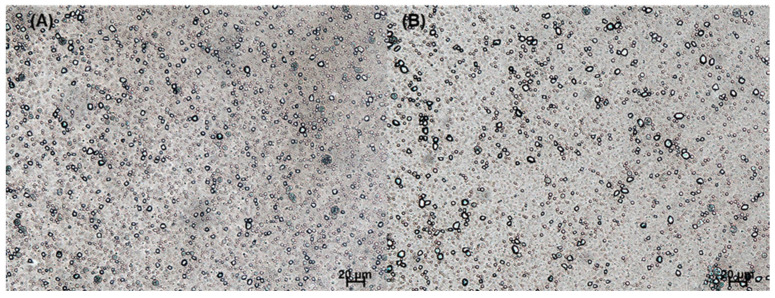
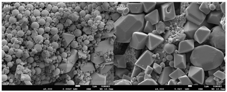
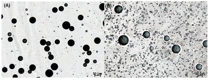
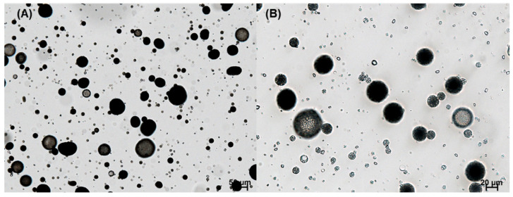
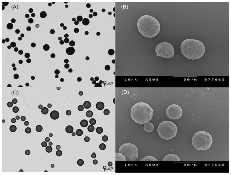
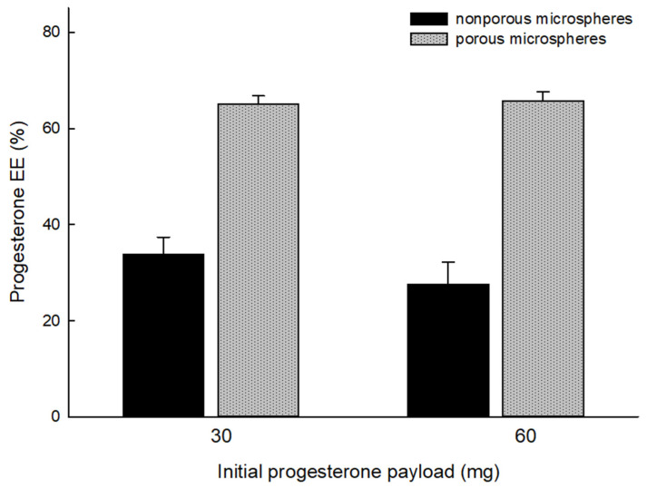
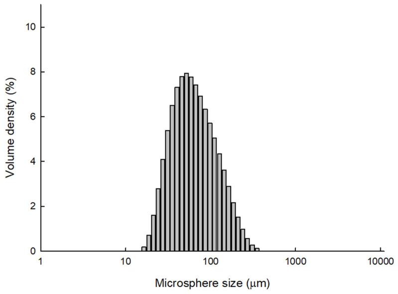
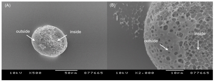
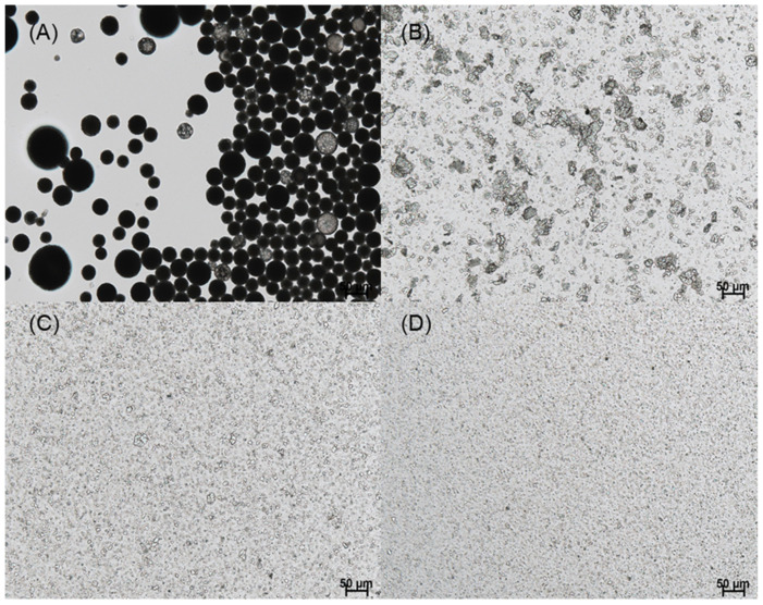
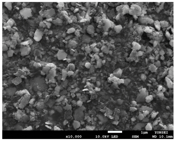
Currently, emulsification-templated nanoencapsulation techniques (e.g., nanoprecipitation) have been most frequently used to prepare poly-d,l-lactide-co-glycolide (PLGA) nanoparticles. This study aimed to explore a new top-down process to produce PLGA nanoparticles. The fundamental strategy was to prepare spongelike PLGA microspheres with a highly porous texture and then crush them into submicron-sized particles via wet milling. Therefore, an ethyl formate-based ammonolysis method was developed to encapsulate progesterone into porous PLGA microspheres. Compared to a conventional solvent evaporation process, the ammonolysis technique helped reduce the tendency of drug crystallization and improved drug encapsulation efficiency accordingly (solvent evaporation, 27.6 ± 4.6%; ammonolysis, 65.1 ± 1.7%). Wet milling was performed on the highly porous microspheres with a D50 of 64.8 μm under various milling conditions. The size of the grinding medium was the most crucial factor for our wet milling. Milling using smaller zirconium oxide beads (0.3~1 mm) was simply ineffective. However, when larger beads with diameters of 3 and 5 mm were used, our porous microspheres were ground into submicron-sized particles. The quality of the resultant PLGA nanoparticles was demonstrated by size distribution measurement and field emission scanning electron microscopy. The present top-down process that contrasts with conventional bottom-up approaches might find application in manufacturing drug-loaded PLGA nanoparticles.

Keywords: microspheres; nanoparticles; poly-d,l-lactide-co-glycolide; wet milling.


Conflict of interest statement

The authors declare that they have no conflict of interest.

Figures

Figure 1
Figure 1
Light microscopy (LM) photographs showing the appearance of non-spherical progesterone crystals formed in the nanoprecipitation process. After injecting a polymeric dispersed phase into an aqueous phase, the status of the resultant mixture was observed at (A) 5 min and (B) 4 h. The bar size is 20 μm.
Figure 2
Figure 2
SEM images of particles collected by ultracentrifugation. (A) PLGA particles are contaminated with non-spherical progesterone crystals of various sizes. (B) is a close-up micrograph showing progesterone crystals. The bar size is 1 μm. The initial progesterone payload used for nanoencapsulation was 30 mg.
Figure 3
Figure 3
LM photographs of the emulsions that were obtained by emulsifying a dispersed phase (PLGA 0.3 g, progesterone 30 mg, ethyl formate 6 mL) in an aqueous phase. The emulsion was stirred for (A) 10 min and (B) 2 h before LM observation. The bar sizes in (A,B) are 50 and 20 μm, respectively.
Figure 4
Figure 4
LM photographs of a microsphere suspension that was obtained by the ammonolysis-based microencapsulation process. The initial progesterone payload was 30 mg. Just before wet sieving, the microsphere suspension was sampled for LM observation. The bar sizes in (A,B) are 50 and 20 μm, respectively.
Figure 5
Figure 5
LM photographs of the microsphere suspensions that were prepared by (A) ammonolysis and (C) solvent evaporation. (B) SEM image of the dried microspheres prepared by ammonolysis, and (D) produced by solvent evaporation. The bar size in all micrographs is 50 μm.
Figure 6
Figure 6
Effect of microencapsulation methods (ammonolysis vs. solvent evaporation) on progesterone EE%. The initial progesterone payload used for microencapsulation varied from 30 to 60 mg, while the PLGA amount was set at 0.3 g.
Figure 7
Figure 7
The size distribution of PLGA microspheres that were produced by the ammonolysis-based microencapsulation process. PLGA (0.3 g) and progesterone (30 mg) were used for the microencapsulation process.
Figure 8
Figure 8
SEM images of the surface and internal morphology of PLGA microspheres prepared by ammonolysis. The initial progesterone payload was 30 mg. The bar sizes in (A,B) micrographs are 50 and 10 μm, respectively. A tape was used to peel off a portion of the microsphere surface.
Figure 9
Figure 9
LM micrographs show dynamic changes in the size of spongelike PLGA microspheres as a function of milling time. Beads of 5 mm diameter were used as a grinding medium for (A) 0, (B) 1, (C) 5, and (D) 10 h. The bar size is 50 μm.
Figure 10
Figure 10
(A) The size distribution of PLGA nanoparticles that were prepared by milling spongelike PLGA microspheres. (B) Measurements of the Z-average size and PDI of the PLGA nanosuspension while stored at room temperature for a week.
Figure 11
Figure 11
An FE-SEM image of PLGA nanoparticles that were produced by milling pre-formed spongelike PLGA microspheres. The bar size is 1 μm.

References

    1. Danhier F., Ansorena E., Silva J.M., Coco R., Le Breton A., Préat V. PLGA-based nanoparticles: An overview of biomedical applications. J. Control. Release. 2012;161:505–522. doi: 10.1016/j.jconrel.2012.01.043. - DOI - PubMed
    1. Rocha C.V., Gonçalves V., da Silva M.C., Bañobre-López M., Gallo J. PLGA-based composites for various biomedical applications. Int. J. Mol. Sci. 2022;23:2034. doi: 10.3390/ijms23042034. - DOI - PMC - PubMed
    1. Muddineti O.S., Omri A. Current trends in PLGA based long-acting injectable products: The industry perspective. Expert Opin. Drug Deliv. 2022;19:559–576. doi: 10.1080/17425247.2022.2075845. - DOI - PubMed
    1. Operti M.C., Bernhardt A., Grimm S., Engel A., Figdor C.G., Tagit O. PLGA-based nanomedicines manufacturing: Technologies overview and challenges in industrial scale-up. Int. J. Pharm. 2021;605:120807. doi: 10.1016/j.ijpharm.2021.120807. - DOI - PubMed
    1. Ding D., Zhu Q. Recent advances of PLGA micro/nanoparticles for the delivery of biomacromolecular therapeutics. Mater. Sci. Eng. C Mater. Biol. Appl. 2018;92:1041–1060. doi: 10.1016/j.msec.2017.12.036. - DOI - PubMed

LinkOut - more resources