. 2022 Jul 25;14(8):1546.
doi: 10.3390/pharmaceutics14081546.

Localized Controlled Release of Kynurenic Acid Encapsulated in Synthetic Polymer Reduces Implant-Induced Dermal Fibrosis

Affiliations

Localized Controlled Release of Kynurenic Acid Encapsulated in Synthetic Polymer Reduces Implant-Induced Dermal Fibrosis

Layla Nabai et al. Pharmaceutics. .

Abstract

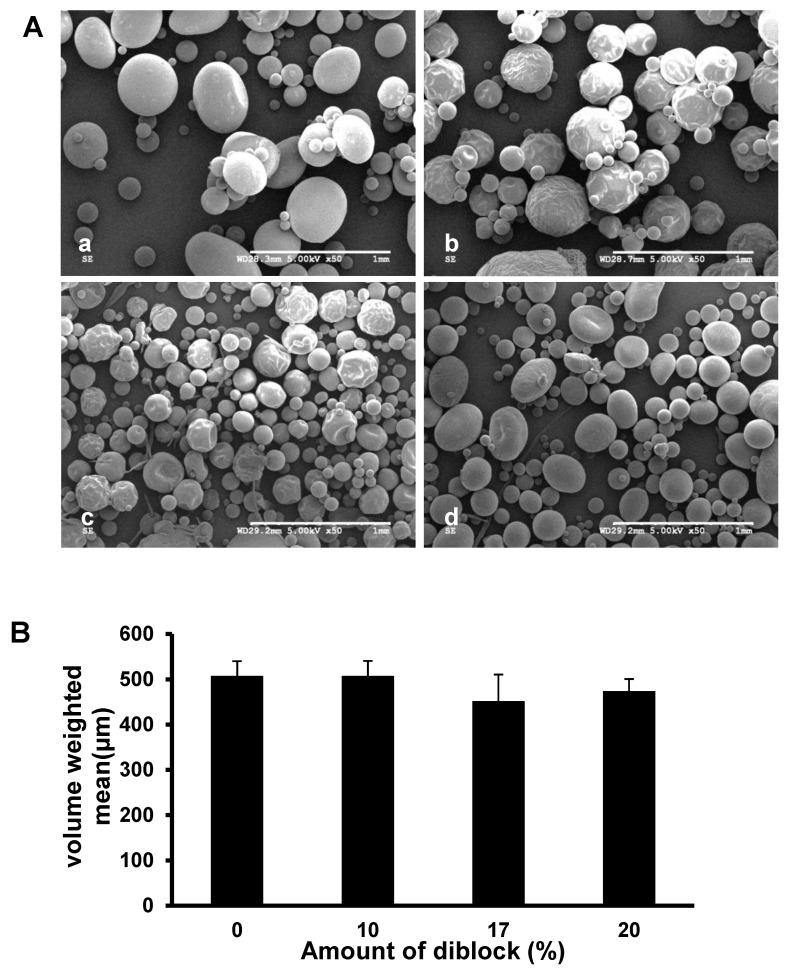
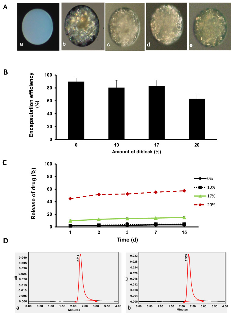
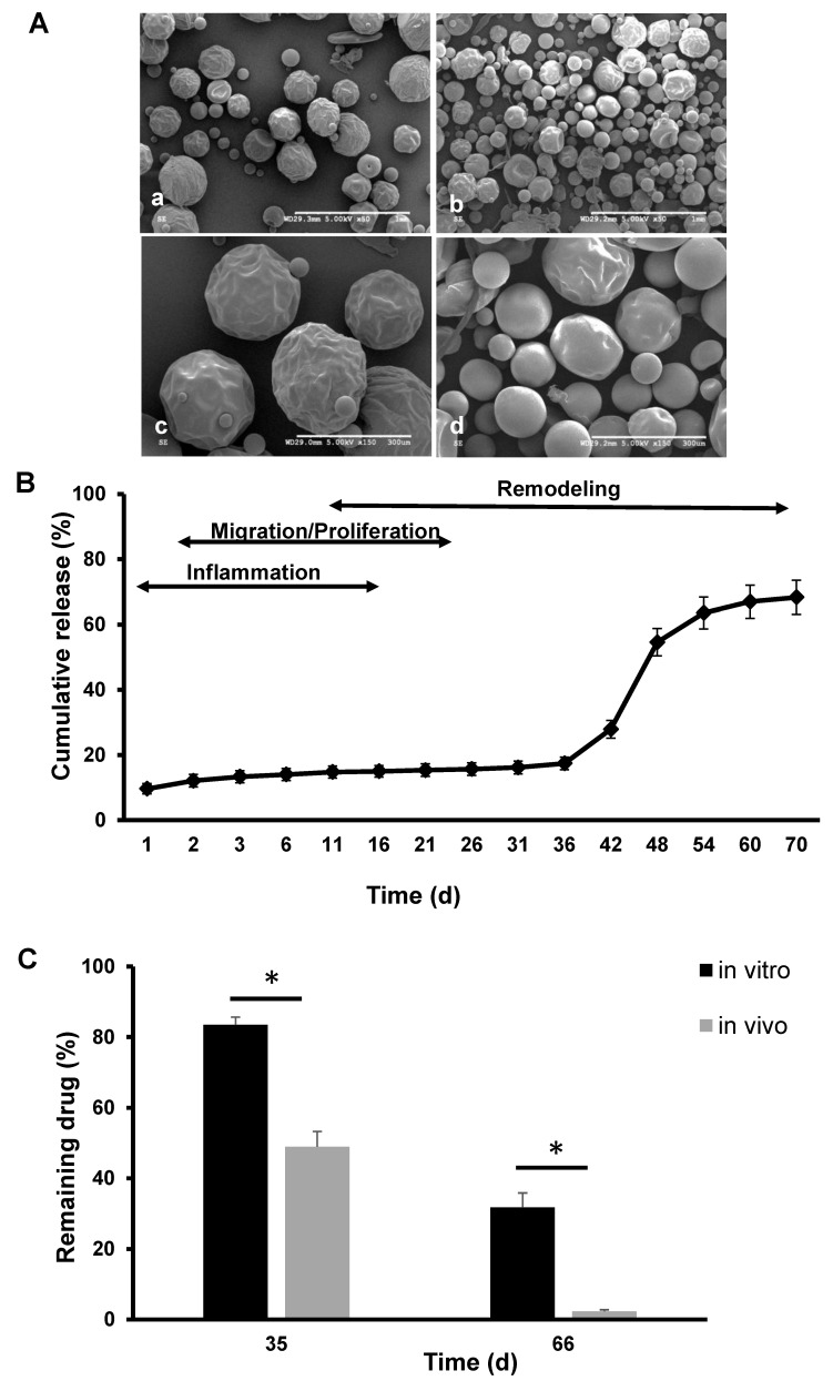
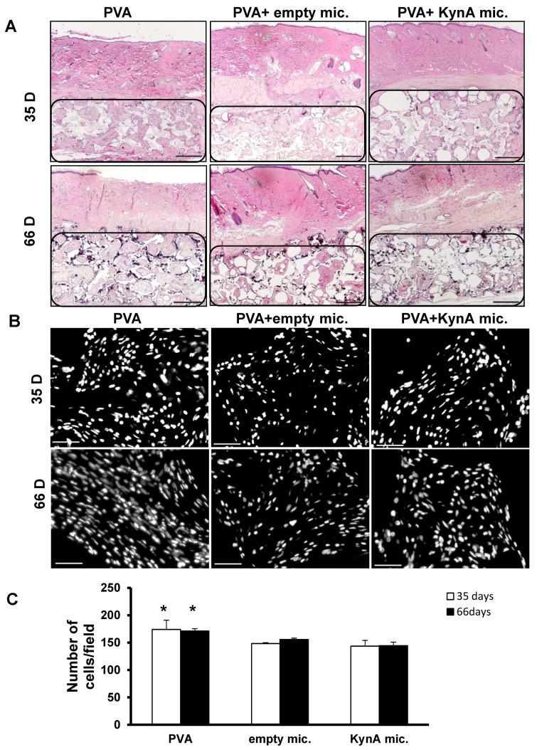
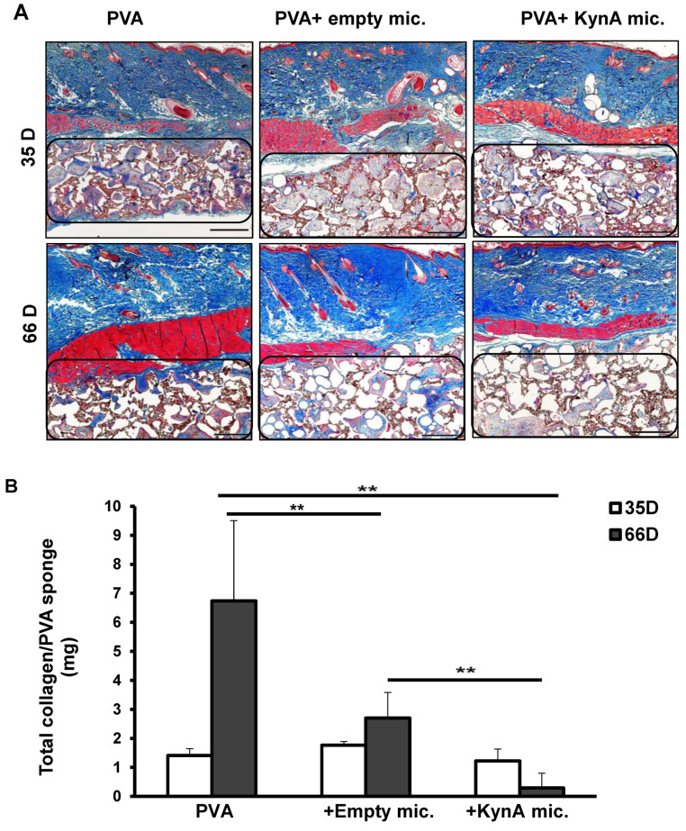
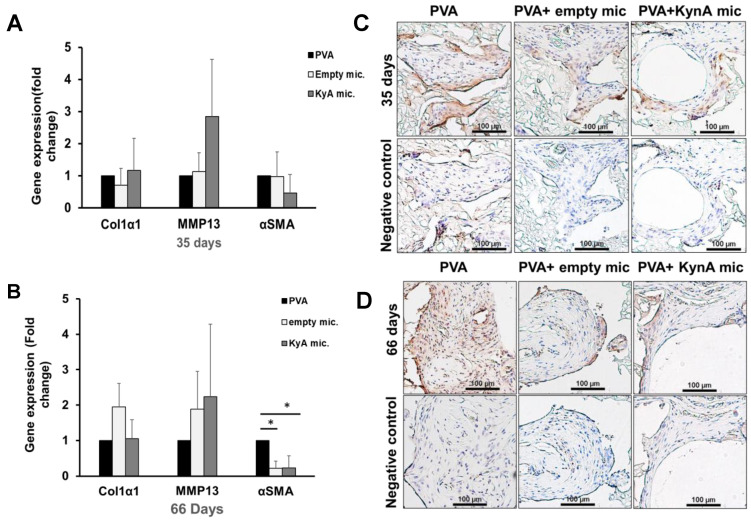
Excessive fibrosis following surgical procedures is a challenging condition with serious consequences and no effective preventive or therapeutic option. Our group has previously shown the anti-fibrotic effect of kynurenic acid (KynA) in vitro and as topical cream formulations or nanofiber dressings in open wounds. Here, we hypothesized that the implantation of a controlled release drug delivery system loaded with KynA in a wound bed can prevent fibrosis in a closed wound. Poly (lactic-co-glycolic acid) (PLGA), and a diblock copolymer, methoxy polyethylene glycol-block-poly (D, L-lactide) (MePEG-b-PDLLA), were used for the fabrication of microspheres which were evaluated for their characteristics, encapsulation efficiency, in vitro release profile, and in vivo efficacy for reduction of fibrosis. The optimized formulation exhibited high encapsulation efficiency (>80%), low initial burst release (~10%), and a delayed, gradual release of KynA. In vivo evaluation of the fabricated microspheres in the PVA model of wound healing revealed that KynA microspheres effectively reduced collagen deposition inside and around PVA sponges and α-smooth muscle actin expression after 66 days. Our results showed that KynA can be efficiently encapsulated in PLGA microspheres and its controlled release in vivo reduces fibrotic tissue formation, suggesting a novel therapeutic option for the prevention or treatment of post-surgical fibrosis.

Keywords: PLGA; fibrosis; kynurenic acid; microsphere.


Conflict of interest statement

Aziz Ghahary holds patent on kynurenic acid.

Figures

Figure 1
Figure 1
Scanning electron microscopy images and size distribution of different formulations of PLGA microspheres. (A) SEM images of KynA loaded PLGA microspheres fabricated without diblock (a) or with 10% (b), 17% (c), and 20% (d) MePEG- diblock. Scale bar: 1 mm. (B) Average size of the KynA loaded PLGA microspheres fabricated without or with 10%, 17%, and 20% MePEG-diblock. Particle size analysis of the microspheres was performed using laser diffraction particle size analyzer on four separate preparations of each formulation and the average of corresponding particle size was expressed as the volume weighted mean. Data represent the mean ± SD for n = 4.
Figure 2
Figure 2
Encapsulation efficiency, in vitro drug release profile from the microspheres, and stability of KynA in the process of encapsulation (A) Polarized light microscopy images of empty (a), KynA loaded PLGA (b), PLGA+ 10% (c), PLGA + 17% (d), PLGA + 20% (e) MePEG-diblock microspheres, showed successful KynA encapsulation with all four formulations. (B) Quantitative analysis of the encapsulation efficiency of KynA loaded PLGA, PLGA + 10%, PLGA + 17%, and PLGA + 20% MePEG-diblock. Data represent the mean ± SD of independent batches (n = 4) for each formulation. (C) In vitro release profile of KynA from PLGA alone, PLGA+ 10%, 17%, and 20% MePEG-diblock microspheres. Cumulative release % was plotted vs. time (day) (mean ± SD, n = 4). (D) HPLC peak shape and retention time (tR) of the freshly prepared, standard solution of KynA (a) with KynA released from fabricated microspheres (b) showed identical results.
Figure 3
Figure 3
Scanning electron microscopy images of polymer only and KynA loaded microspheres, extended-release kinetics in vitro, and residual KynA inside microspheres in vitro and in vivo at different time points. (A) SEM images of empty PLGA + 17% MePEG-diblock (a,c) and KyA loaded PLGA + 17% MePEG-diblock microspheres (b,d). Scale bar: 1 mm (a,b), 300 μm (c,d). (B) Extended-release profile of KynA from PLGA + 17% MePEG-diblock microspheres in vitro up to 70 days, comparative to distinct phases of wound healing (schematic illustration). (C) Quantitative analysis of the residual KynA in microspheres at 35 and 66 days in vivo and in vitro. Data represent the mean ± SD for n = 4, * statistical significance, p < 0.05.
Figure 4
Figure 4
New tissue growth and total tissue cellularity inside PVA sponges of skin samples. (A) Representative sections of skin samples harvested after 35 and 66 days and stained with H&E. Granulation tissue grown inside of the PVA sponges (region marked by drawing) in all three groups: (i) PVA alone, (ii) PVA + empty microspheres, and (iii) PVA + KynA microspheres at ×20 magnification. Scale bar = 1 mm. (B) Tissue cellularity inside PVA sponges in sections of PVA alone, PVA + empty microspheres, and PVA + KynA microspheres at 35 and 66 days, stained with DAPI, at ×200 magnification. Scale bar = 50 μm. (C) Average number of total cells per field at day 35 (open bars) and day 66 (solid bars) in the PVA alone, PVA + empty, and PVA + KynA microspheres. Data represent the mean ± SD for n = 4, * Statistical significance, p < 0.01.
Figure 5
Figure 5
Biological effect of controlled release KynA on collagen deposition inside PVA sponges. (A) Masson’s trichrome staining of skin samples harvested at two time points (35 and 66 days) shows deposition of collagen (blue color) inside the PVA sponges (region marked by drawing) of three groups: (i) PVA alone, (ii) PVA + empty microspheres and (iii) PVA + KynA microspheres at ×20 magnification, scale bar: 1 mm. (B) Quantitative analysis of the collagen inside PVA sponges using hydroxyproline assay. The amount of collagen was measured in whole PVA sponges of three groups (i) PVA alone, (ii) PVA + empty microspheres, and (iii) PVA + KynA microspheres, harvested after 35 (open bars) and 66 days (solid bars). Data represent the mean ± SD, n = 4 for each group, ** statistical significance, p = 0.009.
Figure 6
Figure 6
Expression of ECM components in tissue grown inside PVA sponges. (A,B) The levels of expression of Col1α1, MMP-13, and α-SMA genes in tissue grown inside PVA sponges of three groups: (i) PVA alone, (ii) PVA +empty microspheres, and (iii) PVA + KynA microspheres analyzed by qPCR at day 35 (A) and 66 (B). The results were normalized to β-actin as an internal control. Data represent as fold change relative to PVA alone group (mean ± SD, n = 3, * statistical significance, p = 0.009). (C,D) Immunohistochemical staining of skin samples from PVA alone, PVA + empty microspheres, and PVA + KynA microspheres groups for α-SMA. Cells expressing α-SMA in tissue grown inside PVA sponges at 35 (C) and 66 days (D), scale bar: 100 μm.

Similar articles

Cited by

References

    1. Mahdavian Delavary B., van der Veer W.M., Ferreira J.A., Niessen F.B. Formation of hypertrophic scars: Evolution and susceptibility. J. Plast. Surg. Hand Surg. 2012;46:95–101. doi: 10.3109/2000656X.2012.669184. - DOI - PubMed
    1. Adams W.P.J. Capsular contracture: What is it? What causes it? How can it be prevented and managed? Clin. Plast. Surg. 2009;36:119–126. doi: 10.1016/j.cps.2008.08.007. - DOI - PubMed
    1. Brüggmann D., Tchartchian G., Wallwiener M., Münstedt K., Tinneberg H.-R., Hackethal A. Intra-abdominal adhesions: Definition, origin, significance in surgical practice, and treatment options. Dtsch Arztebl Int. 2010;107:769–775. - PMC - PubMed
    1. Menzies D., Ellis H. Intestinal obstruction from adhesions—How big is the problem? Ann. R. Coll. Surg. Engl. 1990;72:60–63. - PMC - PubMed
    1. Beyene R.T., Kavalukas S.L., Barbul A. Intra-abdominal adhesions: Anatomy, physiology, pathophysiology, and treatment. Curr. Probl. Surg. 2015;52:271–319. doi: 10.1067/j.cpsurg.2015.05.001. - DOI - PubMed

LinkOut - more resources