. 2022 Jul 25;14(15):3042.
doi: 10.3390/nu14153042.

Ginsenoside Rh4 Suppresses Metastasis of Esophageal Cancer and Expression of c-Myc via Targeting the Wnt/β-Catenin Signaling Pathway

Affiliations

Ginsenoside Rh4 Suppresses Metastasis of Esophageal Cancer and Expression of c-Myc via Targeting the Wnt/β-Catenin Signaling Pathway

Jun Chen et al. Nutrients. .

Abstract

The metastasis of esophageal squamous cell carcinoma (ESCC) is a leading cause of death worldwide, however, it has a poor prognosis. Ginsenoside Rh4 is a rare saponin that has been shown to have potential antitumor effectiveness in ESCC. However, the utility of Rh4 in ESCC metastasis and its undiscovered mode of action has not yet been explored. In this study, we found that Rh4 could inhibit ESCC metastasis by regulating the Wnt/β-catenin signaling pathway and the level of c-Myc, which is an important transcription factor in cancer. In in vitro experiments, Rh4 could inhibit the migration and invasion of ESCC cells without affecting cell viability. In in vivo experiments, Rh4 restrained ESCC metastasis to the lymph nodes and lungs via the suppression of epithelial-mesenchymal transition (EMT). The Wnt agonist HLY78 promoted EMT and migration of ESCC cells, whereas treatment of Rh4 can attenuate the promotion effect of HLY78. The siRNA knocking out c-Myc can also significantly reduce the expression of EMT-related marker proteins. This study illustrates a new concept for further research on the mechanism of Rh4 in ESCC.

Keywords: ESCC; Wnt/β-catenin; c-Myc; ginsenoside Rh4; metastasis.


Conflict of interest statement

The authors declare no conflict of interest.

Figures
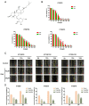
Figure 1
Figure 1
(A)The chemical structure of ginsenoside Rh4. (B) KYSE30, KYSE150, and KYSE410 were measured with designated content (0–140 µM) of Rh4 cell viability (C) Migration images of Rh4 cells with or without the addition of Rh4 cells at 0, 12, and 24 h. The yellow line is the migration edge. Scale bar = 100 µm (D) Percent wound healing, treatment group vs. control group. All data are presented as mean ± SD, * p < 0.05, ** p < 0.01, and *** p < 0.001.
Figure 2
Figure 2
(A) Migration and invasion images of KYSE30, KYSE150, and KYSE410 cells. (B) Counting cells after crystal violet staining (C,D) Protein level of E-cadherin, N-cadherin, Vimentin, snail, MMP2, and MMP9 in KYSE30, KYSE150, and KYSE410 after Rh4 treatment (10 and 20 µM). All data are presented as mean ± SD, * p < 0.05, ** p < 0.01, and *** p < 0.001.
Figure 3
Figure 3
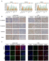
(A) Transcript of E-cadherin, N-cadherin, Vimentin, snail, wnt, β-catenin, and c-Myc after Rh4 treatment (10 and 20 µM). (B) Immunohistochemical images of E-cadherin, N-cadherin, Vimentin, and snail. In lymph nodes and lungs. Scale bar = 100 µm. (C) Immunofluorescence analysis showed that Rh4 promoted E-cadherin in lymph nodes and lungs and inhibited the expression of N-cadherin. Scale bar = 10 µm. All data are presented as mean ± SD, * p < 0.05, ** p < 0.01, *** and p < 0.001.
Figure 4
Figure 4
(A,B) Protein level of E-cadherin, N-cadherin, Vimentin, MMP2, MMP9, Wnt, β-catenin, and c-Myc in the lymph node. (C) Body weight change curve of six groups of mice. (D) Representative image of KYSE30 popliteal lymph nodes of measured groups. (E) Popliteal lymph nodes were measured after sacrifice. All data are presented as mean ± SD, * p < 0.05, ** p < 0.01, and *** p < 0.001.
Figure 5
Figure 5
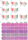
(A) The contents of peripheral blood white blood cells (WBC), lymphocytes (LYM), and granulocytes (GRAN) in each group. Liver and kidney function indexes, consisting of (B) urea, uric acid, and CRE. (C) ALT and AST. (D) Images of major organs in each group, consisting of heart, liver, spleen, lung, and kidney. Scale bar = 100 µm. All data are presented as mean ± SD, * p < 0.05, and ** p < 0.01.
Figure 6
Figure 6
(A,B) Relative protein levels of Wnt, β-catenin, and p-β-catenin after KYSE30, KYSE150, and KYSE410 were treated by Rh4. (C) Immunohistochemical analysis showed that Rh4 attenuated Wnt, β-catenin, and p-β-catenin in lymph nodes and lungs. Scale bar = 100 µm. (D) Immunofluorescence analysis showed that Rh4 also attenuated the level of Wnt and β-catenin in lymph nodes and lungs. Scale bar = 10 µm. All data are presented as mean ± SD, * p < 0.05, and ** p < 0.01.
Figure 7
Figure 7
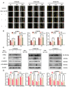
(A) Migration images with or without the addition of Rh4(HLY78) at 0, 12, and 24 h. The yellow line is the migration edge. Scale bar = 100 µm (B) Percent wound healing, treatment group vs. control group. (C) Protein levels after Rh4 and HLY-78 combined treatment of KYSE30, KYSE150, and KYSE410. (D) Bar graphs showing protein expression level. All data are presented as mean ± SD, * p < 0.05 and ** p < 0.01.
Figure 8
Figure 8
(A,B) Addition of c-Myc-siRNA enhanced Rh4 inhibition of E-cadherin, N-cadherin, Vimentin, and snail (C) Rh4 reduces c-Myc expression in lymph nodes and lungs in vivo. Scale bar = 100 µm. All data are presented as mean ± SD, * p < 0.05, ** p < 0.01, *** p < 0.001.
Figure 9
Figure 9
Proposed molecular mechanism of Rh4 anti-metastasis of ESCC.

References

    1. Bray F., Ferlay J., Soerjomataram I., Siegel R.L., Torre L.A., Jemal A. Global cancer statistics 2018: GLOBOCAN estimates of incidence and mortality worldwide for 36 cancers in 185 countries. CA A Cancer J. Clin. 2018;68:394–424. doi: 10.3322/caac.21492. - DOI - PubMed
    1. Siegel R., Miller K.D., Jemal A. Cancer statistics 2016. CA A Cancer J. Clin. 2015;66:7–30. doi: 10.3322/caac.21332. - DOI - PubMed
    1. Ohashi S., Miyamoto S.i., Kikuchi O., Goto T., Amanuma Y., Muto M. Recent Advances From Basic and Clinical Studies of Esophageal Squamous Cell Carcinoma. Gastroenterology. 2015;149:1700–1715. doi: 10.1053/j.gastro.2015.08.054. - DOI - PubMed
    1. Lin D.-C., Wang M.-R., Koeffler H.P. Genomic and Epigenomic Aberrations in Esophageal Squamous Cell Carcinoma and Implications for Patients. Gastroenterology. 2018;154:374–389. doi: 10.1053/j.gastro.2017.06.066. - DOI - PMC - PubMed
    1. Kai F., Drain A.P., Weaver V.M. The Extracellular Matrix Modulates the Metastatic Journey. Dev. Cell. 2019;49:332–346. doi: 10.1016/j.devcel.2019.03.026. - DOI - PMC - PubMed

MeSH terms