. 2022 Jul 25;14(15):3053.
doi: 10.3390/nu14153053.

KYMASIN UP Natural Product Inhibits Osteoclastogenesis and Improves Osteoblast Activity by Modulating Src and p38 MAPK

Affiliations

KYMASIN UP Natural Product Inhibits Osteoclastogenesis and Improves Osteoblast Activity by Modulating Src and p38 MAPK

Laura Salvadori et al. Nutrients. .

Abstract

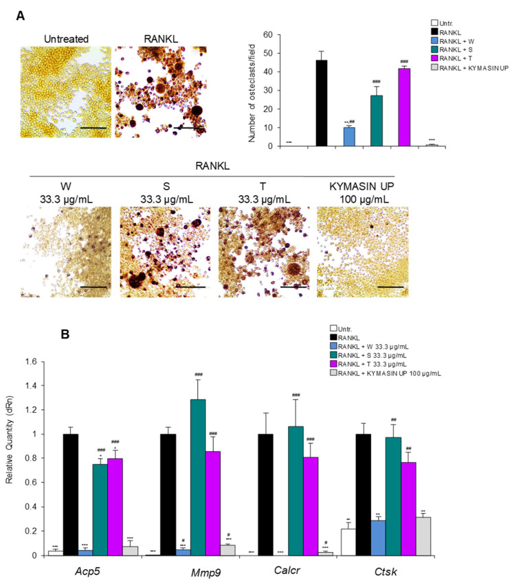
The imbalance in osteoblast (OB)-dependent bone formation in favor of osteoclast (OC)-dependent bone resorption is the main cause of loss of tissue mineral mass during bone remodeling leading to osteoporosis conditions. Thus, the suppression of OC activity together with the improvement in the OB activity has been proposed as an effective therapy for maintaining bone mass during aging. We tested the new dietary product, KYMASIN UP containing standardized Withania somnifera, Silybum marianum and Trigonella foenum-graecum herbal extracts or the single extracts in in vitro models mimicking osteoclastogenesis (i.e., RAW 264.7 cells treated with RANKL, receptor activator of nuclear factor kappa-Β ligand) and OB differentiation (i.e., C2C12 myoblasts treated with BMP2, bone morphogenetic protein 2). We found that the dietary product reduces RANKL-dependent TRAP (tartrate-resistant acid phosphatase)-positive cells (i.e., OCs) formation and TRAP activity, and down-regulates osteoclastogenic markers by reducing Src (non-receptor tyrosine kinase) and p38 MAPK (mitogen-activated protein kinase) activation. Withania somnifera appears as the main extract responsible for the anti-osteoclastogenic effect of the product. Moreover, KYMASIN UP maintains a physiological release of the soluble decoy receptor for RANKL, OPG (osteoprotegerin), in osteoporotic conditions and increases calcium mineralization in C2C12-derived OBs. Interestingly, KYMASIN UP induces differentiation in human primary OB-like cells derived from osteoporotic subjects. Based on our results, KYMASIN UP or Withania somnifera-based dietary supplements might be suggested to reverse the age-related functional decline of bone tissue by re-balancing the activity of OBs and OCs, thus improving the quality of life in the elderly and reducing social and health-care costs.

Keywords: RANKL; age-related diseases; natural product; osteoblast; osteoclast; signaling pathways.


Conflict of interest statement

T.M., C.E. and L.C. of Biokyma Laboratories were not involved in the design, data acquisition and interpretation, data management and statistical analysis. The other authors declare that they have no competing interests.

Figures

Figure 1
Figure 1
RAW 264.7 cells were treated for 5 days with RANKL (receptor activator of nuclear factor-kappa Β ligand) in the absence (black bars) or presence of KYMASIN UP (12.5–100 µg/mL) (grey bars). The untreated control was also shown (white bars). Real-time PCR analysis of the osteoclast (OC) markers, Acp5 (acid phosphatase 5, tartrate resistant), Calcr (calcitonin receptor), Mmp9 (matrix metallopeptidase 9), and Ctsk (cathepsin K) was performed. Gene expressions were normalized to Gapdh. Six independent experiments were performed. Statistical analysis was conducted using the two-tailed t-test. * p < 0.05, ** p < 0.01, and *** p < 0.001, significantly different from RANKL. # p < 0.05, ## p < 0.01, and ### p < 0.001, significantly different from untreated control. $ p < 0.05, significantly different.
Figure 2
Figure 2
(AE) RAW 264.7 cells were treated for 5 days with RANKL in the absence (black bars) or presence of KYMASIN UP (12.5–100 µg/mL) (grey bars). The untreated control was also shown (white bars). (A) TRAP (tartrate-resistant acid phosphatase) staining was performed and TRAP-positive OCs (≥3 nuclei) were counted. Representative images and high-magnification insets are reported (arrows mark OCs). (B) The supernatants of cells in A were collected, and TRAP activity was measured by ELISA and reported as fold change vs. RANKL treatment (black bar). (C) The F-actin ring (red) formation in RANKL-induced OCs was assessed using phalloidin fluorescence staining. DAPI (4′,6-diamidino-2-phenylindole, blue) was used to stain nuclei. (D) The number of F-actin rings positive cells containing ≥3 nuclei was determined. (E) Conditioned media were collected, trichloroacetic acid precipitated and subjected to Western blotting for detection of released OPG (osteoprotegerin). GAPDH relative to cell lysates is included as a loading control. Results are means ± standard error of the mean (A) or standard deviation (B,D). Six (A,B) or three (CE) independent experiments were performed. Statistical analysis was conducted using the two-tailed t-test (A,B), and the one-way ANOVA Tukey’s test (D). * p < 0.05, ** p < 0.01, and *** p < 0.001 significantly different from RANKL. # p < 0.05, ## p < 0.01, and ### p < 0.001, significantly different from untreated control. $ p < 0.05, significantly different. Scale bars (A,C), 100 μm.
Figure 3
Figure 3
(A,B) RAW 264.7 cells were treated for 5 days with RANKL in the absence or presence of W. somnifera (W), S. marianum (S), T. foenum-graecum (T) or KYMASIN UP at the indicated concentrations. (A) TRAP staining was performed and representative images are reported. The numbers of TRAP-positive OCs (≥3 nuclei) were determined. (B) Real-time PCR analysis of Acp5, Mmp9, Calcr, and Ctsk was performed. Gene expressions were normalized to Gapdh. Results are means ± standard deviation (A,B). Three independent experiments were performed. Statistical analysis was conducted using the two-tailed t-test. * p < 0.05, ** p < 0.01, and *** p < 0.001, significantly different from RANKL. # p < 0.05, ## p < 0.01, and ### p < 0.001, significantly different from untreated control (Untr). Scale bars (A), 100 μm.
Figure 4
Figure 4
(AD) C2C12 cells were treated with BMP2 (bone morphogenetic protein 2) with or without KYMASIN UP (100 µg/mL) for 3 or 6 days. (A) Phase-contrast images and immunofluorescence staining for the myogenic marker, desmin (red), were performed after 6 days of culture. DAPI (4′,6-diamidino-2-phenylindole, blue) was used to stain nuclei. The percentages of desmin-positive areas were evaluated. (B) The expressions of the late markers of myogenic differentiation, myogenin and MyHC-II (adult isoform II of myosin heavy chain), were analyzed by Western blotting at 3 or 6 days, respectively. Reported are the relative densities with respect to GAPDH or tubulin. (C) ALP (alkaline phosphatase) staining was performed, and ALP activity was measured by ELISA. (D) Real-time PCR analysis of the osteoblast (OB) markers, Osx (Sp7 transcription factor, Osterix), Col1a1 (collagen type I alpha 1 chain), Bglap (bone gamma-carboxyglutamate protein), and Runx2 (RUNX family transcription factor 2) was performed after 3 or 6 days of treatment. Gene expressions were normalized to Gapdh. Results are means ± standard error of the mean (A,C) or standard deviation (B,D). Three independent experiments were performed (AD). Statistical analysis was conducted using the two-tailed t-test. *** p < 0.001, significantly different from BMP2. # p < 0.05, ## p < 0.01, and ### p < 0.001, significantly different from untreated control (Untr). Reported are representative images (AC). Scale bars, 200 μm (A,C) or 100 μm (insets in (A)).
Figure 5
Figure 5
(A,B) C2C12 cells treated with BMP2 with or without KYMASIN UP (100 µg/mL) were cultured for 12 days in the presence of ascorbic acid (A.A.), β-glycerophosphate (β-gly) and dexamethasone (Dex) to induce transdifferentiation and deposition of calcium nodules. (A) Phase contrast images were reported. Calcium nodule deposition was observed in fluorescence after calcein (green) staining. (B) The quantification of calcein-positive nodules was performed and reported as calcein optical density. Results are means ± standard deviation. Three independent experiments were performed. Statistical analysis was conducted using the two-tailed t-test. * p < 0.05, and *** p < 0.001, significantly different from BMP2. # p < 0.05, significantly different from untreated control. Reported are representative images. Scale bars (A), 400 μm.
Figure 6
Figure 6
(A,B) Pre-OB cells derived from healthy and osteoporotic subjects (n = 3 each group) were treated for 12 days with KYMASIN UP (10 µg/mL) or with Dex as positive OB differentiation control. (A) Representative images of ALP staining are reported. (B) ALP activity was measured and normalized to ATP (adenosine triphosphate) as an index of cell numbers and reported as percentages with respect to Dex treatment. Results are means ± standard error of the mean. Statistical analysis was conducted using the two-tailed t-test. * p < 0.05, and *** p < 0.001, significantly different from Dex. # p < 0.05, significantly different from untreated control (Untr). Scale bars (A), 200 μm.
Figure 7
Figure 7
(A,B) The expressions of phosphorylated (p) Src (non-receptor tyrosine kinase) and p38 MAPK (p38 mitogen-activated protein kinase) were analyzed by Western blotting in RAW 264.7 cells treated with RANKL for 5 days (A) or in C2C12 cells treated with BMP2 for 6 days (B), in the absence or presence of KYMASIN UP (100 µg/mL). Representative images and the relative densities with respect to the total forms of Src and p38 are reported. Results are means ± standard error of the mean. Six (A,B) independent experiments were performed. Statistical analysis was conducted using the two-tailed t-test. * p < 0.05, ** p < 0.01, and *** p < 0.001, significantly different from RANKL (A) or BMP2 (B). # p < 0.05, and ## p < 0.01, significantly different from untreated control (Untr).

References

    1. Raisz L.G. Pathogenesis of osteoporosis: Concepts, conflicts, and prospects. J. Clin. Investig. 2005;115:3318–3325. doi: 10.1172/JCI27071. - DOI - PMC - PubMed
    1. Choi M.H., Yang J.H., Seo J.S., Kim Y.-J., Kang S.-W. Prevalence and diagnosis experience of osteoporosis in postmenopausal women over 50: Focusing on socioeconomic factors. PLoS ONE. 2021;16:e0248020. doi: 10.1371/journal.pone.0248020. - DOI - PMC - PubMed
    1. Song S., Guo Y., Yang Y., Fu D. Advances in pathogenesis and therapeutic strategies for osteoporosis. Pharmacol. Ther. 2022;237:108168. doi: 10.1016/j.pharmthera.2022.108168. - DOI - PubMed
    1. Corrado A., Cici D., Rotondo C., Maruotti N., Cantatore F.P. Molecular Basis of Bone Aging. Int. J. Mol. Sci. 2020;21:3679. doi: 10.3390/ijms21103679. - DOI - PMC - PubMed
    1. Noh J.-Y., Yang Y., Jung H. Molecular Mechanisms and Emerging Therapeutics for Osteoporosis. Int. J. Mol. Sci. 2020;21:7623. doi: 10.3390/ijms21207623. - DOI - PMC - PubMed