Review
. 2022 Jun 22;8(4):1649-1665.
doi: 10.3390/tomography8040137.

Tuberculosis of the Heart: A Diagnostic Challenge

Affiliations
Review

Tuberculosis of the Heart: A Diagnostic Challenge

Karuna M Das et al. Tomography. .

Abstract

Tuberculosis of the heart is relatively rare and presents a significant diagnostic difficulty for physicians. It is the leading cause of death from infectious illness. It is one of the top 10 leading causes of death worldwide, with a disproportionate impact in low- and middle-income nations. The radiologist plays a pivotal role as CMR is a non-invasive radiological method that can aid in identifying potential overlap and differential diagnosis between tuberculosis, mass lesions, pericarditis, and myocarditis. Regardless of similarities or overlap in observations, the combination of clinical and certain particular radiological features, which are also detected by comparison to earlier and follow-up CMR scans, may aid in the differential diagnosis. CMR offers a significant advantage over echocardiography for detecting, characterizing, and assessing cardiovascular abnormalities. In conjunction with clinical presentation, knowledge of LGE, feature tracking, and parametric imaging in CMR may help in the early detection of tuberculous myopericarditis and serve as a surrogate for endomyocardial biopsy resulting in a quicker diagnosis and therapy. This article aims to explain the current state of cardiac tuberculosis, the diagnostic utility of CMR in tuberculosis (TB) patients, and offer an overview of the various imaging and laboratory procedures used to detect cardiac tuberculosis.

Keywords: irregular thickening of pericardium; magnetic resonance imaging; transmural mesocardial and epicardial fat late enhancement; tuberculous myopericarditis.


Conflict of interest statement

The authors declare no conflict of interest.

Figures
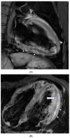
Figure 1
Figure 1
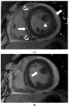
(A) A 35-year-old male with a recent history of treated tuberculous pericarditis for six months had CMR for ventricular tachycardia. 2CH T2w STIR pictures demonstrate increased epicardial and transmural signals in the inferior wall (arrows). (B) A 35-year-old male with a recent history of treated tuberculous pericarditis for six months had CMR for ventricular tachycardia. 4CH T2w STIR pictures demonstrate irregular thickening of the pericardium (small arrows) with the increased transmural signal of the mid-lateral wall (thick arrow).
Figure 2
Figure 2
(A) A 35-year-old male with a recent history of treated tuberculous pericarditis for six months had CMR for ventricular tachycardia. Short axis mid myocardial slice, late enhancement images, shows subepicardial anterolateral (notched arrow), mesocardial mid and inferior septal (white arrow), anterior and posterior RV insertion points (curve arrow) with thickened pericardium and epicardial fat enhancement (thin arrows). Enlarged AL papillary muscle (arrowhead). (B) A 35-year-old male with a recent history of treated tuberculous pericarditis for six months had CMR for ventricular tachycardia. Short axis mid myocardial slice late enhancement images show all the features of (A) and asymmetrical enlargement of the AL papillary muscle (arrow). (C) A 35-year-old male with a recent history of treated tuberculous pericarditis for six months had CMR for ventricular tachycardia. Short axis mid myocardial slice late enhancement images show the RV lateral and inferior wall (arrow), superior and inferior insertion (white arrow) with epicardial fat enhancement (thin arrows). PM papillary muscle is with central enhancement (arrowhead).
Figure 2
Figure 2
(A) A 35-year-old male with a recent history of treated tuberculous pericarditis for six months had CMR for ventricular tachycardia. Short axis mid myocardial slice, late enhancement images, shows subepicardial anterolateral (notched arrow), mesocardial mid and inferior septal (white arrow), anterior and posterior RV insertion points (curve arrow) with thickened pericardium and epicardial fat enhancement (thin arrows). Enlarged AL papillary muscle (arrowhead). (B) A 35-year-old male with a recent history of treated tuberculous pericarditis for six months had CMR for ventricular tachycardia. Short axis mid myocardial slice late enhancement images show all the features of (A) and asymmetrical enlargement of the AL papillary muscle (arrow). (C) A 35-year-old male with a recent history of treated tuberculous pericarditis for six months had CMR for ventricular tachycardia. Short axis mid myocardial slice late enhancement images show the RV lateral and inferior wall (arrow), superior and inferior insertion (white arrow) with epicardial fat enhancement (thin arrows). PM papillary muscle is with central enhancement (arrowhead).
Figure 3
Figure 3
A 35-year-old male with a recent history of treated tuberculous pericarditis for six months had CMR for ventricular tachycardia. 4 CH, late enhancement images, shows RV free wall (epicardial and transmural thin arrows), inferior septal (transmural- thick Arrow), lateral wall LV (transmural-thick arrow), and interatrial septum (thin arrow) enhancement.
Figure 4
Figure 4
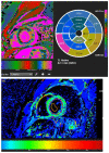
A 35-year-old male with a recent history of treated tuberculous pericarditis for six months had CMR for ventricular tachycardia. A native T1 map of 1132 ± 178 (Figure 4) and ECV of 25 ± 8%.
Figure 5
Figure 5
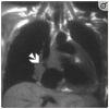
T2W dark-blood coronal MRI image shows diffuse myopericardial thickening. The thickening is hypointense on T2W images and is causing attenuation of the proximal SVC (arrow). The image was adapted from [53].
Figure 6
Figure 6
Cardiac magnetic resonance images. (a,b) Axial T1-weighted images showing isointense masses (arrows) along the anterior right atrium (*), right ventricular outflow tract (#), and along both ventricles (+ indicates left ventricle). (c) Short axis T2-weighted image showing that the lesions are mildly hyperintense. (d) Steady-state free precession image revealing the infiltrative nature of the lesion along the left ventricle. The delayed enhanced short-axis image (e) shows heterogeneous enhancement of the mass. The image was adapted from [47].
Figure 7
Figure 7
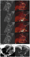
(A) Cardiac magnetic resonance (CMR) shows multifocal subepicardial to mid-myocardial linear enhancement along the right ventricular insertion site, mid-anterolateral, and inferior segments (arrows) with corresponding focal myocardial edema. (B) Fused cardiac positron emission tomography CMR shows patchy areas of increased 18F-flurodeoxyglycose (FDG) uptake in the apical to mid-anterolateral, mid-to-basal anteroseptal at the right ventricular insertion site, and mildly increased FDG uptake in the apical inferior segments of the left ventricular myocardium corresponding to the regions of myocardial enhancement seen on CMR. (C,D) T2 black blood image showing T2 hyperintense changes in the left ventricular myocardium. The image was adapted from [61].

Similar articles

Cited by

References

    1. Frieden T.R., Brudney K.F., Harries A.D. Global tuberculosis: Perspectives, prospects, and priorities. JAMA. 2014;312:1393–1394. doi: 10.1001/jama.2014.11450. - DOI - PubMed
    1. Syed F.F., Ntsekhe M., Gumedze F., Badri M., Mayosi B.M. Myopericarditis in tuberculous pericardial effusion: Prevalence, predictors and outcome. Heart. 2014;100:135–139. doi: 10.1136/heartjnl-2013-304786. - DOI - PubMed
    1. Horn H., Saphir O. The Involvement of the Myocardium in Tuberculosis, A Review of the Literature and Report of 3 Cases. Am. Rev. Tuberc. 1935;32:492–506.
    1. Fowler N.O. Tuberculous pericarditis. JAMA. 1991;266:99–103. doi: 10.1001/jama.1991.03470010103039. - DOI - PubMed
    1. Njovane X. Intramyocardial tuberculosis: A rare underdiagnosed entity. SAMJ S. Afr. Med. J. 2009;99:152–153. - PubMed