Review
. 2022 Jul 25;11(8):831.
doi: 10.3390/pathogens11080831.

CD4+ Cytotoxic T Cells Involved in the Development of EBV-Associated Diseases

Affiliations
Review

CD4+ Cytotoxic T Cells Involved in the Development of EBV-Associated Diseases

Manuel Ruiz-Pablos. Pathogens. .

Abstract

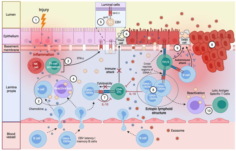
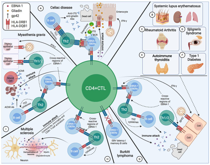
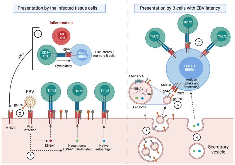
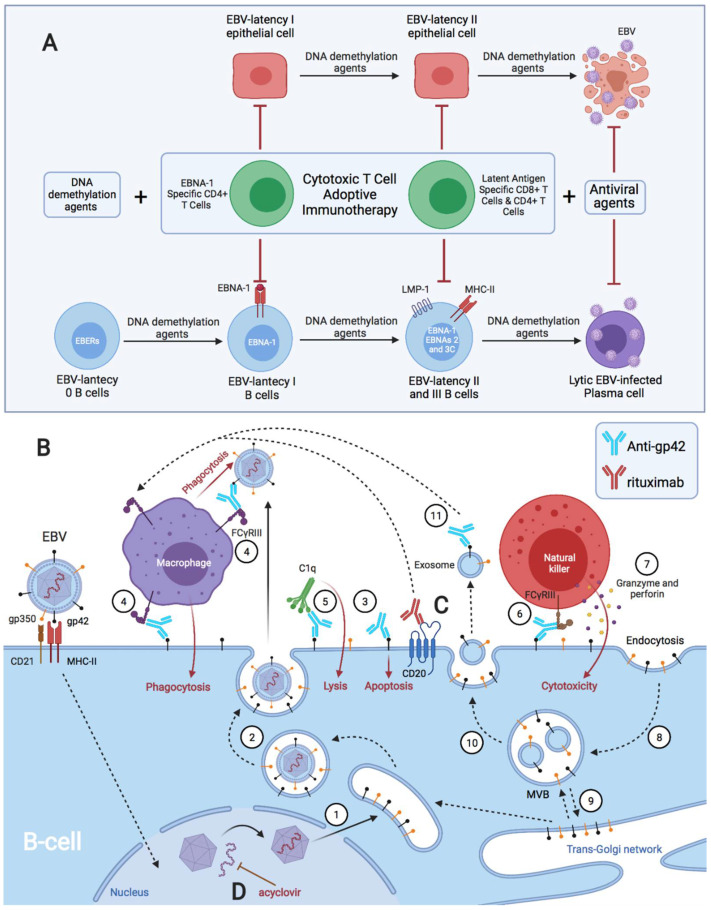
Activated cytotoxic CD4 T cells (HLA-DR+) play an important role in the control of EBV infection, especially in cells with latency I (EBNA-1). One of the evasion mechanisms of these latency cells is generated by gp42, which, via peripherally binding to the β1 domain of the β chain of MHC class II (HLA-DQ, -DR, and -DP) of the infected B lymphocyte, can block/alter the HLA class II/T-cell receptor (TCR) interaction, and confer an increased level of susceptibility towards the development of EBV-associated autoimmune diseases or cancer in genetically predisposed individuals (HLA-DRB1* and DQB1* alleles). The main developments predisposing the factors of these diseases are: EBV infection; HLA class II risk alleles; sex; and tissue that is infiltrated with EBV-latent cells, forming ectopic lymphoid structures. Therefore, there is a need to identify treatments for eliminating cells with EBV latency, because the current treatments (e.g., antivirals and rituximab) are ineffective.

Keywords: CD4+ CTL; DQB1; DRB1; EBV EBNA-1; Gp42; HLA; autoimmunity; cancer.


Conflict of interest statement

The author declares no conflict of interest.

Figures

Figure 1
Figure 1
Sequence of EBV infection, chronic inflammation, autoimmunity, and/or cancer in the mucosa of genetically predisposed patients. (1) An infection or any inflammatory stimulus recruit leukocytes in the mucosa, including both latency I B cells (EBNA-1) and healthy B cells. (2) In the mucosa, B cells form ectopic lymphoid aggregates that allow for the generation of antigen-specific immune responses. These ectopic lymphoid structures generate a favorable environment for the transformation of EBV-latent B cells into proliferating blasts, to become memory B cells. (3) In addition, NK cell activation occurs, both in response to the first inflammatory stimulus, and to restrict B cell transformation by EBV. Exposure to foreign antigens from the first stimulus or to viral antigens from EBV leads to activation of CD4 T cells and release of IFN-γ, followed by upregulation of MHC-II on epithelial cells, which favors the acquisition of a nonprofessional antigen-presenting cell phenotype. (4) In addition, the presence of foreign antigens could also lead to terminal differentiation and activation of EBV-latent B lymphocytes, allowing the transition from the latent to the lytic phase of the virus. (5) The newly generated viral particles then infect more epithelial cells through gp42/MHC-II interaction, leading to further inflammation and ultimately to latent EBV infection. Furthermore, this chronic inflammation elicits a cytokine response, leading to increased B-cell recruitment and perpetuation of the viral infection. (6) Latent EBV epithelial cells could enter a lytic phase, releasing new virions, lyse as a consequence of the T-cell response, or undergo neoplastic transformation. (7) The mechanisms of immune evasion of EBV latency (epithelial cells and B cells) involve decreased activation and decreased cytotoxic capacity of EBNA-1-specific CD4 T cells through the release of IL-10 and EBV miRNAs contained in exosomes, which could suppress the expression of target genes in the viral or host genome to maintain latent EBV infection. (8) This altered immunosurveillance leads to increased proliferation of EBV-latent B- and epithelial cells, which increases the risk of neoplastic transformation or autoimmune disease in genetically predisposed patients with EBV-susceptible MHC-II ancestral alleles. (9) Presentation through MHC-II/gp42 of native cellular autoantigens or viral EBNA-1, which can undergo posttranslational modifications, such as citrullination, and form neoantigens, could trigger the activation of autoreactive CD4 T cells and the formation of autoantibodies against tissue cells. (10) Other phases of virus latency or the lytic phase would be controlled by NK cells, and CD4 and CD8 T cells, with specificity for EBV lytic proteins.
Figure 2
Figure 2
CD4 CTLs involved in the development of EBV-associated diseases. (A) In celiac disease, the infiltration of EBV-latent B cells into tissue, and infection of enterocytes via virus transfer from infected B cells increases IFN-γ levels as a response of CD4 CTLs. The increase in IFN-γ increases the expression of MHC-II by enterocytes, further enabling infection of these cells via gp42/MHC-II interaction, fusing the viral lipid bilayer with the cellular lipid bilayer. Gliadin from the diet is internalized into enterocytes by endocytosis, and can cross the epithelium via transcytosis, where they are deaminated by tissue transglutaminase-2 (tTG), thus interacting with antigen-presenting cells. In this case, B cells with EBV latency capture the tTG–gluten complex or deamidated gliadin through the BCR, where they process and present them in MHC-II/gp42 to CD4 T cells, activating them, and generating a cellular autoimmune response against gliadin, and a humoral autoimmune response (Th2) against gliadin and tTG. (B) In systemic lupus, erythematosus latency I B cells present EBNA-1 on gp42/MHC-II, activating EBNA-1-specific CD4 T lymphocytes that are cross-reactive to common lupus antigens (Ro, Sm B/B′, and Sm D1) by molecular mimicry, activating Th1 and Th2 cells that generate the autoimmune response. (C) In rheumatoid arthritis, as EBNA-1 undergoes citrullination by presentation on MHC class II molecules after macroautophagy in EBV-latent B cells, neoantigens can be formed, activating Th1 and Th2 cells with an autoimmune response against citrulline. (D) In Sjögren’s syndrome, EBNA-1 presentation by EBV-latent B cells infiltrating the glands, as well as by infected glandular epithelial cells, activate Th1 and Th2 cells, with an autoimmune response against the glandular epithelial cells, causing cell death and generating severe ocular and oral dryness. (E) In Graves’ disease, the infiltration of cells with EBV latency into thyroid tissue increases IFN-γ levels in that tissue as a response of CD4 CTLs, increasing MHC-II expression in thyrocytes, and, thus, allowing infection of these cells by EBV. Both EBNA-1 presentation by EBV-latent B cells infiltrating this tissue, and by infected thyrocytes, activate Th2 cells that are cross-reactive to thyroid antigens. These Th2 cells would help B cells to secrete thyroid-stimulating immunoglobulins against the thyroid-stimulating hormone receptor, resulting in rampant thyroid hormone production and hyperthyroidism. Subsequently, there would be a compensatory increase in TSH secretion that maintains the thyroid with a sufficient reserve until it is overwhelmed by massive thyroid follicle destruction and fibrosis, both by cytotoxic cells (Th1 and CD8) and by the rise of antibodies against thyroid peroxidase and thyroglobulin, which arise after chronic immune stimulation over the years by infiltrating EBV latency B cells, as well as via infected thyrocytes, leading to thyroid insufficiency and, thus, to the development of Hashimoto’s thyroiditis. (F) In type 1 diabetes, EBNA-1 presentation by EBV latency B cells infiltrating the pancreas, as by infected pancreatic β cells, activate Th1 and Th2 cells with an autoimmune response against pancreatic β cells, causing cell death and insulin deficiency. (G) EBV latency I B cells in Burkitt’s lymphoma are defective in gp42/MHC-II-mediated antigenic presentation, preventing activation and recognition by CD4+ T cells. (H) In multiple sclerosis, latency I B cells present EBNA-1 in gp42/MHC-II class II, activating EBNA-1-specific CD4 T lymphocytes that are cross-reactive to myelin, activating Th1 and Th2 cells. (I) In myasthenia gravis, EBV latency B cells infiltrating the thymus causes increased levels of IFN-γ released by CD4 CTLs, increasing MHC-II expression in thymic epithelial cells, and, thus, allowing infection of these cells by EBV. Both EBNA-1 presentation by EBV-latent B cells and thymic epithelial cells would activate Th1 and Th2 cells, cross-reacting against the acetylcholine receptor (AChR) by targeting AChR-expressing myoid cells and the neuromuscular junction, ultimately leading to skeletal muscle weakness and fatigue.
Figure 3
Figure 3
Model of EBV-associated autoimmune disease development. Leukocytes circulating in the peripheral blood, including EBV-latent B cells, are recruited to the affected tissues by inflammatory stimuli (infectious, autoimmune, or neoplastic processes). (1) Both the inflammation caused by the first stimulus and that caused by B cells with EBV latency allow for the activation of CD4 T lymphocytes and NK cells, releasing both IFN-γ. (2) The increase in IFN-γ induces the expression of MHC class II molecules in the cells of that tissue, converting them into nonprofessional antigen-presenting cells and allowing infection through gp42/MHC-II interaction. (3) Through MHC-II, they can present EBNA-1, peptides that undergo post-translational modifications and that can form neoantigens, such as citrullinated EBNA-1, or self-antigens that are native to the cell itself, activating CD4 T lymphocytes and generating Th1 and/or Th2 cells with an autoimmune response against the tissue cells, ultimately causing cell death. (4) Both the neoantigens and native autoantigens can be released by exocytosis, or in exosomes, and be taken up by antigen-presenting cells. (5) Exosomes can have EBV latency proteins, such as LMP-1/2A, and viral glycoproteins, such as gp350 and gp42, in their membrane, since these glycoproteins are present in the cell membrane. Upon internalization of part of the membrane, an endocytic vesicle is formed, which subsequently fuses with the early endosome. After inward budding of the endosome membrane, the intraluminal vesicles will form and give rise to exosomes. They may also contain messenger RNA (mRNA), microRNA (miRNA), and other products of EBV, such as EBV DNA. (6) Exosomes bind to EBV-latent B lymphocytes or other uninfected B lymphocytes through the interaction of gp350/CD21 and/or g42/MHC-II, releasing their contents into the cellular interior. (7) EBV-latent B cells can process and present these antigens in the MHC-II/gp42 complex, activating Th1 and/or Th2 cells with an autoimmune response against the tissue cells.
Figure 4
Figure 4
Different treatments against EBV latent cells. (A) Schematic model of treatment with DNA demethylation agents followed by adoptive immunotherapy of EBV-specific T cells and antiviral agents. Administration of low-dose DNA demethylation agents restores the expression of MHC class II molecules and induces the expression of LMP1, EBNA-2, EBNA3A, and EBNA-3C, allowing the transformation of EBV latency 0 and I B cells into latency II and III B cells. They also induce the transformation of EBV latency I epithelial cells to latency II. In this way, the recognition of these cells by EBV-specific T cells is improved. EBNA-1-specific CD4 T cells can only recognize latent I cells exhibiting EBNA-1 in MHC class II molecules, since EBNA-1 is poorly immunogenic. DNA demethylation agents induce lytic infection and apoptosis in EBV-transformed B cells and epithelial cells. Antiviral agents prevent viral replication. (B) Model of anti-gp42 antibody treatment. (1) Schematic representation of packaging into EBV virions, where the viral nucleocapsid acquires its final lipid envelope by budding in the trans-Golgi network (TGN). (2) It is then transported to the plasma membrane in secretory vesicles and released from the cell. Finally, after this whole process, viral glycoproteins, such as gp42 and gp350, from the secretory vesicles remain in the plasma membrane of the cell. (3) These glycoproteins can be detected by specific antibodies, such as anti-gp42. Following anti-gp42 binding, cells with gp42 on their membrane can undergo: (4) antibody-dependent phagocytosis by activated macrophages, (5) complement-mediated cytotoxicity leading to cell lysis, (6) direct death mediated by natural killer cells, (7) and antibody-dependent cellular cytotoxicity mediated by perforin and granzyme cytokines. (8) Exosome formation begins via endocytosis, where part of the membrane, together with membrane receptors and viral glycoproteins, are internalized, forming an endocytic vesicle, which subsequently fuses with the early endosome. (9) During the maturation process of the early endosome, it communicates with the Golgi apparatus through the exchange of vesicles in a bidirectional manner, forming the late endosome or multivesicular body (MVB). (10) Inward budding of the endosome membrane forms the intraluminal vesicles that will be released into the extracellular space as exosomes. (11) In the case of exosomes with viral glycoproteins present on their membrane, anti-gp42 binding triggers antibody-dependent phagocytosis by activated macrophages. (C) Anti-CD20 monoclonal antibodies (rituximab) also act through complement-dependent cytotoxicity, antibody-dependent cellular phagocytosis, antibody-dependent cellular cytotoxicity, and the induction of apoptosis. (D) Antivirals prevent viral replication by inhibiting viral DNA synthesis.

Similar articles

Cited by

References

    1. Tian Y., Sette A., Weiskopf D. Cytotoxic CD4 T Cells: Differentiation, Function, and Application to Dengue Virus Infection. Front. Immunol. 2016;7:531. doi: 10.3389/fimmu.2016.00531. - DOI - PMC - PubMed
    1. Marshall N.B., Swain S.L. Cytotoxic CD4 T Cells in Antiviral Immunity. J. Biomed. Biotechnol. 2011;2011:954602. doi: 10.1155/2011/954602. - DOI - PMC - PubMed
    1. Hintzen R.Q., de Jong R., Lens S.M., Brouwer M., Baars P., van Lier R.A. Regulation of CD27 Expression on Subsets of Mature T-Lymphocytes. [(accessed on 8 January 2021)];J. Immunol. 1993 151:2426–2435. Available online: https://pubmed.ncbi.nlm.nih.gov/7689607/ - PubMed
    1. Globerson A., Effros R.B. Ageing of Lymphocytes and Lymphocytes in the Aged. Immunol. Today. 2000;21:515–521. doi: 10.1016/S0167-5699(00)01714-X. - DOI - PubMed
    1. van de Berg P.J., van Leeuwen E.M., ten Berge I.J., van Lier R. Cytotoxic Human CD4+ T Cells. Curr. Opin. Immunol. 2008;20:339–343. doi: 10.1016/j.coi.2008.03.007. - DOI - PubMed