

Influence of dilution on arterial-phase artifacts and signal intensity on gadoxetic acid-enhanced liver MRI
- PMID: 35895119
- PMCID: PMC9755096
- DOI: 10.1007/s00330-022-08984-0
Influence of dilution on arterial-phase artifacts and signal intensity on gadoxetic acid-enhanced liver MRI
Abstract
Objectives: To investigate the effect of saline-diluted gadoxetic acid, done for arterial-phase (AP) artifact reduction, on signal intensity (SI), and hence focal lesion conspicuity on MR imaging.
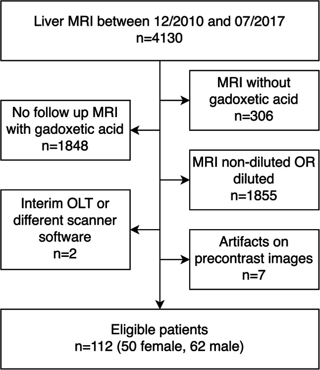
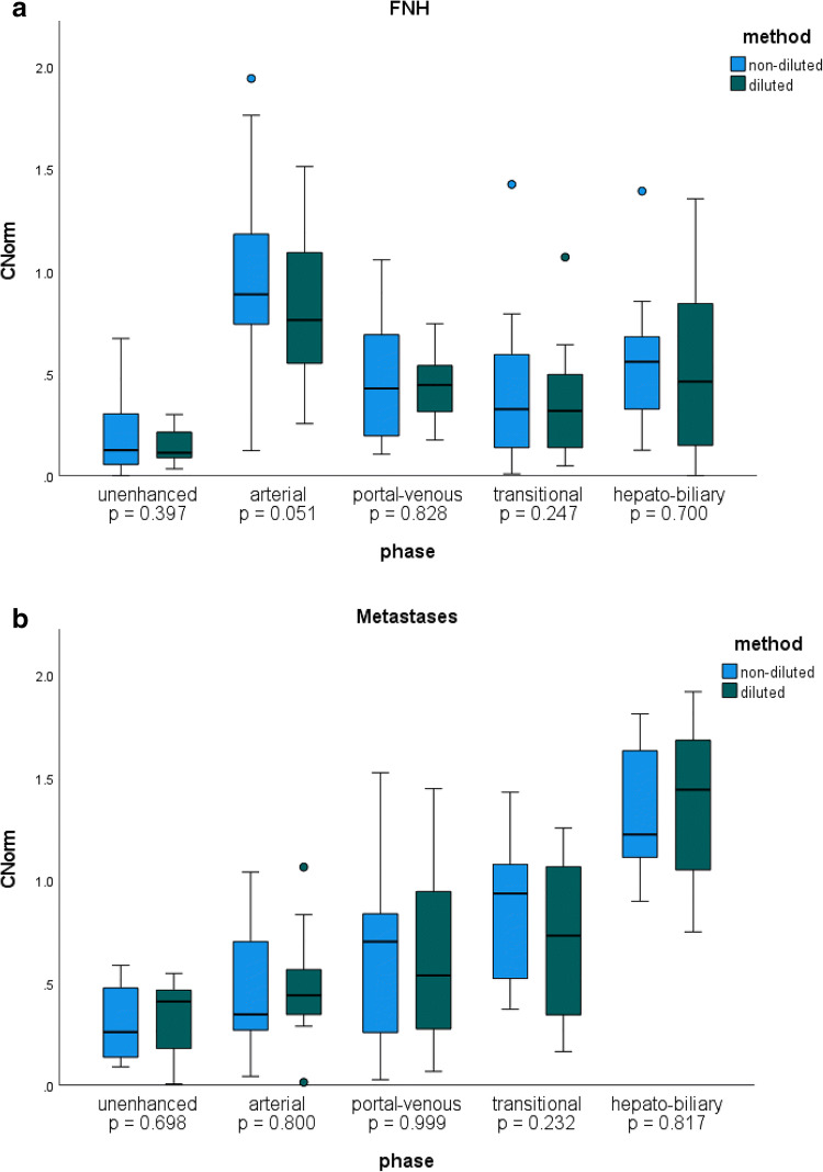
Methods: We retrospectively examined 112 patients who each had at least two serial gadoxetic acid-enhanced liver MRIs performed at 1 ml/s, first with non-diluted (ND), then with 1:1 saline-diluted (D) contrast. Two blinded readers independently analyzed the artifacts and graded dynamic images using a 5-point scale. The absolute SI of liver parenchyma, focal liver lesions (if present), aorta, and portal vein at the level of the celiac trunk and the SI of the paraspinal muscle were measured in all phases. The signal-to-norm (SINorm) of the vascular structures, hepatic parenchyma and focal lesions, and the contrast-to-norm (CNorm) of focal liver lesions were calculated.
Results: AP artifacts were significantly reduced with dilution. Mean absolute contrast-enhanced liver SI was significantly higher on the D exams compared to the ND exams. Likewise, SINorm of liver parenchyma was significantly higher in all contrast-enhanced phases except transitional phase on the D exams. SINorm values in the AP for the aorta and in the PVP for portal vein were significantly higher on the diluted exams. The CNorm was not significantly different between ND and D exams for lesions in any imaging phase. The interclass correlation coefficient was excellent (0.89).
Conclusion: Gadoxetic acid dilution injected at 1ml/s produces images with significantly fewer AP artifacts but no significant loss in SINorm or CNorm compared to standard non-diluted images.
Key points: • Diluted gadoxetic acid at slow injection (1 ml/s) yielded images with higher SINorm of the liver parenchyma and preserved CNorm for focal liver lesions. • Gadoxetic acid-enhanced MRI injected at 1 ml/s is associated with arterial-phase (AP) artifacts in 31% of exams, which may degrade image quality and limits focal liver lesion detection. • Saline dilution of gadoxetic acid 1:1 combined with a slow injection rate of 1 ml/s significantly reduced AP artifacts from 31 to 9% and non-diagnostic AP artifacts from 16 to 1%.
Keywords: Artifacts; Gadolinium ethoxybenzyl DTPA; Liver; Magnetic resonance imaging; Signal intensity.
© 2022. The Author(s).
Conflict of interest statement
Ahmed Ba-Ssalamah has received consultant fees and honorarium for lectures from Bayer.
All other authors of this manuscript declare no relationships with any companies whose products or services may be related to the subject matter of the article.
Figures


References
-
- Bashir MR, Castelli P, Davenport MS, et al. Respiratory motion artifact affecting hepatic arterial phase MR imaging with gadoxetate disodium is more common in patients with a prior episode of arterial phase motion associated with gadoxetate disodium. Radiology. 2015;274:141–148. doi: 10.1148/radiol.14140386. - DOI - PubMed
MeSH terms
Substances
LinkOut - more resources
Full Text Sources
Medical
