. 2022 Jul 27;79(8):452.
doi: 10.1007/s00018-022-04480-2.

Endoplasmic reticulum stress contributes to cisplatin-induced chronic kidney disease via the PERK-PKCδ pathway

Affiliations

Endoplasmic reticulum stress contributes to cisplatin-induced chronic kidney disease via the PERK-PKCδ pathway

Shaoqun Shu et al. Cell Mol Life Sci. .

Abstract

Background: Cisplatin is an effective chemotherapeutic drug, but it may induce both acute and chronic kidney problems. The pathogenesis of chronic kidney disease (CKD) associated with cisplatin chemotherapy remains largely unclear.

Methods: Mice and renal tubular cells were subjected to repeated low-dose cisplatin (RLDC) treatment to induce CKD and related pathological changes. The roles of endoplasmic reticulum (ER) stress, PERK, and protein kinase C-δ (PKCδ) were determined using pharmacological inhibitors and genetic manipulation.

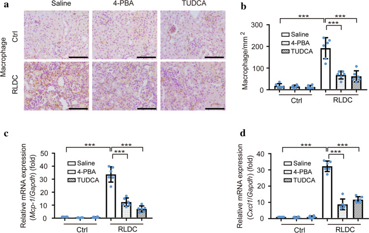
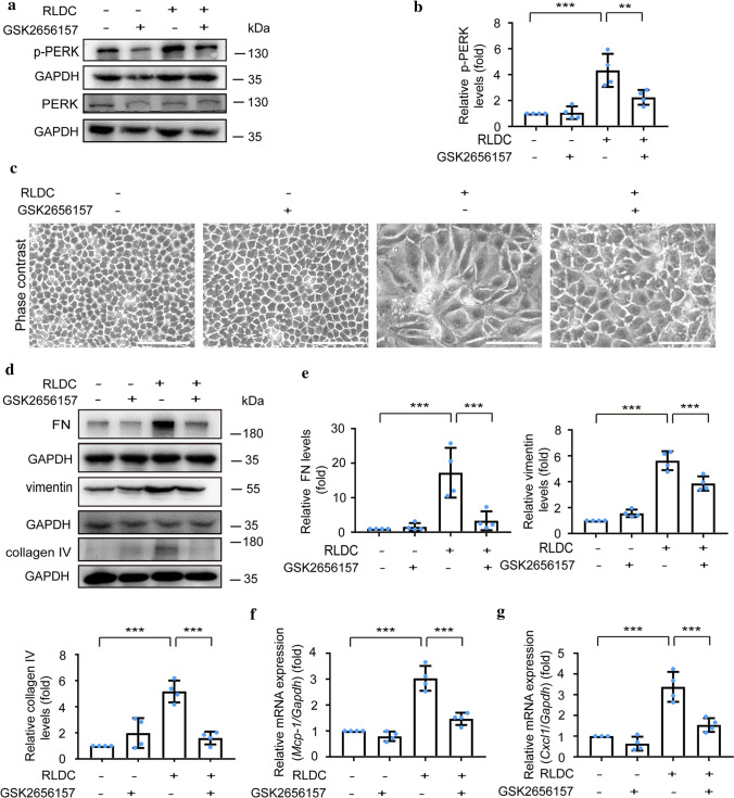
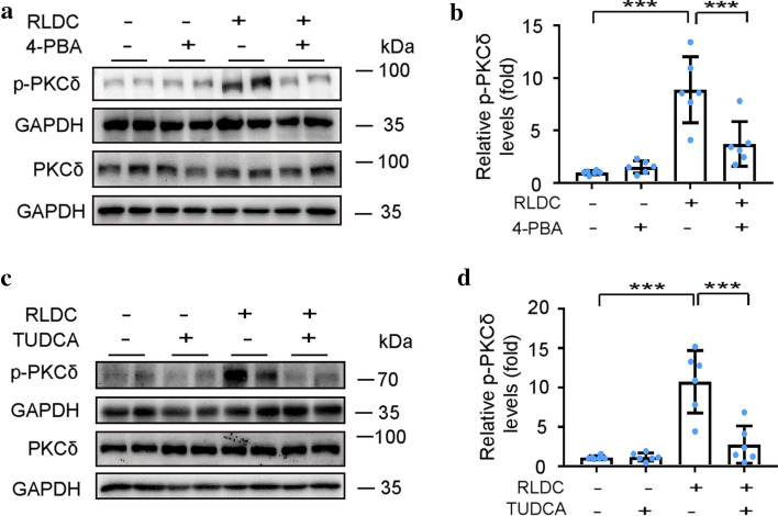
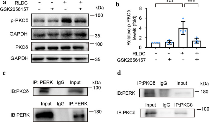
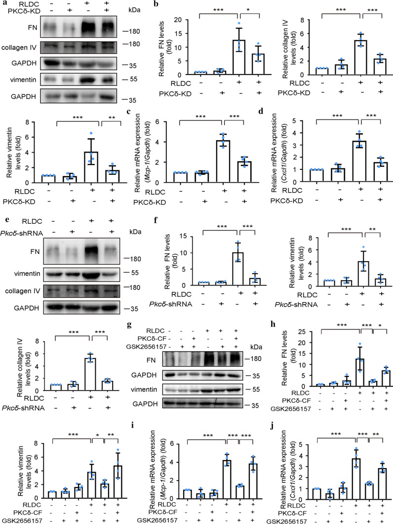
Results: ER stress was induced by RLDC in kidney tubular cells in both in vivo and in vitro models. ER stress inhibitors given immediately after RLDC attenuated kidney dysfunction, tubular atrophy, kidney fibrosis, and inflammation in mice. In cultured renal proximal tubular cells, inhibitors of ER stress or its signaling kinase PERK also suppressed RLDC-induced fibrotic changes and the expression of inflammatory cytokines. Interestingly, RLDC-induced PKCδ activation, which was blocked by ER stress or PERK inhibitors, suggesting PKCδ may act downstream of PERK. Indeed, suppression of PKCδ with a kinase-dead PKCδ (PKCδ-KD) or Pkcδ-shRNA attenuated RLDC-induced fibrotic and inflammatory changes. Moreover, the expression of active PKCδ-catalytic fragment (PKCδ-CF) diminished the beneficial effects of PERK inhibitor in RLDC-treated cells. Co-immunoprecipitation assay further suggested PERK binding to PKCδ.

Conclusion: These results indicate that ER stress contributes to chronic kidney pathologies following cisplatin chemotherapy via the PERK-PKCδ pathway.

Keywords: Chronic nephrotoxicity; Kidney repair; Protein kinase C-δ; Unfolded protein response.


Conflict of interest statement

All authors declared no competing interests.

Figures

Fig. 1
Fig. 1
4-PBA and TUDCA attenuate renal dysfunction, tubular atrophy, and kidney fibrosis in post-repeated low-dose cisplatin (RLDC) mouse kidneys. C57BL/6 male mice were subjected to four weekly injections of cisplatin (8 mg/kg). 4-PBA, TUDCA, or saline was given daily after the last injection of cisplatin for 1 week. FITC-Sinistrin was injected via tail vein before sacrifice to measure glomerular filtration rate (GFR). Blood and kidney tissues were collected 1 month after the last injection of cisplatin. a Quantitative analysis of GFR. b Concentration of serum creatinine. c Concentration of blood urea nitrogen (BUN). d Ratio of kidney weight to body weight. e Representative HE staining images. Bar = 100 μm. f Pathological tubular atrophy score. gj Immunoblot analysis of FN, vimentin, collagen IV, α-SMA, and GAPDH. For quantification, the protein was analyzed through densitometry and then normalized with GAPDH. k, l Representative images of Masson trichrome staining and quantitative analysis. Bar = 100 μm. N = 6 mice. *p < 0.05; **p < 0.01; ***p < 0.001
Fig. 2
Fig. 2
4-PBA and TUDCA reduce kidney interstitial inflammation post-RLDC treatment in mouse kidneys. C57BL/6 male mice were subjected to four weekly injections of cisplatin (8 mg/kg). 4-PBA, TUDCA, or saline was given daily from the last injection of cisplatin for 1 week. Kidney tissues were collected 1 month after the last injection of cisplatin. a, b Representative immunohistochemical staining images and quantitative analysis of F4/80. Bar = 100 μm. c, d Quantitative real-time PCR analysis of Mcp-1 and Cxcl1 normalized with Gapdh as an internal control. N = 6 mice. ***p < 0.001
Fig. 3
Fig. 3
4-PBA attenuates RLDC-induced fibrotic changes and cytokines production in cultured BUMPT cells. BUMPT cells were incubated with or without cisplatin (2 μM) for 7 h daily for 4 days and then treated with or without 5 mM 4-PBA for 17 h post last cisplatin treatment. a, b Immunoblot analysis of p-PERK, PERK, p-eIF2α, eIF2α, and GAPDH. For quantification, the protein was analyzed through densitometry and then normalized with GAPDH. c Representative images of phase contrast. Bar = 100 μm. d, e Immunoblot analysis of FN, collagen I, vimentin, collagen IV, and GAPDH. f, g Quantitative real-time PCR analysis of Mcp-1 and Cxcl1 with Gapdh as an internal control. n = 4. *p < 0.05; **p < 0.01; ***p < 0.001
Fig. 4
Fig. 4
GSK2656157 attenuates fibrotic changes and cytokines production in BUMPT cells following RLDC treatment. BUMPT cells were incubated with or without cisplatin (2 μM) for 7 h daily for 4 days and then treated with or without 1 μM GSK2656157 for 17 h post last cisplatin treatment. a, b Immunoblot analysis of p-PERK, PERK, and GAPDH. For quantification, the protein was analyzed through densitometry and then normalized with GAPDH. c Representative images of phase contrast. Bar = 100 μm. d, e Immunoblot analysis of FN, vimentin, collagen IV, and GAPDH. f, g Quantitative real-time PCR analysis of Mcp-1 and Cxcl1 with Gapdh as an internal control. n = 4. **p < 0.01; ***p < 0.001
Fig. 5
Fig. 5
4-PBA and TUDCA suppress RLDC-induced PKCδ activation in mouse kidneys. C57BL/6 male mice were subjected to four weekly injections of cisplatin (8 mg/kg). 4-PBA, TUDCA, or saline was given daily from the last injection of cisplatin for 1 week. Kidney tissues were collected 1 month after the last injection of cisplatin. ad Immunoblot analysis of p-PKCδ, PKCδ, and GAPDH. For quantification, the protein was analyzed through densitometry and then normalized with GAPDH. N = 6 mice. ***p < 0.001
Fig. 6
Fig. 6
Role of PERK in PKCδ activation in RLDC-treated BUMPT cells: effect of GSK2656157 and co-immunoprecipitation analysis. BUMPT cells were incubated with or without cisplatin (2 μM) for 7 h daily for 4 days and then treated with or without 1 μM GSK2656157 for 17 h post last cisplatin treatment (a, b). BUMPT cells were incubated with cisplatin (2 μM) for 7 h daily for 4 days (c, d). a, b Immunoblot analysis of p-PKCδ, PKCδ, and GAPDH. For quantification, the protein was analyzed through densitometry and then normalized with GAPDH. c, d Reciprocal co-immunoprecipitation of endogenous PERK and PKCδ. n = 4. ***p < 0.001
Fig. 7
Fig. 7
PKCδ contributes to RLDC-induced fibrotic changes and inflammation in BUMPT cells. BUMPT cells were transfected with PKCδ-KD or PC-DNA3.1b vector, and then incubated with or without cisplatin (2 μM) for 7 h daily for 4 days (ad). Or BUMPT cells were transfected with or without Pkcδ-shRNA, and then incubated with or without 2 μM cisplatin for 7 h daily for 4 days (ef). Or BUMPT cells were transfected with or without PKCδ-CF, and then incubated with or without cisplatin (2 μM) for 7 h daily for 4 days followed by treatment with or without 1 μM GSK2656157 for 17 h post last cisplatin treatment (gj). a, b, eh Immunoblot analysis of FN, collagen IV, vimentin, and GAPDH. For quantification, the protein was analyzed through densitometry and then normalized with GAPDH. c, d, i, j Quantitative real-time PCR analysis of Mcp-1, Cxcl1, and Gapdh (internal control). n = 4. *p < 0.05; **p < 0.01; ***p < 0.001

Similar articles

Cited by

References

    1. Cao W, Yuan Y, Liu X, Li Q, An X, Huang Z, Wu L, Zhang B, Zhang A, Xing C. Adenosine kinase inhibition protects against cisplatin-induced nephrotoxicity. Am J Physiol Renal Physiol. 2019;317(1):F107–F115. doi: 10.1152/ajprenal.00385.2018. - DOI - PubMed
    1. Cao X, Nie X, Xiong S, Cao L, Wu Z, Moore PK, Bian JS. Renal protective effect of polysulfide in cisplatin-induced nephrotoxicity. Redox Biol. 2018;15:513–521. doi: 10.1016/j.redox.2018.01.012. - DOI - PMC - PubMed
    1. Deng B, Lin Y, Ma S, Zheng Y, Yang X, Li B, Yu W, Xu Q, Liu T, Hao C, He R, Ding F. The leukotriene B4-leukotriene B4 receptor axis promotes cisplatin-induced acute kidney injury by modulating neutrophil recruitment. Kidney Int. 2017;92(1):89–100. doi: 10.1016/j.kint.2017.01.009. - DOI - PubMed
    1. Galgamuwa R, Hardy K, Dahlstrom JE, Blackburn AC, Wium E, Rooke M, Cappello JY, Tummala P, Patel HR, Chuah A, Tian L, McMorrow L, Board PG, Theodoratos A. Dichloroacetate prevents cisplatin-induced nephrotoxicity without compromising cisplatin anticancer properties. J Am Soc Nephrol. 2016;27(11):3331–3344. doi: 10.1681/ASN.2015070827. - DOI - PMC - PubMed
    1. Gu X, Yang H, Sheng X, Ko YA, Qiu C, Park J, Huang S, Kember R, Judy RL, Park J, Damrauer SM, Nadkarni G, Loos RJF, My VTH, Chaudhary K, Bottinger EP, Paranjpe I, Saha A, Brown C, Akilesh S, Hung AM, Palmer M, Baras A, Overton JD, Reid J, Ritchie M, Rader DJ, Susztak K. Kidney disease genetic risk variants alter lysosomal beta-mannosidase (MANBA) expression and disease severity. Sci Transl Med. 2021 doi: 10.1126/scitranslmed.aaz1458. - DOI - PMC - PubMed