. 2022 Oct;32(5):421-437.
doi: 10.1089/nat.2022.0009. Epub 2022 Jul 27.

Host-Directed Targeting of LincRNA-MIR99AHG Suppresses Intracellular Growth of Mycobacterium tuberculosis

Affiliations

Host-Directed Targeting of LincRNA-MIR99AHG Suppresses Intracellular Growth of Mycobacterium tuberculosis

Lorna Gcanga et al. Nucleic Acid Ther. 2022 Oct.

Abstract

Tuberculosis (TB) caused by Mycobacterium tuberculosis (Mtb) kills 1.6 million people worldwide every year, and there is an urgent need for targeting host-pathogen interactions as a strategy to reduce mycobacterial resistance to current antimicrobials. Noncoding RNAs are emerging as important regulators of numerous biological processes and avenues for exploitation in host-directed therapeutics. Although long noncoding RNAs (lncRNAs) are abundantly expressed in immune cells, their functional role in gene regulation and bacterial infections remains understudied. In this study, we identify an immunoregulatory long intergenic noncoding RNA, lincRNA-MIR99AHG, which is upregulated in mouse and human macrophages upon IL-4/IL-13 stimulation and downregulated after clinical Mtb HN878 strain infection and in peripheral blood mononuclear cells from active TB patients. To evaluate the functional role of lincRNA-MIR99AHG, we used antisense locked nucleic acid (LNA) GapmeR-mediated antisense oligonucleotide (ASO) lncRNA knockdown experiments. Knockdown of lincRNA-MIR99AHG with ASOs significantly reduced intracellular Mtb growth in mouse and human macrophages and reduced pro-inflammatory cytokine production. In addition, in vivo treatment of mice with MIR99AHG ASOs reduced the mycobacterial burden in the lung and spleen. Furthermore, in macrophages, lincRNA-MIR99AHG is translocated to the nucleus and interacts with high affinity to hnRNPA2/B1 following IL-4/IL-13 stimulation and Mtb HN878 infection. Together, these findings identify lincRNA-MIR99AHG as a positive regulator of inflammation and macrophage polarization to promote Mtb growth and a possible target for adjunctive host-directed therapy against TB.

Keywords: M. tuberculosis; host-directed therapy; inflammation; long noncoding RNAs; macrophages.


Conflict of interest statement

No competing financial interests exist.

Figures
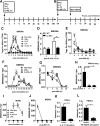
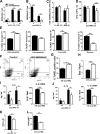
FIG. 1.
FIG. 1.
MIR99AHG is upregulated in M2 (IL-4/IL-13) in murine and human macrophages, downregulated following Mtb HN878 infection, and dependent on the IL-4Rα pathway. Bone marrow cells were differentiated for 10 days into BMDMs and stimulated with IFNγ or IL-4, IL-13, IL-4/IL-13. At 24 h poststimulation, BMDMs were infected with Mtb HN878 for 4, 24, and 48 h. RNA was extracted from lysed cells at different time points poststimulation and post-Mtb HN878 infection. (A, B) Timeline of mouse macrophage cytokine stimulation and Mtb HN878 infection. (C, E) CAGE analysis of MIR99AHG TPM kinetic expression in cytokine stimulated and Mtb HN878 infected BMDMs. Data are representative of three pooled independent experiments. (D) MIR99AHG mRNA expression by RT-qPCR from IL-4Rα–/– BMDMs stimulated with IL-4/IL-13 for 4 and 6 h. Data are representative of three pooled independent experiments. (F, G) RT-qPCR analysis of MIR99AHG kinetic mRNA expression in cytokine stimulated and Mtb HN878 infected BMDMs. Data are representative of three pooled independent experiments. (H) RT-qPCR analysis of MIR99AHG mRNA expression on whole lung from wild-type BALB/c mice infected with Mtb HN878 (100 CFU/mouse) for 11 and 21 days. Data are representative of three independent experiments. (I) RT-qPCR analysis of MIR99AHG kinetic expression in cytokine stimulated human MDMs. Data are representative of three pooled independent experiments. (J) RT-qPCR analysis of MIR99AHG kinetic expression in Mtb HN878 infected human MDMs. (K, L) Human MIR99AHG mRNA expression by RT-qPCR following IL-4/IL-13 and heat killed Mtb HN878 at 4 h poststimulation in PBMCs isolated from healthy and active TB patients. The fold change in gene expression was determined by RT-qPCR and was normalized to the housekeeping gene HPRT. Data are expressed as mean ± SD of triplicates. Data are representative of three independent experiments. P values represented as *P < 0.05, **P < 0.01, and ***P < 0.001; Two-way ANOVA (C–F) and Bonferroni post hoc test and Student's t-test (F). ANOVA, analysis of variance; BMDMs, bone marrow-derived macrophages; CAGE, cap analysis of gene expression; MDMs, monocyte-derived macrophages; PBMCs, peripheral blood mononuclear cells; RT-qPCR, quantitative real-time polymerase chain reaction; SD, standard deviation; TB, tuberculosis.
FIG. 2.
FIG. 2.
MIR99AHG downregulation by Mtb in murine macrophages is mediated by the NF-κB but not the p38 signaling pathway, and MIR99AHG expression is differentially regulated by TLR agonists. (A–C) RT-qPCR of MIR99AHG mRNA expression in BMDMs stimulated with TLR-9 agonist CpG (500 nM), TLR-4 agonist LPS (100 ng/mL), and TLR-2 agonist Pam3Csk4 (100 ng/mL). Data are representative of three pooled independent experiments. (D) BMDMs were pretreated for 1 h with selective pharmacological inhibitors for NF-κB (Bay11–7082; 10 μM), p38 (SB203580; 5 μM) and infected with Mtb HN878 for 4 h. Expression of MIR99AHG mRNA was detected by RT-qPCR. Data are representative of three independent experiments. Data are expressed as mean ± SD of triplicates. P values are represented as *P < 0.05, **P < 0.01, and ***P < 0.001, Student's t-test. LPS, lipopolysaccharide; NS, not significant; TLR, toll like receptor.
FIG. 3.
FIG. 3.
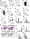
Knockdown of MIR99AHG by ASOs reduces intracellular Mtb growth, necrosis, pro-inflammatory cytokines and increases early apoptosis in murine macrophages. BMDMs were ASO transfected with locked nucleic acid control and ASO-MIR99AHG, stimulated with IL-4/IL-13 for 4 h and infected with Mtb HN878. (A) MIR99AHG mRNA expression by RT-qPCR in ASO-treated BMDMs post IL-4/IL-13 stimulation (B) and post Mtb HN878 infection. Data are representative of three independent experiments. (C) Cell viability was measured in BMDMs at 4 and 72 h post Mtb HN878 infection by CellTiter Blue. Data are representative of three independent experiments. (D) BMDMs were ASO transfected with control and ASO-MIR99AHG, prestimulated with IL-4/IL-13 for 4 h, and infected with Mtb HN878. BMDMs were lysed at 4 h uptake and 72 h post-Mtb HN878 infection to measure mycobacterial growth by CFU counting. Data are representative of three pooled independent experiments. (E–H) BMDMs were ASOs treated with control and ASO-MIR99AHG for 48 h, then stimulated with IL-4/IL-13 for 4 h and infected with Mtb HN878 for 24 h to measure early apoptotic, late apoptotic, necrotic, and live cells with Annexin V and 7-AAD staining by flow cytometry. Data are representative of three independent experiments. (F) Representative flow dot plots by flow cytometry. (G) At 24 h post Mtb HN878 infection, BMDMs were labeled with the TUNEL reaction mixture and the percentages of TUNEL positive apoptotic cells analyzed by fluorescent microscopy. Data are representative of two independent experiments. (H) BMDMs were ASO transfected with control and ASO-MIR99AHG, prestimulated with IL-4/IL-13 for 4 h, infected with Mtb HN878, and RNA collected at 24 h post Mtb. Bax mRNA expression by RT-qPCR in BMDMs infected with Mtb HN878 at 24 h postinfection. Data are representative of three independent experiments. (I, J) Protein and mRNA cytokine levels of IL-6 and IL-1β measured by ELISA and RT-qPCR. Data are representative of three independent experiments. (K, L) Nitrite production measured by Griess reagent assay and CD86 gMFI by flow cytometry. Data are representative of three independent experiments. Data are expressed as mean ± SD of triplicates (A, B, C, E, H, K, L) and quadruplets (D, I, J). P values represented as *P < 0.05, **P < 0.01, ***P < 0.001, and ****P < 0.0001; Student's t-test. ASO, antisense oligonucleotide; CFU, colony forming unit; ELISA, enzyme-linked immunosorbent assay; Mtb, Mycobacterium tuberculosis; TUNEL, terminal deoxynucleotidyl transferase dUTP nick-end labeling.
FIG. 4.
FIG. 4.
Knockdown of MIR99AHG by ASOs reduces intracellular Mtb growth, IL-6, and Bax in Mtb-infected human macrophages. (A) MDMs were ASOs transfected with control and ASO-MIR99AHG, prestimulated with IL-4/IL-13 and infected with Mtb HN878. MIR99AHG mRNA expression by RT-qPCR in ASO-treated MDMs. Data are representative of three independent experiments. (B) Cell viability of ASO transfected human MDMs prestimulated with IL-4/IL-13 for 4 h and infected with Mtb HN878. Data are representative of three independent experiments. (C) MDMs were ASO transfected with control and ASO-MIR99AHG, prestimulated with IL-4/IL-13 for 4 h and infected with Mtb HN878. Cells were lysed at 4 h for uptake and 24 and 48 h post-Mtb HN878 infection to measure bacterial growth by CFU counting. Data are representative of three pooled independent experiments. (D) Bax mRNA expression by RT-qPCR in human MDMs infected with Mtb HN878. Data are representative of three independent experiments. (E) IL-6 production in human MDMs prestimulated with IL-/IL-13 for 4 h and infected with Mtb HN878 measured by ELISA. Data are representative of three independent experiments. Data are expressed as mean ± SD of triplicates. P values represented as *P < 0.05, **P < 0.01, and ***P < 0.001, Student's t-test.
FIG. 5.
FIG. 5.
In vivo ASO knockdown of MIR99AHG reduces mycobacterial burden in mice and pro-inflammatory responses in lung macrophages. BALB/c mice (n = 6/mice/group) were ASO treated with 10 mg/kg locked nucleic acid control and ASO-MIR99AHG. (A) MIR99AHG mRNA expression by RT-qPCR at 14 days post ASO treatment. (B, C) Mycobacterial burden of Mtb HN878-infected mice is shown with indicated CFU in lung and spleen at 3 weeks postinfection. (D) Mtb HN878 infected mice were sacrificed at 24 h postinfection to determine the CFU lung uptake (n = 3 mice/group). (E) Percentage of lung neutrophils at 3 weeks postinfection quantified by flow cytometry. (F–I) The percentage of positive MPO, CD3, Caspase-3, and iNOS staining was quantified from two deep cut lung sections per mice at 3 weeks post Mtb HN878 infection (30 μm apart). (J) Alveolar spaces at 3 weeks postinfection were quantified from four deep cut H&E lung sections per mice (30 μm apart). (K) Representative histopathology sections (2 × magnification) at 3 weeks postinfection for H&E and iNOS (scale bar = 1,000 μm). (L–Q) At 3 weeks postinfection, CD64+CD11c+SiglecF+ AlvM, CD11b+CD11c+CD64+ (moDCs), CD64+CD11b+CD11c-SiglecF IntM, and CD11b+LY6G+ neutrophils were sorted by flow cytometry to determine lung mRNA expression of MIR99AHG, IL6, IL1b, Nos2, Arg1, and Mrc1 by RT-qPCR. Data are expressed as mean ± SD. P values represented as *P < 0.05, **P < 0.01, and ****P < 0.0001; Student's t-test. AlvM, alveolar macrophages; iNOS, inducible nitric oxide synthase; IntM, interstitial recruited macrophage, MPO, myeloperoxidase; N.D., not detected.
FIG. 6.
FIG. 6.
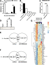
MIR99AHG is translocated from the cytoplasm to the nucleus following IL-4/IL-13 stimulation and interacts with hnRNPA2/B1 in murine macrophages. (A) RT-qPCR analysis of RNAs purified from nuclear and cytoplasm compartments in BMDMs stimulated with IL-4/IL-13 for 4h. Data are representative of three independent experiments. (B) The six most enriched proteins associated with MIR99AHG identified by affinity purification and mass spectrometry in ASO treated BMDMs prestimulated with IL-4/IL-13 for 4 h and infected with Mtb HN878 for 24 h. Data are representative of three independent experiments. (C) hnRNPA2/B1 RIP followed by mRNA expression of MIR99AHG by RT-qPCR in anti-hnRNPA2/B1 antibody or control IgG immunoprecipitates from nuclear lysates of macrophages prestimulated with IL-4/IL-13 for 4 h and infected with Mtb HN878 for 24 h. (D) Western blot of hnRNPA2/B1 in Input, anti-hnRNPA2/B1 antibody, and control IgG immunoprecipitates from nuclear lysates of macrophages prestimulated with IL-4/IL-13 for 4 h and infected with Mtb HN878 for 24 h. (E–G) BMDMs were ASO transfected with locked nucleic acid control and ASO-MIR99AHG for 48 h and infected with Mtb HN878 for 4 and 24 h. (E, F) Venn diagrams displaying up- and downregulated genes between control ASO-MIR99AHG at 4 and 24 h post Mtb infection. Data are representative of three independent experiments. (G) Heatmap representative of genes commonly upregulated and downregulated at 4 and 24 h post Mtb infection. Each row (gene) was centered on the mean expression value across all samples. Data are representative of three independent experiments. Data shown are mean of three replicates. Data are expressed as mean ± SD (A, C) of triplicates. P values represented as ***P < 0.001 and ****P < 0.0001, Student's t-test. RIP, RNA immunoprecipitation.

Similar articles

Cited by

References

    1. WHO. (2019). Global Tuberculosis Report. WHO Press.
    1. Flynn JL and Chan J. (2001). Immunology of tuberculosis. Annu Rev Immunol 19:93–129. - PubMed
    1. Guler R and Brombacher F. (2015). Host-directed drug therapy for tuberculosis. Nat Chem Biol 11:748–751. - PubMed
    1. Fitzgerald KA and Caffrey DR. (2014). Long noncoding RNAs in innate and adaptive immunity. Curr Opin Immunol 26:140–146. - PMC - PubMed
    1. Consortium EP, Birney E, Stamatoyannopoulos JA, Dutta A, Guigo R, Gingeras TR, Margulies EH, Weng Z, Snyder Met al. (2007). Identification and analysis of functional elements in 1% of the human genome by the ENCODE pilot project. Nature 447:799–816. - PMC - PubMed

Publication types