. 2022 Jul 29;8(30):eabm6376.
doi: 10.1126/sciadv.abm6376. Epub 2022 Jul 27.

A quantitative biology approach correlates neuronal toxicity with the largest inclusions of TDP-43

Affiliations

A quantitative biology approach correlates neuronal toxicity with the largest inclusions of TDP-43

Roberta Cascella et al. Sci Adv. .

Abstract

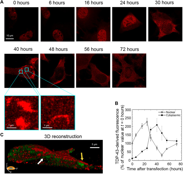
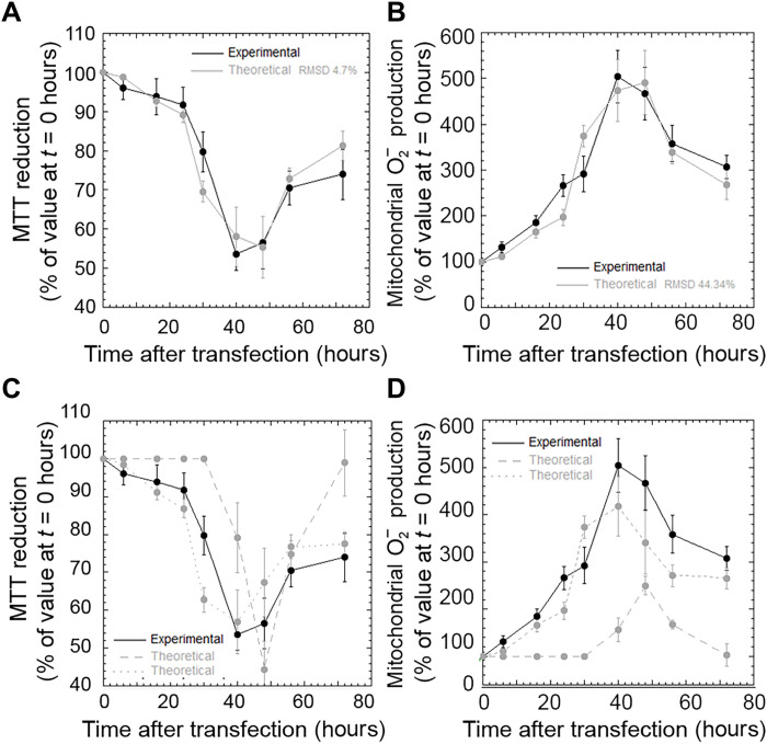
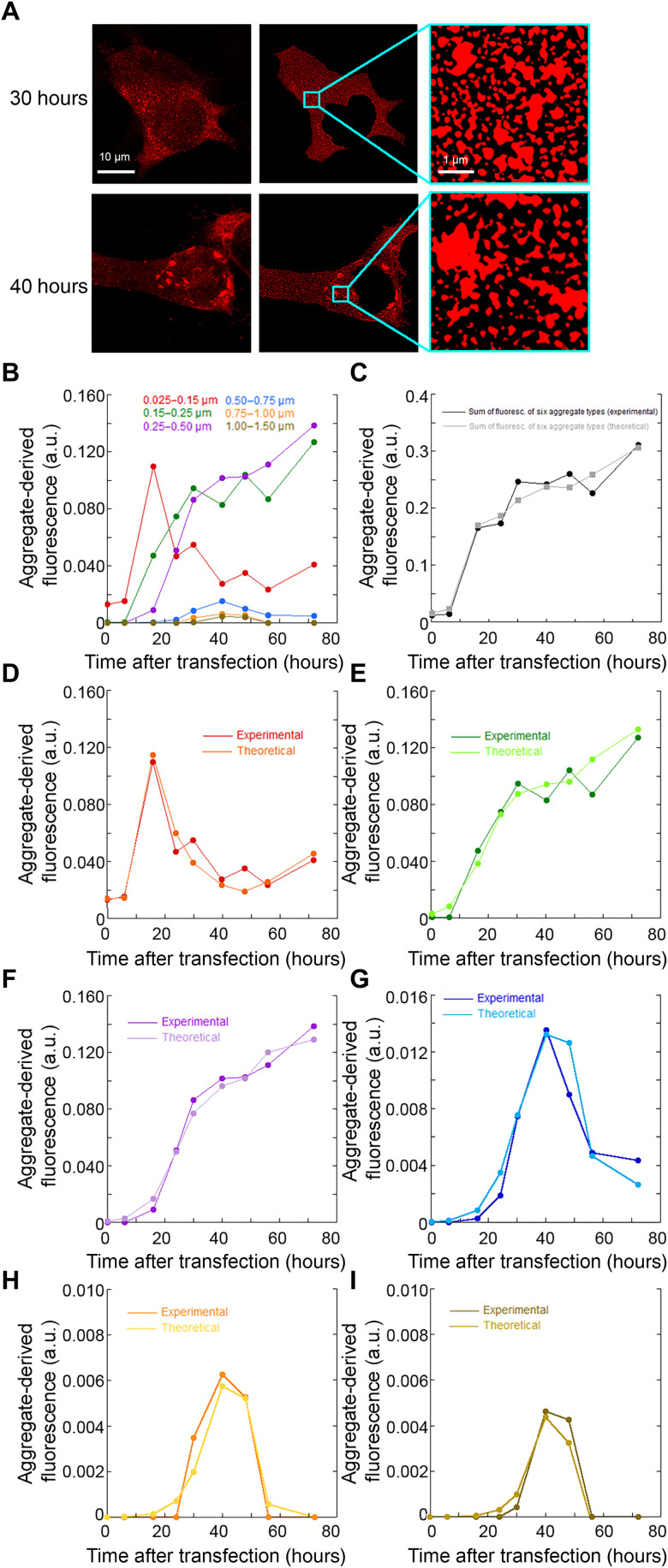
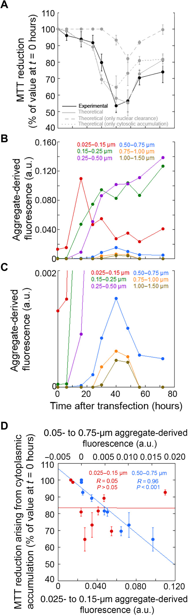
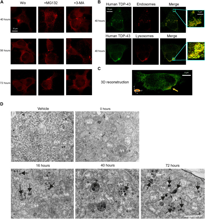
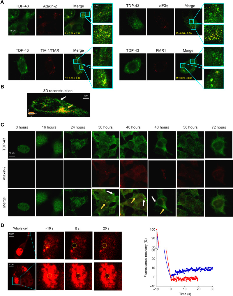
A number of neurodegenerative conditions are associated with the formation of cytosolic inclusions of TDP-43 within neurons. We expressed full-length TDP-43 in a motoneuron/neuroblastoma hybrid cell line (NSC-34) and exploited the high-resolution power of stimulated emission depletion microscopy to monitor the changes of nuclear and cytoplasmic TDP-43 levels and the formation of various size classes of cytoplasmic TDP-43 aggregates with time. Concomitantly, we monitored oxidative stress and mitochondrial impairment using the MitoSOX and MTT reduction assays, respectively. Using a quantitative biology approach, we attributed neuronal dysfunction associated with cytoplasmic deposition component to the formation of the largest inclusions, independently of stress granules. This is in contrast to other neurodegenerative diseases where toxicity is attributed to small oligomers. Using specific inhibitors, markers, and electron microscopy, the proteasome and autophagy were found to target mainly the largest deleterious inclusions, but their efficiency soon decreases without full recovery of neuronal viability.


Figures

Fig. 1.
Fig. 1.. Overexpression of TDP-43 caused a redistribution of TDP-43 between the nucleus and the cytoplasm.
(A) Representative STED microscopy images of NSC-34 cells (n = 3) transfected with 20 μg of pCI-neo plasmid expressing human TDP-43 and analyzed at different lengths of time after transfection. Red fluorescence, total TDP-43 (endogenous and exogenous). Higher magnifications show round (left) and filamentous (right) TDP-43 inclusions. (B) Time courses of nuclear and cytoplasmic TDP-43–derived fluorescence determined over 200 to 250 NSC-34 cells and expressed as a percentage of the value for cells transfected for 0 hours, taken as 100% (n = 3). (C) 3D reconstruction of the z-stack analysis (5-μm-thick slices) of the specimens shown in (A) (40 hours). Red and green fluorescence, cell membrane (WGA) and exogenous (human) TDP-43, respectively. An NSC-34 cell was virtually dissected on the zy plane to show more clearly the intracellular TDP-43 inclusions. Yellow and white arrows, round and filamentous inclusions, respectively.
Fig. 2.
Fig. 2.. Both nuclear depletion and cytoplasmic accumulation of TDP-43 are relevant for neuronal dysfunction.
(A to D) Experimental (black) and theoretical (gray) time courses of MTT reduction (A and C) (n = 3) and mitochondrial superoxide production (B and D) (n = 3) in NSC-34 cells treated as in (A). The theoretical traces refer to nuclear depletion and cytoplasmic accumulation together (gray solid lines), only nuclear depletion (gray dashed lines), and only cytoplasmic accumulation (gray dotted lines). Experimental errors are SEMs. The degree of agreement [root mean square deviation (RMSD)] between experimental and theoretical analyses is also reported.
Fig. 3.
Fig. 3.. Time courses of different size classes of TDP-43 inclusions.
(A) Approach used to identify size classes of TDP-43 inclusions: STED microscopy images of NSC-34 cells (n = 3) transfected with pCI-neo plasmid expressing human TDP-43 (left) were treated to exclude the nucleus area, after background subtraction, to obtain a better definition of the cytoplasmic inclusions (middle) and magnified to identify inclusions (right) and count them, as described in Materials and Methods. (B) Time courses of fluorescence density [F(t)/A] associated with each of the six size classes of cytoplasmic TDP-43 inclusions. (C) Experimental and theoretical time courses of total fluorescence density [F(t)/A] for the six aggregate types together. (D to I) Experimental and theoretical time courses of fluorescence density [F(t)/A] for the individual inclusion classes: diameters of 0.025 to 0.15 μm (D), 0.15 to 0.25 μm (E), 0.25 to 0.50 μm (F), 0.50 to 0.75 μm (G), 0.75 to 1.00 μm (H), and 1.00 to 1.50 μm (I). a.u., arbitrary units.
Fig. 4.
Fig. 4.. Identification of the mechanism of TDP-43 inclusion formation and toxic inclusions by a quantitative analytical approach.
(A) Experimental (black line) and theoretical (gray solid line) time course analysis of MTT reduction (n = 3), expressed as the percentage of the value for cells transfected at 0 hours (taken as 100%). The theoretical contributions of nuclear depletion (dashed line) and cytoplasmic accumulation (dotted line) are also shown. Experimental errors are SEMs. (B and C) Experimental time courses for the individual inclusion classes (same as those in Fig. 3B). Classes 4 to 6 are magnified in the second panel. (D) Dependence of the contribution of TDP-43 cytoplasmic accumulation to MTT reduction on the aggregate-derived fluorescence of the indicated size classes. Experimental errors are SEMs.
Fig. 5.
Fig. 5.. Large inclusions are degraded by proteasome and autophagy.
(A) Representative STED microscopy images of NSC-34 cells (n = 3) with 20 μg of pCI-neo plasmid expressing human TDP-43, treated at 38 hours following cell transfection with 5 μM MG132 or 10 mM 3-MA and analyzed 40, 56, and 72 hours after transfection. Red fluorescence, total TDP-43 (human and murine). (B) Representative STED microscopy images of NSC-34 cells (n = 3) transfected with 20 μg of pCI-neo plasmid expressing human TDP-43 and analyzed 40 hours after transfection. Green and red fluorescence, human TDP-43 (exogenous) and endosomes (top) or lysosomes (bottom), respectively. Magnification boxes, areas with TDP-43 assemblies with a high degree of colocalization with endosomes (top) or lysosomes (bottom). (C) 3D reconstruction of the z-stack analysis (5-μm-thick slices) of the specimens shown in (B), bottom right. An NSC-34 cell was virtually dissected on the zy plane to show more clearly the intracellular TDP-43 inclusions colocalizing with lysosomes. (D). Representative TEM images of NSC-34 cells (n = 3) transfected with vehicle or 20 μg of pCI-neo plasmid expressing TDP-43 and analyzed 0, 16, 40, and 72 hours after transfection. Labels indicate lysosomes (lys), mitochondria (mit), and rough endoplasmic reticulum (ER). Black arrows, cytoplasmic TDP-43 inclusions. Scale bar, 1 μm.
Fig. 6.
Fig. 6.. TDP-43 is recruited to SGs, but the formation of TDP-43 inclusions is driven by different pathways.
(A) Representative STED microscopy images of NSC-34 cells (n = 3) transfected with 20 μg of pCI-neo plasmid expressing human TDP-43 and observed at 40 hours after transfection. The green and red fluorescence indicate total TDP-43 (endogenous and exogenous) and the indicated SG markers, respectively. The top and bottom magnification boxes indicate areas with TDP-43 assemblies with a low and high degree of colocalization with SG markers, respectively. R values are indicated in merge images. (B) 3D reconstruction of the z-stack analysis (5-μm-thick slices) of the specimens shown in (A). A cell was virtually dissected on the zy plane to show the intracellular TDP-43 inclusions (green) with (yellow arrow) and without (blank arrow) colocalization with the SG marker ataxin-2 (red). (C) Representative confocal scanning microscopy images of NSC-34 cells (n = 3) transfected with 20 μg of pCI-neo plasmid expressing total TDP-43 (endogenous and exogenous) and analyzed after different lengths of time after transfection. The green and red fluorescence indicate TDP-43 and ataxin-2, respectively. Yellow and white arrows indicate TDP-43 inclusions with or without colocalization with ataxin-2, respectively. (D) Representative images of two TDP-43 inclusions in NSC-34 cells (n = 3) transfected with 25 μg of wtTDP43tdTOMATOHA plasmid expressing tdTOMATO-labeled human TDP-43 and observed at 40 hours after transfection. The yellow circled regions of interest (ROIs) were photobleached starting at time −10 s for 10 s, and FRAP was then monitored continuously for 20 to 30 s starting at time 0 s. FRAP was plotted over time (blue and red curves correspond to top and bottom panels, respectively). Fluorescence values were normalized to the average intensity of each ROI before photobleaching (−10 s).

Similar articles

Cited by

References

    1. Arai T., Hasegawa M., Akiyama H., Ikeda K., Nonaka T., Mori H., Mann D., Tsuchiya K., Yoshida M., Hashizume Y., Oda T., TDP-43 is a component of ubiquitin-positive tau-negative inclusions in frontotemporal lobar degeneration and amyotrophic lateral sclerosis. Biochem. Biophys. Res. Commun. 351, 602–611 (2006). - PubMed
    1. Neumann M., Sampathu D. M., Kwong L. K., Truax A. C., Micsenyi M. C., Chou T. T., Bruce J., Schuck T., Grossman M., Clark C. M., McCluskey L. F., Miller B. L., Masliah E., Mackenzie I. R., Feldman H., Feiden W., Kretzschmar H. A., Trojanowski J. Q., Lee V. M. Y., Ubiquitinated TDP-43 in frontotemporal lobar degeneration and amyotrophic lateral sclerosis. Science 314, 130–133 (2006). - PubMed
    1. Mackenzie I. R., Bigio E. H., Ince P. G., Geser F., Neumann M., Cairns N. J., Kwong L. K., Forman M. S., Ravits J., Stewart H., Eisen A., McClusky L., Kretzschmar H. A., Monoranu C. M., Highley J. R., Kirby J., Siddique T., Shaw P. J., Lee V. M., Trojanowski J. Q., Pathological TDP-43 distinguishes sporadic amyotrophic lateral sclerosis from amyotrophic lateral sclerosis with SOD1 mutations. Ann. Neurol. 61, 427–434 (2007). - PubMed
    1. Brettschneider J., Del Tredici K., Toledo J. B., Robinson J. L., Irwin D. J., Grossman M., Suh E., Van Deerlin V. M., Wood E. M., Baek Y., Kwong L., Lee E. B., Elman L., McCluskey L., Fang L., Feldengut S., Ludolph A. C., Lee V. M., Braak H., Trojanowski J. Q., Stages of pTDP-43 pathology in amyotrophic lateral sclerosis. Ann. Neurol. 74, 20–38 (2013). - PMC - PubMed
    1. Hasegawa M., Arai T., Nonaka T., Kametani F., Yoshida M., Hashizume Y., Beach T. G., Buratti E., Baralle F., Morita M., Nakano I., Oda T., Tsuchiya K., Akiyama H., Phosphorylated TDP-43 in frontotemporal lobar degeneration and amyotrophic lateral sclerosis. Ann. Neurol. 64, 60–70 (2008). - PMC - PubMed