. 2022 Sep 29;140(13):1549-1564.
doi: 10.1182/blood.2022016424.

The Gab2-MALT1 axis regulates thromboinflammation and deep vein thrombosis

Affiliations

The Gab2-MALT1 axis regulates thromboinflammation and deep vein thrombosis

Vijay Kondreddy et al. Blood. .

Abstract

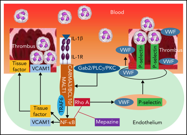
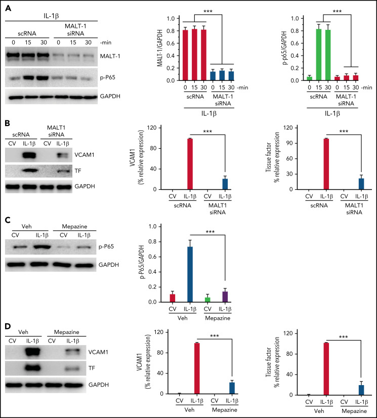
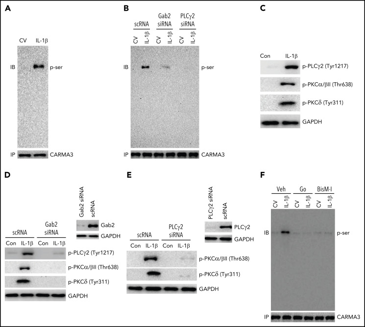
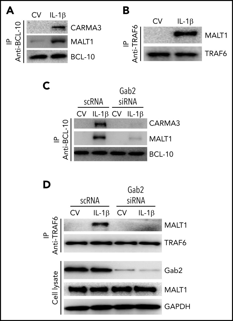
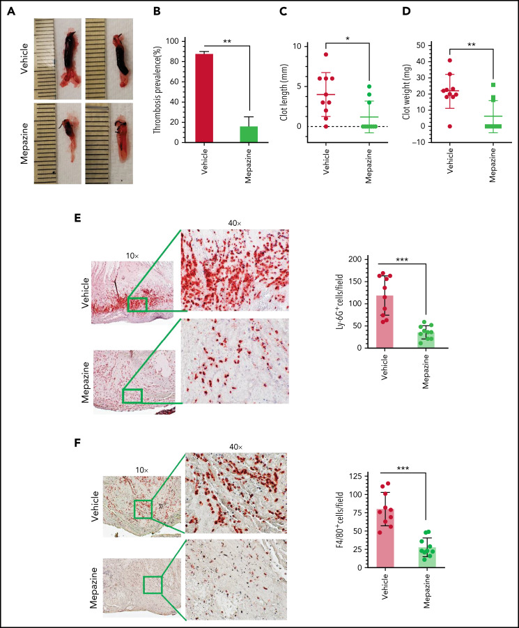
Deep vein thrombosis (DVT) is the third most common cause of cardiovascular mortality. Several studies suggest that DVT occurs at the intersection of dysregulated inflammation and coagulation upon activation of inflammasome and secretion of interleukin 1β (IL-1β) in restricted venous flow conditions. Our recent studies showed a signaling adapter protein, Gab2 (Grb2-associated binder 2), plays a crucial role in propagating inflammatory signaling triggered by IL-1β and other inflammatory mediators in endothelial cells. The present study shows that Gab2 facilitates the assembly of the CBM (CARMA3 [CARD recruited membrane-associated guanylate kinase protein 3]-BCL-10 [B-cell lymphoma 10]-MALT1 [mucosa-associated lymphoid tissue lymphoma translocation protein 1]) signalosome, which mediates the activation of Rho and NF-κB in endothelial cells. Gene silencing of Gab2 or MALT1, the effector signaling molecule in the CBM signalosome, or pharmacological inhibition of MALT1 with a specific inhibitor, mepazine, significantly reduced IL-1β-induced Rho-dependent exocytosis of P-selectin and von Willebrand factor (VWF) and the subsequent adhesion of neutrophils to endothelial cells. MALT1 inhibition also reduced IL-1β-induced NF-κB-dependent expression of tissue factor and vascular cell adhesion molecule 1. Consistent with the in vitro data, Gab2 deficiency or pharmacological inhibition of MALT1 suppressed the accumulation of monocytes and neutrophils at the injury site and attenuated venous thrombosis induced by the inferior vena cava ligation-induced stenosis or stasis in mice. Overall, our data reveal a previously unrecognized role of the Gab2-MALT1 axis in thromboinflammation. Targeting the Gab2-MALT1 axis with MALT1 inhibitors may become an effective strategy to treat DVT by suppressing thromboinflammation without inducing bleeding complications.


Figures

None
Graphical abstract
Figure 1.
Figure 1.
Gab2 is essential for IL-1β–induced exocytosis of P-selectin and VWF in endothelial cells and the adhesion of neutrophils to activated endothelial cells. (A) IL-1β induces P-selectin expression on the endothelial cell surface. HUVECs were treated with IL-1β (10 ng/mL) or TNFα (10 ng/mL) alone or in combination for 1 hour. At the end of 1 hour, the cells were fixed in 2% paraformaldehyde (PFA), and the surface expression of P-selectin was measured by cell surface ELISA. Since the inclusion of TNFα in IL-1β treatment gave a robust increase in P-selectin translocation to the cell surface, in all experiments described in this and other figures that examined the exocytosis of P-selectin and VWF, IL-1β was supplemented with TNFα in treating endothelial cells. (B) Gab2 silencing does not affect total P-selectin concentrations. HUVECs were transfected with 200 nM scrambled RNA (scRNA) or Gab2 small interfering RNA(siRNA). After 48 hours, the cells were treated with IL-1β for 1 hour. The cells were lysed, and the concentrations of Gab2 and P-selectin were analyzed by immunoblotting. Densitometric analysis of immunoblots showed that Gab2 silencing knocked down around 85% of Gab2 and no change in the total P-selectin. (C) Gab2 silencing impairs the translocation of P-selectin to the cell surface. HUVECs transfected with scRNA or Gab2 siRNA were treated with IL-1β for 1 hour. The cell surface expression of P-selectin was measured by cell surface ELISA. (D-E) Gab2 silencing impairs IL-1β–induced expression of P-selectin at the cell surface without impairing the total cellular expression of P-selectin. HUVECs, cultured on cover glasses, were transfected with 200 nM of scRNA or Gab2 siRNA. After 48 hours, the cells were treated with IL-1β for 1 hour. P-selectin expression was analyzed in (D) unpermeabilized or (E) permeabilized cells by immunofluorescence microscopy. (F) Gab2 silencing suppresses IL-1β–induced secretion of VWF. HUVECs transfected with scRNA or Gab2 siRNA were treated with IL-1β for 1 hour. At the end of 1 hour, cell supernatants were removed and precipitated with trichloroacetic acid (TCA) (6%) to concentrate proteins. The suspension of TCA precipitates was subjected to immunoblot analysis to measure secreted (S) VWF. As a control, VWF in cell lysates was also analyzed by immunoblot analysis. Concentrations of secreted VWF were quantified by densitometric analysis of VWF (S) band, and the value obtained in control scRNA transfected cells was arbitrarily assigned as 1. The top panel shows representative immunoblot, and the bottom panel shows densitometric quantification of the VWF (S) band. (G) P-selectin-dependent adhesion of neutrophils to activated endothelial cells. HUVECs cultured on cover glasses were treated with IL-1β for 1 or 6 hours. The cells were washed and incubated with a P-selectin–blocking monoclonal antibody or control IgG (10 µg/mL) for 1 hour. After that, monolayers of HUVECs were layered with PKH-labeled human neutrophils (5 × 10⁵/mL). Neutrophils were allowed to adhere for 30 minutes. Then, unbound neutrophils were removed, the endothelial cell monolayers were washed, and the adhered neutrophils to endothelial cells were fixed in 4% PFA for 15 minutes. The cover glass was mounted with antifade Fluro gel, and the cells were visualized under a confocal microscope at 20× magnification. The micrographs shown were the representative images of 3 independent experiments. The adhered cells were enumerated at 3 random locations in each cover glass, and these data were presented in the right-side panel. (H) Gab2 silencing in endothelial cells attenuates neutrophil adhesion to activated endothelial cells. HUVECs transfected with scRNA or Gab2 siRNA were treated with IL-1β, and the adhesion of neutrophils to activated endothelial cells was analyzed as described in panel (G). (I) IL-1β–induced expression of VCAM1. HUVECs were treated with IL-1β for 1 or 6 hours. The cell lysates were probed for VCAM1 by immunoblot analysis. Data are the mean ± standard deviation of 3 independent experiments. *P < .05, **P < .01, and ***P < .001. ns, no statistically significant difference.
Figure 2.
Figure 2.
IL-1β–induced and Gab2-dependent mobilization of P-selectin and VWF was Rho-dependent and independent of TAK1 and NF-κB. (A) IL-1β activates Rho kinase in endothelial cells. HUVECs were treated with a control vehicle or Rho-specific inhibitor, rhosin, for 1 hour. After that, the cells were stimulated with IL-1β for indicated periods. Rho activation was measured as GTP-bound Rho using the Rho activation assay kit (left panel). Rho-GTP signals were quantified by densitometric analysis, and the signal intensity in cells not treated with IL-1β or rhosin was taken as 1. (B) Gab2 silencing blocks IL-1β–induced Rho activation in endothelial cells. HUVECs were transfected with 200 nM of scRNA or Gab2 siRNA. After 48 hours, the transfected cells were treated with IL-1β for indicated time periods. Rho activation was measured and quantified as described in (A). (C) Pharmacological inhibition of Rho activation attenuates IL-1β–induced translocation of P-selectin to the cell surface and VWF secretion. HUVECs were treated with a control vehicle or Rho-specific inhibitors, rhosin or Y27632 (10 µM), for 1 hour. Then, the cells were stimulated with IL-1β. After 1 hour, the cell supernatants were collected, and cell lysates were harvested. Cell lysates were probed for total P-selectin, VWF, and GAPDH. Cell supernatants were precipitated with TCA to concentrate proteins and probed for VWF by immunoblot analysis to assess secreted (S) VWF (left panel). The translocation of P-selectin to the cell surface was measured by cell surface ELISA (middle panel). VWF secretion was quantified by densitometric analysis of VWF (S) immunoblots (right panel). (D) TAK1 silencing does not affect IL-1β–induced translocation of P-selectin to the cell surface or VWF secretion. HUVECs were transfected with scRNA or TAK1 siRNA (200 nM) for 48 hours. The transfected cells were stimulated with IL-1β for 1 hour. P-selectin translocation to the cell surface and VWF secretion were evaluated as described in (C). (E) Pharmacological inhibition of NF-κB does not curtail IL-1β–induced translocation of P-selectin to the cell surface or VWF secretion. HUVECs were treated with an NF-κB–specific inhibitor, BAY117082 (20 µM), or a control vehicle for 1 hour. Thereafter, the cells were stimulated with IL-1β for 1 hour, and P-selectin translocation to the cell surface and VWF secretion were evaluated as described in (C). *P < .05, **P < .01, and ***P < .001. ns, no statistically significant difference.
Figure 3.
Figure 3.
MALT-1 regulates IL-1β–induced Rho activation, mobilization of P-selectin and VWF, and neutrophil adhesion to activated endothelial cells. (A) MALT1 silencing suppresses IL-1β–induced Rho activation in endothelial cells. HUVECs were transfected with 200 nM of scRNA or MALT1 siRNA. After 48 hours, the transfected cells were treated with IL-1β for indicated time periods. Rho activation was measured as GTP-bound Rho as described in the Methods section. The left panel shows a representative blot (t-Rho in the blot indicates total Rho). The right panel shows the quantification of Rho-GTP concentrations from densitometric analysis of signals of immunoblots. Rho-GTP concentrations measured in scRNA-transfected cells and not treated with IL-1β were taken as 1, and other values were shown relative to this value. (B) MALT1 silencing inhibits IL-1β–induced translocation of P-selectin to the cell surface and secretion of VWF. HUVECs transfected with scRNA or MALT1 siRNA were treated with a control vehicle or IL-1β for 1 hour. After 1 hour, the cell supernatants were collected, and cell lysates were harvested. Cell supernatants were precipitated with TCA to concentrate proteins. Cell lysates were probed for MALT1, P-selectin, and VWF by immunoblot analysis; cell supernatants were probed for VWF by immunoblot analysis to assess the concentrations of secreted (S) VWF (left panel). The translocation of P-selectin to the cell surface was measured by cell surface ELISA (middle panel). VWF secretion was quantified by densitometric analysis of VWF (S) immunoblots (right panel). Concentrations of P-selectin on the cell surface and secreted VWF measured in cells transfected with scRNA and treated with a control vehicle were taken as 1.0, and other values were shown relative to these. (C) Pharmacological inhibition of MALT1 attenuates IL-1β–induced P-selectin translocation to the cell surface and secretion of VWF. HUVECs were treated with MALT1 inhibitor, mepazine (20 µM), or vehicle (veh) for 1 hour. After that, the cells were treated with a control vehicle (CV) or IL-1β for 1 hour. The assessment of P-selectin translocation to the cell surface and VWF secretion was performed as described in (B). (D-E) (D) MALT1 silencing or (E) pharmacological inhibition attenuates neutrophil adhesion to IL-1β–activated endothelial cells. HUVECs cultured on cover glasses were transfected with scRNA, MALT1 siRNA, or treated with MALT1 inhibitor, mepazine, as described in the figure legends of (B-C). HUVECs were stimulated with IL-1β for 1 or 6 hours. After washing the cells, PKH-labeled human neutrophils (5 × 10⁵/mL) were added to endothelial cells. After 30 minutes, the unbound neutrophils were removed, and endothelial cells were washed. Neutrophils adhered to endothelial cells were fixed in 4% PFA for 15 minutes. The cells were visualized and imaged under 20× magnification using a confocal microscope (left panel). The cell count enumerated at 3 random locations of each cover glass of 3 independent experiments was shown in the bar graph (right panel). **P < .01 and ***P < .001.
Figure 4.
Figure 4.
MALT1 inhibition suppresses IL-1β–induced thromboinflammatory gene expression. (A) MALT1 silencing suppresses IL-1β–induced NF-κB activation. HUVECs were transfected with scRNA or MALT1 siRNA (200 nM). After 48 hours, the transfected cells were stimulated with IL-1β for varying times. At the end of treatment, cell lysates were harvested, and the phosphorylation of the P65 subunit of NF-κB was analyzed by immunoblot analysis. The blot was also probed for MALT1 to confirm MALT1 knockdown and GAPDH as a loading control. The left panel shows a representative immunoblot, and the middle and right panels show quantification of MALT1 knockdown and p65 phosphorylation by densitometric analysis of immunoblots. (B) MALT1 silencing suppresses IL-1β–induced expression of TF and VCAM1. HUVECs transfected with scRNA or MALT1 siRNA were treated with a control vehicle (CV) or IL-1β for 6 hours. The cell lysates were probed for VCAM1 and TF expression by immunoblot analysis. The left panel shows a representative immunoblot, and the middle and right panels show quantification of VCAM1 and TF concentrations by densitometric analysis of immunoblots. The concentrations of VCAM1 and TF measured in IL-1β–stimulated cells were taken as 100%, and other values were shown relative to these values. (C-D) (C) MALT1 inhibitor attenuates IL-1β–induced NF-κB activation and (D) expression of VCAM1 and TF. HUVEC were treated with MALT1-specific inhibitor mepazine (20 µM) for 1 hourand then the cells were treated with a control vehcile (CV) or IL-1β for 30 minutes (C) or 6 hours (D). The cell lysates were probed for the phosphorylation of p65 (C) or the expression of VCAM1 and TF (D). The left panel shows a representative immunoblot, and the middle and right panels show densitometric quantification of immunoblot band intensities. The concentrations of p65 were normalized to GAPDH (C). VCAM1 and TF concentrations measured in IL-1β–stimulated cells were taken as 100%, and other values were shown relative to these values. Data are the mean ± standard deviation of 3 independent experiments. ***P < .001.
Figure 5.
Figure 5.
Gab2/PLCγ2/PKC axis mediates IL-1β–induced activation of CARMA3. (A) IL-1β treatment induces the phosphorylation of CARMA3 in endothelial cells. HUVEC were treated with a control vehicle or IL-1β for 5 minutes. The cells were lysed in radioimmunoprecipitation assay (RIPA) buffer, and the CARMA3 was immunoprecipitated using polyclonal CARMA3 antibodies. The immunoprecipitates were subjected to immunoblot analysis and probed with monoclonal antibodies against phosphoserine. (B) Gab2 or PLCγ2 silencing blocks IL-1β–induced phosphorylation of CARMA3. HUVECs transfected with scRNA, Gab2 siRNA, or PLCγ2 siRNA were treated with a control vehicle or IL-1β for 5 minutes. The cell lysates were immunoprecipitated with CARMA3 antibodies, and the immunoprecipitates were probed for the presence of phosphorylated CARMA3, as described in the legend to panel (A). (C) IL-1β induces the activation of PLCγ2 and PKC isomers in endothelial cells. HUVECs were treated with a control vehicle or IL-1β for 5 minutes. At the end of the 5-minute treatment, the cell lysates were harvested and probed for the phosphorylation of PLCγ2, PKCα/β, and PKCδ by immunoblot analysis using antibodies against phosphorylated PLCγ2, PKCα/β, and PKCδ. (D) Gab2 silencing inhibits IL-1β–induced activation of PLCγ2 and PKC isomers in endothelial cells. HUVECs were transfected with scRNA or Gab2 siRNA (200 nM). After 48 hours, the Gab2 knockdown was confirmed by immunoblot analysis. HUVECs transfected with scRNA or Gab2 siRNA were treated with a control vehicle or IL-1β for 5 minutes. The cell lysates were probed for the presence of phosphorylated PLCγ2, PKCα/β, and PKCδ by immunoblot analysis. (E) PLCγ2 silencing blocks the IL-1β–induced activation of PKC isomers. HUVECs were transfected with scRNA or PLCγ2 siRNA. After 48 hours, PLCγ2 knockdown was confirmed by immunoblotting. HUVECs transfected with scRNA or PLCγ2 siRNA were treated with a control vehicle or IL-1β for 5 minutes. The phosphorylation of PKCα/β and PKCδ were analyzed by immunoblot analysis. (F) Inhibition of PKC attenuates IL-1β–induced phosphorylation of CARMA3. HUVECs were treated with PKC α/β inhibitor Go6976 (Go; 100 nM), pan-PKC inhibitor bisindolylmaleimide 1 (BisM-1; 500 nM), or DMSO vehicle for 1 hour. After that, the cells were treated with a control vehicle or IL-1β for 5 minutes. The cell lysates were processed as described in the panel (D) and probed for the phosphorylation of CARMA3. The immunoblots shown were the representative blots of 3 independent experiments with similar results.
Figure 6.
Figure 6.
Gab2 facilitates IL-1β–induced assembly of the CBM signalosome and MALT1 interaction with TRAF6. (A) IL-1β induces the formation of the CBM signalosome. HUVECs were treated with a control vehicle or IL-1β for 5 minutes. The cell lysates were immunoprecipitated with BCL-10 antibodies, and the immunoprecipitates were probed for the presence of CARMA3, MALT1, and BCL-10 by immunoblot analysis. (B) IL-1β induces MALT1 association with TRAF6. HUVECs were treated with a control vehicle or IL-1β for 5 minutes, and the cell lysates were immunoprecipitated with TRAF6 antibodies. The immunoprecipitates were probed for the presence of MALT1 and TRAF6. (C) Gab2 silencing prevents IL-1β–induced CBM signalosome formation. HUVECs were transfected with scRNA or Gab2 siRNA (200 nM). After 48 hours, the cells were treated with a control vehicle or IL-1β for 5 minutes. The cell lysates were immunoprecipitated with BCL-10 antibodies, and the immunoprecipitates were probed for the presence of CARMA3, MALT1, and BCL-10 by immunoblot analysis. (D) Gab2 silencing abolishes IL-1β–induced MALT1 association with TRAF6. HUVECs transfected with scRNA or Gab2 siRNA were treated with a control vehicle of IL-1β for 5 minutes. The cell lysates were immunoprecipitated with TRAF6 antibodies, and the immunoprecipitates were probed for the presence of MALT1 and TRAF6. In addition, cell lysates were probed for Gab2, MALT1, and GAPDH.
Figure 7.
Figure 7.
MALT1 inhibition protects against venous thrombosis induced by flow restriction. C57/BL 6J wild-type mice were administered 2 doses of MALT1 inhibitor, mepazine (12.5 mg/kg body weight each dose, intraperitoneally), the first dose 2 hours before surgery, and a second dose 24 hours after the IVC ligation. Control mice were administered with a control vehicle (DMSO). Mice were subjected to the IVC ligation-induced stenosis. Forty-eight hours following the IVC ligation, mice were killed, and thrombus formation in the ligated vein was evaluated. (A) Representative images of thrombus; (B) thrombosis prevalence; (C) thrombus length; (D) thrombus weight. Thrombi collected were processed for tissue sectioning, and sections were stained with antibodies against (E) Ly-6G or (F) F4/80 antigens. The sections were visualized under a bright field microscope at 10×, and selected areas (boxed) were viewed at 40× magnification. The number of LY-6G or F4/80-positive cells was analyzed at 10 randomly chosen areas of the high-power field covering the entire section of 10 different mice. Data are mean ± standard deviation of 3 independent experiments. *P < .05, **P < .01, and ***P < .001.

Comment in

  • Endothelial cell-mediated venous thrombosis.
    Henke P. Henke P. Blood. 2022 Sep 29;140(13):1459-1460. doi: 10.1182/blood.2022017938. Blood. 2022. PMID: 36173663 No abstract available.
  • What the neighbors say.
    [No authors listed] [No authors listed] J Thromb Haemost. 2022 Dec;20(12):2721. doi: 10.1111/jth.15904. J Thromb Haemost. 2022. PMID: 36372446 No abstract available.

Similar articles

Cited by

References

    1. Colling ME, Tourdot BE, Kanthi Y. Inflammation, infection and venous thromboembolism. Circ Res. 2021;128(12):2017-2036. - PMC - PubMed
    1. Klemen ND, Feingold PL, Hashimoto B, et al. . Mortality risk associated with venous thromboembolism: a systematic review and Bayesian meta-analysis. Lancet Haematol. 2020;7(8):e583-e593. - PubMed
    1. Søgaard KK, Schmidt M, Pedersen L, Horváth-Puhó E, Sørensen HT. 30-year mortality after venous thromboembolism: a population-based cohort study. Circulation. 2014;130(10):829-836. - PubMed
    1. von Brühl ML, Stark K, Steinhart A, et al. . Monocytes, neutrophils, and platelets cooperate to initiate and propagate venous thrombosis in mice in vivo. J Exp Med. 2012;209(4):819-835. - PMC - PubMed
    1. Esmon CT. Basic mechanisms and pathogenesis of venous thrombosis. Blood Rev. 2009;23(5):225-229. - PMC - PubMed

Publication types

MeSH terms