Review
. 2022 Jul 27;31(165):220056.
doi: 10.1183/16000617.0056-2022. Print 2022 Sep 30.

A pulmonologist's guide to perform and analyse cross-species single lung cell transcriptomics

Affiliations
Review

A pulmonologist's guide to perform and analyse cross-species single lung cell transcriptomics

Peter Pennitz et al. Eur Respir Rev. .

Abstract

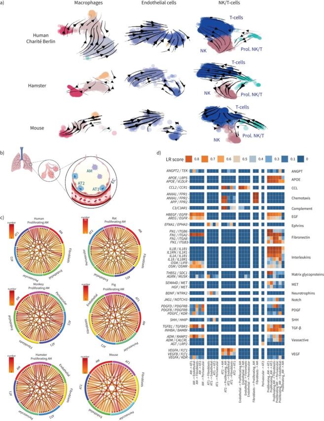
Single-cell ribonucleic acid sequencing is becoming widely employed to study biological processes at a novel resolution depth. The ability to analyse transcriptomes of multiple heterogeneous cell types in parallel is especially valuable for cell-focused lung research where a variety of resident and recruited cells are essential for maintaining organ functionality. We compared the single-cell transcriptomes from publicly available and unpublished datasets of the lungs in six different species: human (Homo sapiens), African green monkey (Chlorocebus sabaeus), pig (Sus domesticus), hamster (Mesocricetus auratus), rat (Rattus norvegicus) and mouse (Mus musculus) by employing RNA velocity and intercellular communication based on ligand-receptor co-expression, among other techniques. Specifically, we demonstrated a workflow for interspecies data integration, applied a single unified gene nomenclature, performed cell-specific clustering and identified marker genes for each species. Overall, integrative approaches combining newly sequenced as well as publicly available datasets could help identify species-specific transcriptomic signatures in both healthy and diseased lung tissue and select appropriate models for future respiratory research.


Conflict of interest statement

Conflict of interest: H. Kirsten reports support for the present manuscript from German Federal Ministry of Education and Research (BMBF) grants e:Med CAPSyS (01ZX1304A) and e:Med SYMPATH (01ZX1906B). E. Wyler has received payment or honoraria for lectures, presentations, speakers’ bureaus, manuscript writing or educational events from Podcast Gegenblende by DGB, outside the submitted work. M. Landthaler reports support for the present manuscript from Berlin Institute of Health. M. Scholz has received grants or contracts from Pfizer Inc. for a project not related to this research. W.M. Kuebler reports support for the present manuscript from German Research Foundation (KU 1218/9-1, KU 1218/11-1, CRC TR84 A02, CRC TR84 C09, CRC 1449 B01), Germany Ministry for Research and Education (SYMPATH, PROVID consortia), German Center for Cardiovascular Research (Partner site project Berlin) and Berlin Institute of Health (Focus Area Vascular Biology). M. Witzenrath has received grants or contracts from Deutsche Forschungsgemeinschaft, Bundesministerium für Bildung und Forschung, Deutsche Gesellschaft für Pneumologie, European Respiratory Society, Marie Curie Foundation, Else Kröner Fresenius Stiftung, Capnetz Stiftung, International Max Planck Research School, Quark Pharma, Takeda Pharma, Noxxon, Pantherna, Silence Therapeutics, Vaxxilon, Actelion, Bayer Health Care, Biotest and Boehringer Ingelheim, outside the submitted work. M. Witzenrath has received personal fees for consulting from Noxxon, Pantherna, Silence Therapeutics, Vaxxilon, Aptarion, GlaxoSmithKline, Sinoxa and Biotest, and for lectures, presentations, speakers’ bureaus, manuscript writing or educational events from AstraZeneca, Berlin Chemie, Chiesi, Novartis, Teva, Actelion, Boehringer Ingelheim, GlaxoSmithKline, Biotest and Bayer Health Care, outside the submitted work. M. Witzenrath has the following patents planned, issued or pending: EPO 12181535.1: IL-27 for modulation of immune response in acute lung injury (issued: 2012); WO/2010/094491: Means for inhibiting the expression of Ang-2 (issued: 2010); and DE 102020116249.9: Camostat/Niclosamide cotreatment in SARS-CoV-2 infected human lung cells (issued: 2020/21). G. Nouailles receives funding from Biotest AG for a project not related to this work. All other authors have nothing to disclose.

Figures

FIGURE 1
FIGURE 1
Integration and bioinformatic pre-processing of sequenced lung cells. Schematic depicts single-cell RNA sequencing workflow, created with Biorender.com. FASTQ: text-based format for storing sequence and corresponding quality data; BAM: binary alignment map file format; TSV: tab-separated value file format. #: may require a first round of processing for preliminary clustering; : not for Smart Seq2 data.
FIGURE 2
FIGURE 2
Cross-species comparison of sequenced lung cells. a) Uniform manifold approximation and projection (UMAP) plot of identified cell populations across species including all datasets, with pie charts of lung cell frequencies in indicated species estimated from single-cell ribonucleic acid sequencing (scRNA-seq) data; b) dot plots indicating the stress response of lung cells across species for all unique molecular identifier based datasets using eight stress-related genes: HSPA8, FOS, DUSP1, IER3, EGR1, FOSB, HSPB1 and ATF3. AM: alveolar macrophages; prol. AM: proliferating alveolar macrophages; Mɸ: interstitial macrophages/monocytes; PMN: polymorphonuclear leukocytes; DC: dendritic cells; mast: mast cells; NK: natural killer cells; prol. NK/T: proliferating NK- and T-cells; AT1: alveolar epithelial cells type 1; AT2: alveolar epithelial cells type 2; ciliated: ciliated cells; club: club cells; EC: endothelial cells; ly. EC: lymphatic endothelial cells; fibro: fibroblasts; perivasc: perivascular cells. Samples: human Charité, cells from four fresh lung explants; human Travaglini et al. [24], data of patient 2; mouse, cells of two whole lungs pooled prior analysis; hamster, cells from lobus caudalis of three hamsters, data from [18]; monkey cells from two lungs, data from [21]; rat and pig, cells from two lungs each, data from [25]. All experiments involving animals were approved by institutional and governmental authorities (Charité Universitätsmedizin Berlin or Freie Universität Berlin and LaGeSo Landesamt für Gesundheit und Soziales Berlin, Germany). Human Charité dataset biomaterial was obtained following approval of the Charité ethics commission, application ID: EA2/079/13, while written informed consent was obtained from all patients.
FIGURE 3
FIGURE 3
RNA velocity in pulmonary cells and alveolar niche communication. a) Uniform manifold approximation and projection (UMAP) plots depicting human, hamster and mouse lung cells overlaid with stream arrows derived from the RNA velocity analysis. The colouration indicates chosen cell clusters. b–d) Intercellular communication estimated through ligand–receptor co-expression in the indicated cell types: b) scheme of the healthy alveolus, created with Biorender.com; c) count of relevant receptor–ligand interaction (LRscore ≥0.5) (indicated by arrows) in all species for all unique molecular identifier based datasets. In humans and several animals, proliferating alveolar macrophages appear to show increased intercellular communication. d) Human ligand–receptor pairs found to be conserved (detected in at least one additional nonhuman species) based on the classification of Raredon et al. [25]. Ligand–receptor pairs found only in humans are shown in supplementary figure S4. NK: natural killer cells; prol. NK/T: proliferating NK and T-cells; AM: alveolar macrophages; AT1: alveolar epithelial cells type 1; AT2: alveolar epithelial cells type 2; EC: endothelial cells; ANGPT: angiopoietin; APOE: apolipoprotein E; CCL: C-C motif chemokine ligand; EGF: epidermal growth factor; PDGF: platelet-derived growth factor; SHH: Sonic Hedgehog; TGF: transforming growth factor; VEGF: vascular endothelial growth factor.

References

    1. Cheung P, Khatri P, Utz PJ, et al. . Single-cell technologies – studying rheumatic diseases one cell at a time. Nat Rev Rheumatol 2019; 15: 340–354. doi:10.1038/s41584-019-0220-z - DOI - PMC - PubMed
    1. Paik DT, Cho S, Tian L, et al. . Single-cell RNA sequencing in cardiovascular development, disease and medicine. Nat Rev Cardiol 2020; 17: 457–473. doi:10.1038/s41569-020-0359-y - DOI - PMC - PubMed
    1. Ramachandran P, Matchett KP, Dobie R, et al. . Single-cell technologies in hepatology: new insights into liver biology and disease pathogenesis. Nat Rev Gastroenterol Hepatol 2020; 17: 457–472. doi:10.1038/s41575-020-0304-x - DOI - PubMed
    1. Tang F, Barbacioru C, Wang Y, et al. . mRNA-seq whole-transcriptome analysis of a single cell. Nat Methods 2009; 6: 377–382. doi:10.1038/nmeth.1315 - DOI - PubMed
    1. Guo G, Huss M, Tong GQ, et al. . Resolution of cell fate decisions revealed by single-cell gene expression analysis from zygote to blastocyst. Dev Cell 2010; 18: 675–685. doi:10.1016/j.devcel.2010.02.012 - DOI - PubMed

LinkOut - more resources