

Tracheal bronchus: a rare cause of recurrent pneumonia in adults
- PMID: 35896305
- PMCID: PMC9335028
- DOI: 10.1136/bcr-2022-250715
Tracheal bronchus: a rare cause of recurrent pneumonia in adults
Abstract
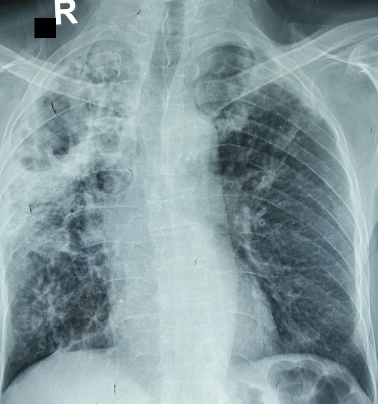
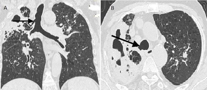
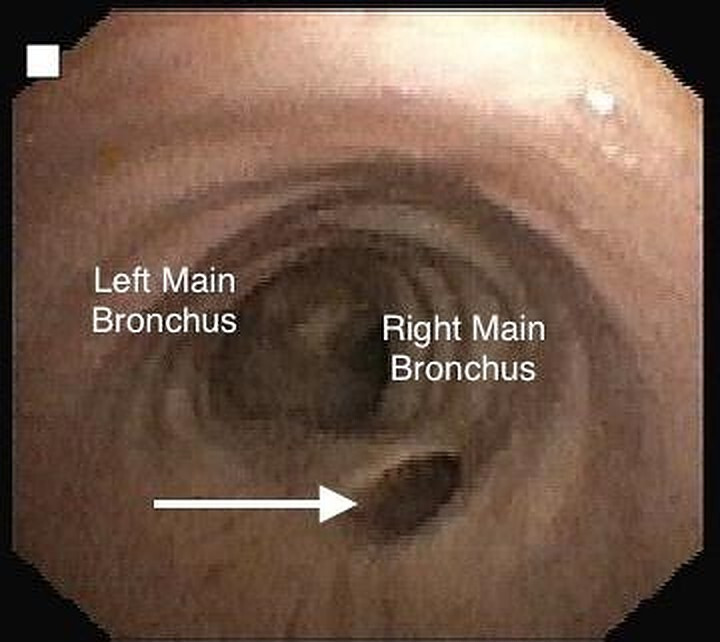
Tracheal bronchus, also known as bronchus suis, is a rare congenital anomaly of the airway where an accessory bronchus originates directly from the trachea. With an estimated incidence of 0.001%-2.0%, this condition is rarely reported in literature. It is usually discovered as an incidental finding in an otherwise asymptomatic individual. However, it can act as a focus of recurrent infection or present as persistent radiographic infiltrates. Multidetector CT imaging and bronchoscopy play a crucial role in the identification of this entity. We hereby report the case of a middle-aged man who presented with recurrent right upper lobe pneumonia, which was found to be due to an underlying tracheal bronchus.
Keywords: Pneumonia (respiratory medicine); Radiology; Respiratory medicine.
© BMJ Publishing Group Limited 2022. No commercial re-use. See rights and permissions. Published by BMJ.
Conflict of interest statement
Competing interests: None declared.
Figures
Similar articles
-
Evaluation of tracheobronchial anomalies in children using low-dose multidetector CT: report of a 13-year-old boy with a tracheal bronchus and recurrent pulmonary infections.Pediatr Pulmonol. 2004 Aug;38(2):168-73. doi: 10.1002/ppul.20077. Pediatr Pulmonol. 2004. PMID: 15211702
-
[A report of 4 cases with tracheal bronchus].Zhonghua Er Ke Za Zhi. 2006 Sep;44(9):698-9. Zhonghua Er Ke Za Zhi. 2006. PMID: 17217667 Chinese. No abstract available.
-
Rare case of tracheal bronchus in a patient posted for minimal invasive cardiac surgery.Ann Card Anaesth. 2020 Jul-Sep;23(3):364-366. doi: 10.4103/aca.ACA_215_18. Ann Card Anaesth. 2020. PMID: 32687102 Free PMC article.
-
Anesthetic Management of One-Lung Ventilation in Patients With Tracheal Bronchus: A Narrative Review.J Cardiothorac Vasc Anesth. 2024 Oct;38(10):2426-2432. doi: 10.1053/j.jvca.2024.05.033. Epub 2024 May 28. J Cardiothorac Vasc Anesth. 2024. PMID: 38918087 Review.
-
Radiology quiz case: Tracheal bronchus.Arch Otolaryngol Head Neck Surg. 2006 Apr;132(4):453, 454. doi: 10.1001/archotol.132.4.453. Arch Otolaryngol Head Neck Surg. 2006. PMID: 16618917 Review. No abstract available.
Cited by
-
Coexisting bilateral tracheal bronchi and accessory cardiac bronchus complicated with pneumonia and empyema: a case report.J Med Case Rep. 2025 Apr 15;19(1):175. doi: 10.1186/s13256-025-05213-2. J Med Case Rep. 2025. PMID: 40229776 Free PMC article.
References
-
- Lodha R, Kabra SK. Recurrent/persistent pneumonia. Indian Pediatr 2000;37:1085–92. - PubMed
Publication types
MeSH terms
LinkOut - more resources
Full Text Sources
Medical