Review
. 2022 Nov;122(11):1858-1868.
doi: 10.1055/a-1910-4538. Epub 2022 Jul 27.

Current View on the Molecular Mechanisms Underlying Fibrin(ogen)-Dependent Inflammation

Affiliations
Review

Current View on the Molecular Mechanisms Underlying Fibrin(ogen)-Dependent Inflammation

Sergiy Yakovlev et al. Thromb Haemost. 2022 Nov.

Abstract

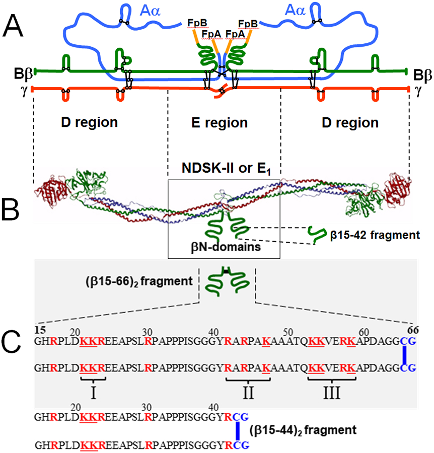
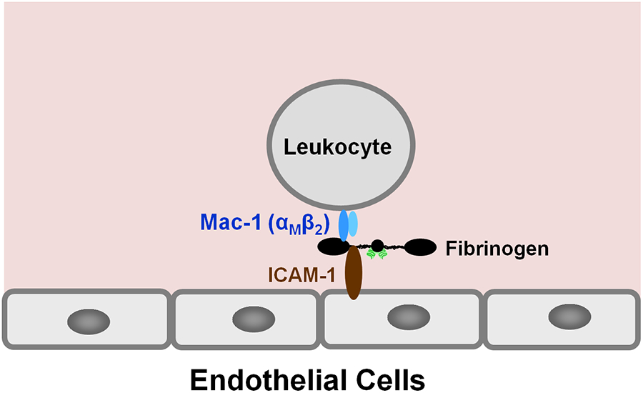
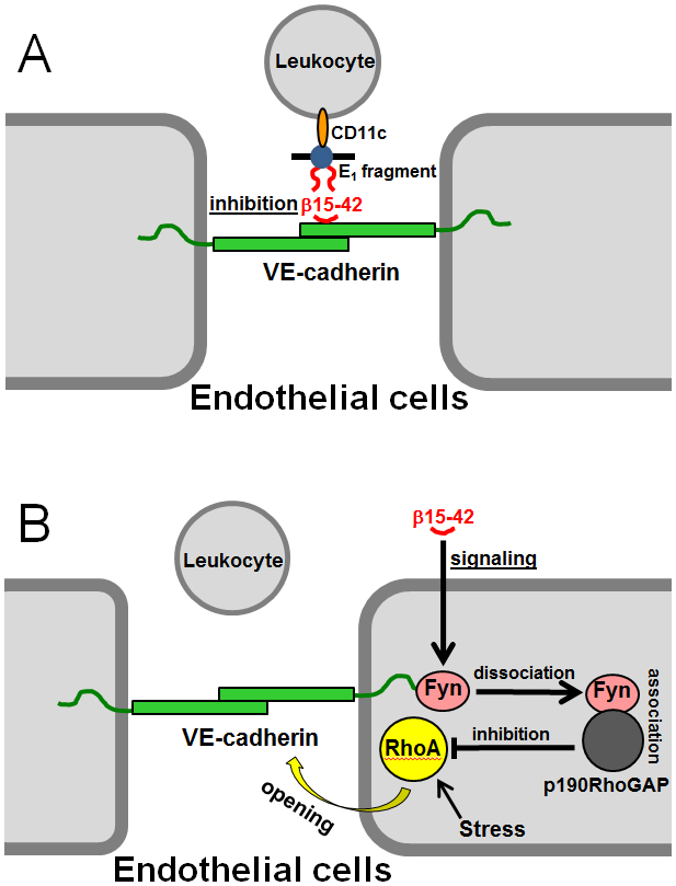
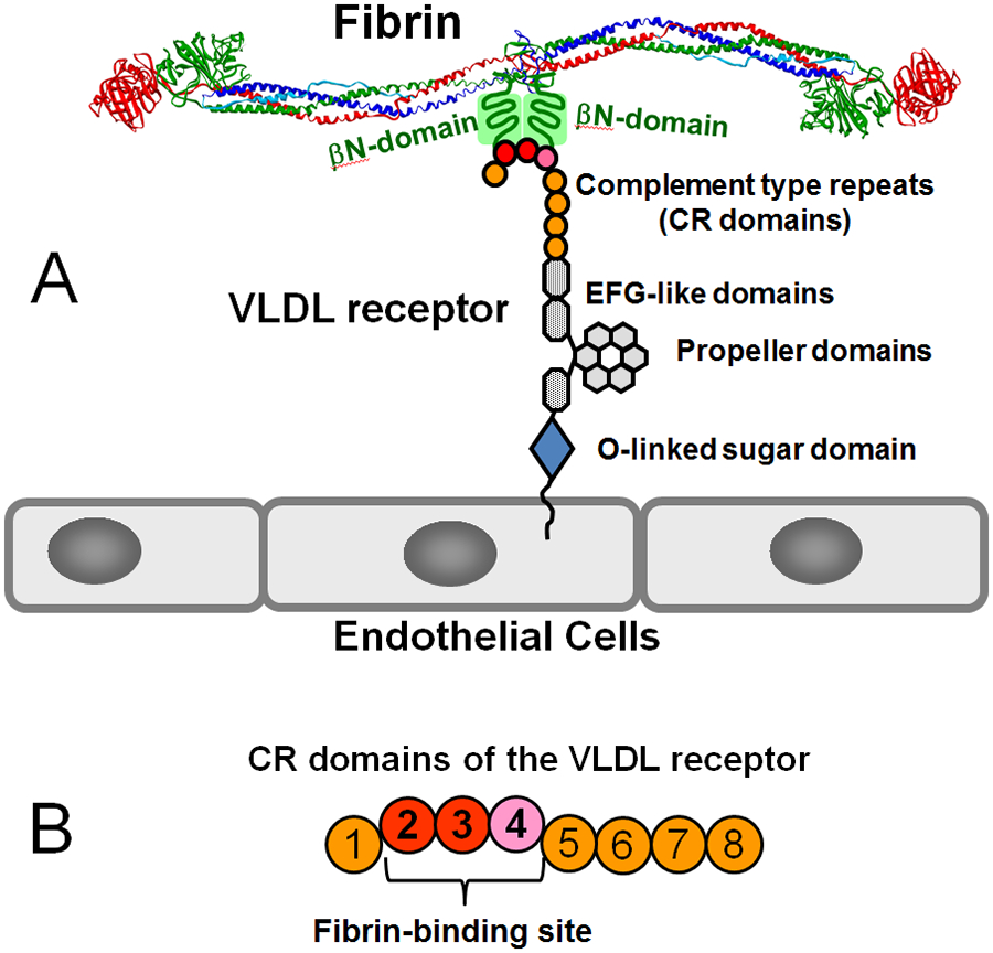
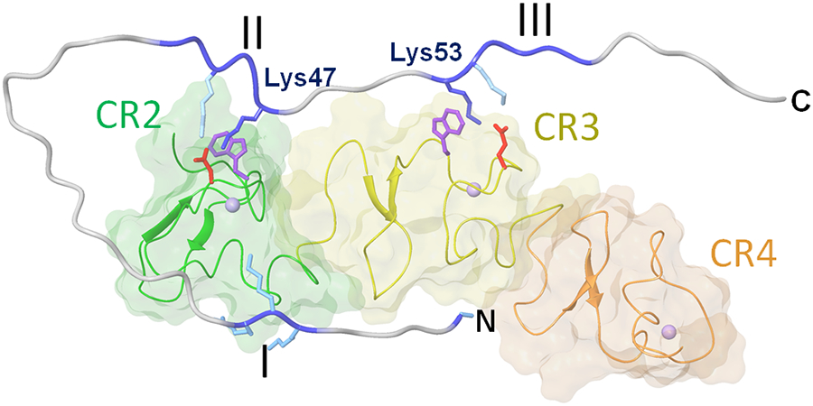
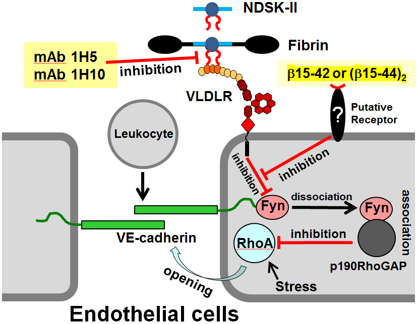
Numerous studies have revealed the involvement of fibrinogen in the inflammatory response. To explain the molecular mechanisms underlying fibrinogen-dependent inflammation, two bridging mechanisms have been proposed in which fibrin(ogen) bridges leukocytes to endothelial cells. The first mechanism suggests that bridging occurs via the interaction of fibrinogen with the leukocyte receptor Mac-1 and the endothelial receptor ICAM-1 (intercellular adhesion molecule-1), which promotes leukocyte transmigration and enhances inflammation. The second mechanism includes bridging of leukocytes to the endothelium by fibrin degradation product E1 fragment through its interaction with leukocyte receptor CD11c and endothelial VE-cadherin to promote leukocyte transmigration. The role of E1 in promoting inflammation is inhibited by the fibrin-derived β15-42 fragment, and this has been suggested to result from its ability to compete for the E1-VE-cadherin interaction and to trigger signaling pathways through the src kinase Fyn. Our recent study revealed that the β15-42 fragment is ineffective in inhibiting the E1- or fibrin-VE-cadherin interaction, leaving the proposed signaling mechanism as the only viable explanation for the inhibitory function of β15-42. We have discovered that fibrin interacts with the very-low-density lipoprotein (VLDL) receptor, and this interaction triggers a signaling pathway that promotes leukocyte transmigration through inhibition of the src kinase Fyn. This pathway is inhibited by another pathway induced by the interaction of β15-42 with a putative endothelial receptor. In this review, we briefly describe the previously proposed molecular mechanisms underlying fibrin-dependent inflammation and their advantages/disadvantages and summarize our recent studies of the novel VLDL receptor-dependent pathway of leukocyte transmigration which plays an important role in fibrin-dependent inflammation.


Conflict of interest statement

None declared.

Figures

Fig. 1
Fig. 1
(A) Polypeptide chain composition of the fibrinogen molecule with the Aα, Bβ, and γ chains are in blue, green, and red colors, respectively, and intra- and inter-chain disulfide bonds are in black color; fibrinopeptides A and B (FpA and FpB, respectively) are in orange color. (B) Ribbon diagram of the fibrinogen molecule based on its crystal structure; the colors of individual polypeptide chains are the same as in panel A, the αC-domains are not shown for simplicity. The βN-domains, whose structure has not been identified, and the β15-42 fragment are shown schematically by green curved lines. The approximate boundaries between the D and E regions are shown by dashed vertical lines between panels A and B; the NDSK-II and E1 fragments derived from the E region of fibrin are shown by box. (C) Schematic representation of the recombinant disulfide-linked (β15-66)2 fragment and its amino acid sequence, as well as the sequence of the disulfide-linked (β15-44)2 fragment. The Cys-Gly (CG) amino acid residues added to dimerize both fragments are in blue color and the disulfide bonds are shown by blue vertical bars. The positively charged Lys (K) and Arg (R) residues are shown in red color. The three clusters of the positively charged residues are indicated by roman numerals.
Fig. 2
Fig. 2
(A) Schematic representation of a fibrin polymer. D and E1 identify the D (grey color) and E (blue color) regions of individual fibrin molecules; the αC-domains are not shown for simplicity. Fibrin polymerization occurs mainly through non-covalent interactions between the complementary polymerization sites located in the D and E regions (the DD:E1 interaction shown by red triangle). Factor XIIIa crosslinks D-D regions by formation covalent bonds (shown by blue bars) between the C-terminal portions of the γ chains of these regions reinforcing fibrin polymers. Plasmin cleavage between the D and E regions of fibrin is shown by red arrows. (B and C) Cleavage of fibrin by plasmin results in high molecular mass fibrin degradation products (HMM-FDP) and the D-D:E1 complex. (D) Major final fibrin degradation products, the D-D dimer, the E3 fragment, and the β15-42 fragment.
Fig. 3
Fig. 3
The proposed bridging mechanism in which fibrinogen bridges leukocytes to the endothelium through the interaction with leukocyte receptor Mac-1 and endothelial receptor ICAM-1.
Fig. 4
Fig. 4
The proposed inhibitory role of the β15-42 fragment in leukocyte transmigration. (A) Fibrin degradation product, the E1 fragment, bridges leukocytes to the endothelium by interacting with endothelial VE-cadherin through its βN-domains (shown by red curved lines) and with leukocyte integrin CD11c, and β15-42 (shown by red curved line) inhibits this bridging by competing for the E1-VE-cadherin interaction., (B) The proposed additional signaling role of the β15-42 fragment in leukocyte transmigration. Treatment of endothelial cells with β15-42 results in dissociation of src kinase Fyn from VE-cadherin followed by association of Fyn with p190RhoGAP30 and this complex inhibits GTPase protein RhoA thereby preventing cell contraction and maintaining endothelial barrier function.
Fig. 5
Fig. 5
(A) Domain structure of the VLDL receptor and its interaction with the βN-domains of the fibrin molecule. (B) CR domains 2-4 of the VLDL receptor forming the fibrin-binding site.
Fig. 6
Fig. 6
Model of the complex between the βN-domain of fibrin and the VLDLR(2-4) fragment of the VLDL receptor based on NMR study. The CR2, CR3 and CR4 domains of VLDLR(2-4) are shown in different colors; the three clusters of the positively charged amino acid residues of the fibrin βN-domain are colored in blue and denoted by roman numerals; Lys47 and Lys 53 interacting with the Lys-binding sites of the VLDLR(2-4) fragment are also shown. N and C indicate N- and C-terminal ends of the βN-domain.
Fig. 7
Fig. 7
Schematic representation of the fibrin-VLDL receptor-dependent pathway of leukocyte transmigration and its inhibition by the β15-42 or (β15-44)2 fragments and by the specific anti-VLDLR monoclonal antibodies, mAb 1H5 and mAb 1H10., Interaction of fibrin or its mimetic NDSK-II with the endothelial VLDL receptor (VLDLR), which occurs through the βN-domains of the former (shown by red curved lines) and CR domains 2-4 of the later, induces intracellular pathway resulting in the inhibition of src kinase Fyn and preventing formation of its complex with p190RhoGAP. This precludes inhibition of GTPase protein RhoA, which in active state promotes stress-induced opening of endothelial adherent junctions and thereby leukocyte transmigration. The two monoclonal antibodies inhibit fibrin-VLDL receptor interaction thereby preventing activation of the fibrin-VLDL receptor-dependent pathway. Binding of the β15-42 or (β15-44)2 fragments to a putative receptor triggers a pathway that inhibits the fibrin-VLDL receptor-dependent pathway. This prevents inhibition of Fyn and results in subsequent inhibition of RhoA by the Fyn-p190RhoGAP complex thereby preventing junction opening and leukocyte transmigration.

References

    1. Riddle JM, Barnhart ML. Ultrastructural study of fibrin dissolution via emigrated polymorphonuclear neutrophils. Am J Pathol 1964;45(5):805–823 - PMC - PubMed
    1. Riddle JM, Barnhart ML. The eosinophil as a source for profibrinolysin in acute inflammation. Blood 1965;25(5):776–794 - PubMed
    1. Barnhart MI, Riddle JM, Bluhm GB, Quintana C. Fibrin promotion and lysis in arthritic joints. Ann Rheum Dis 1967;26(3):206–218 - PMC - PubMed
    1. Colvin RB, Johnson RA, Mihm MC Jr, Dvorak HF. Role of the clotting system in cell mediated hypersensitivity. I. Fibrin deposition in delayed skin reactions in man. J Exp Med 1973;138(3):686–698 - PMC - PubMed
    1. McRitchie DI, Girotti MJ, Glynn MF, Goldberg JM, Rotstein OD. Effect of systemic fibrinogen depletion on intraabdominal abscess formation. J Lab Clin Med 1991;118(1):48–55 - PubMed