

A ricin-based peptide BRIP from Hordeum vulgare inhibits Mpro of SARS-CoV-2
- PMID: 35896605
- PMCID: PMC9326418
- DOI: 10.1038/s41598-022-15977-y
A ricin-based peptide BRIP from Hordeum vulgare inhibits Mpro of SARS-CoV-2
Abstract
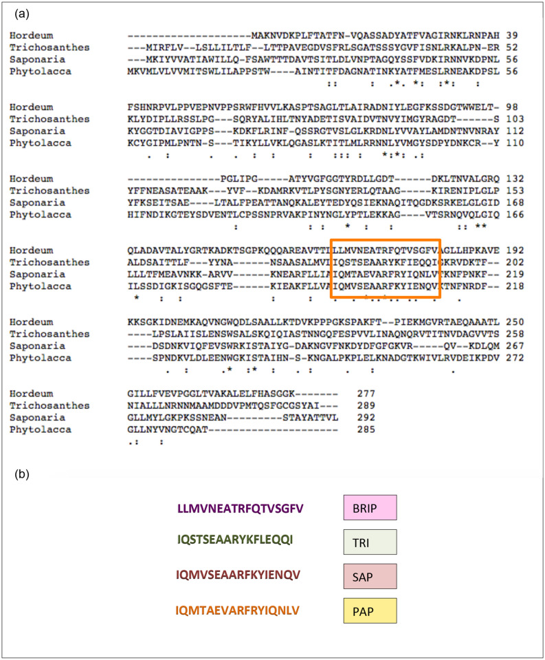
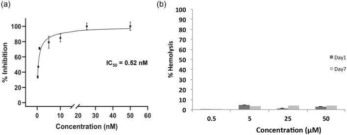
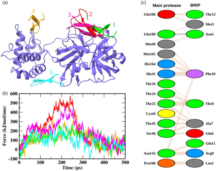
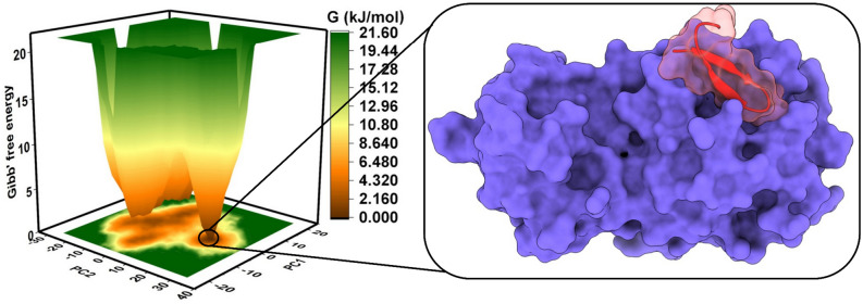
COVID-19 pandemic caused by SARS-CoV-2 led to the research aiming to find the inhibitors of this virus. Towards this world problem, an attempt was made to identify SARS-CoV-2 main protease (Mpro) inhibitory peptides from ricin domains. The ricin-based peptide from barley (BRIP) was able to inhibit Mpro in vitro with an IC50 of 0.52 nM. Its low and no cytotoxicity upto 50 µM suggested its therapeutic potential against SARS-CoV-2. The most favorable binding site on Mpro was identified by molecular docking and steered molecular dynamics (MD) simulations. The Mpro-BRIP interactions were further investigated by evaluating the trajectories for microsecond timescale MD simulations. The structural parameters of Mpro-BRIP complex were stable, and the presence of oppositely charged surfaces on the binding interface of BRIP and Mpro complex further contributed to the overall stability of the protein-peptide complex. Among the components of thermodynamic binding free energy, Van der Waals and electrostatic contributions were most favorable for complex formation. Our findings provide novel insight into the area of inhibitor development against COVID-19.
© 2022. The Author(s).
Conflict of interest statement
The authors declare no competing interests.
Figures
Similar articles
-
Optimization Rules for SARS-CoV-2 Mpro Antivirals: Ensemble Docking and Exploration of the Coronavirus Protease Active Site.Viruses. 2020 Aug 26;12(9):942. doi: 10.3390/v12090942. Viruses. 2020. PMID: 32859008 Free PMC article.
-
Anticoagulants as Potential SARS-CoV-2 Mpro Inhibitors for COVID-19 Patients: In Vitro, Molecular Docking, Molecular Dynamics, DFT, and SAR Studies.Int J Mol Sci. 2022 Oct 13;23(20):12235. doi: 10.3390/ijms232012235. Int J Mol Sci. 2022. PMID: 36293094 Free PMC article.
-
Identification of high-affinity inhibitors of SARS-CoV-2 main protease: Towards the development of effective COVID-19 therapy.Virus Res. 2020 Oct 15;288:198102. doi: 10.1016/j.virusres.2020.198102. Epub 2020 Jul 24. Virus Res. 2020. PMID: 32717346 Free PMC article.
-
Targeting the Dimerization of the Main Protease of Coronaviruses: A Potential Broad-Spectrum Therapeutic Strategy.ACS Comb Sci. 2020 Jun 8;22(6):297-305. doi: 10.1021/acscombsci.0c00058. Epub 2020 May 27. ACS Comb Sci. 2020. PMID: 32402186 Review.
-
The research progress of SARS-CoV-2 main protease inhibitors from 2020 to 2022.Eur J Med Chem. 2023 Sep 5;257:115491. doi: 10.1016/j.ejmech.2023.115491. Epub 2023 May 22. Eur J Med Chem. 2023. PMID: 37244162 Free PMC article. Review.
Cited by
-
L-shaped distribution of the relative substitution rate (c/μ) observed for SARS-COV-2's genome, inconsistent with the selectionist theory, the neutral theory and the nearly neutral theory but a near-neutral balanced selection theory: Implication on "neutralist-selectionist" debate.Comput Biol Med. 2023 Feb;153:106522. doi: 10.1016/j.compbiomed.2022.106522. Epub 2023 Jan 5. Comput Biol Med. 2023. PMID: 36638615 Free PMC article.
-
Dock-able linear and homodetic di, tri, tetra and pentapeptide library from canonical amino acids: SARS-CoV-2 Mpro as a case study.J Pharm Anal. 2023 May;13(5):523-534. doi: 10.1016/j.jpha.2023.04.008. Epub 2023 Apr 15. J Pharm Anal. 2023. PMID: 37275125 Free PMC article.
-
In silico screening and evaluation of antiviral peptides as inhibitors against ORF9b protein of SARS-CoV-2.3 Biotech. 2024 Sep;14(9):192. doi: 10.1007/s13205-024-04032-4. Epub 2024 Aug 6. 3 Biotech. 2024. PMID: 39118822 Free PMC article. Review.
-
In-silico study: docking simulation and molecular dynamics of peptidomimetic fullerene-based derivatives against SARS-CoV-2 Mpro.3 Biotech. 2023 Jun;13(6):185. doi: 10.1007/s13205-023-03608-w. Epub 2023 May 13. 3 Biotech. 2023. PMID: 37193325 Free PMC article.
-
Inhibition of nonstructural protein 15 of SARS-CoV-2 by golden spice: A computational insight.Cell Biochem Funct. 2022 Dec;40(8):926-934. doi: 10.1002/cbf.3753. Epub 2022 Oct 6. Cell Biochem Funct. 2022. PMID: 36203381 Free PMC article.
References
Publication types
MeSH terms
Substances
LinkOut - more resources
Full Text Sources
Miscellaneous
