Review
. 2022 Jul 27;16(1):24.
doi: 10.1186/s13037-022-00333-w.

Trans-sacral screw fixation of posterior pelvic ring injuries: review and expert opinion

Affiliations
Review

Trans-sacral screw fixation of posterior pelvic ring injuries: review and expert opinion

Navid Ziran et al. Patient Saf Surg. .

Abstract

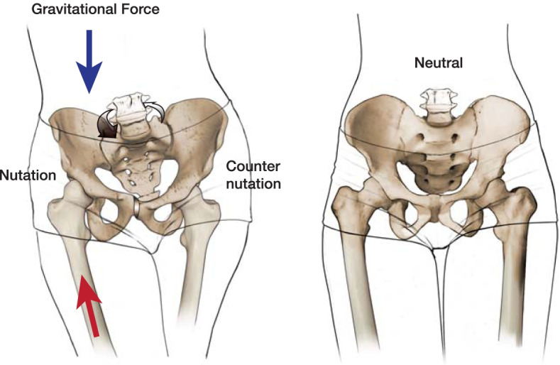
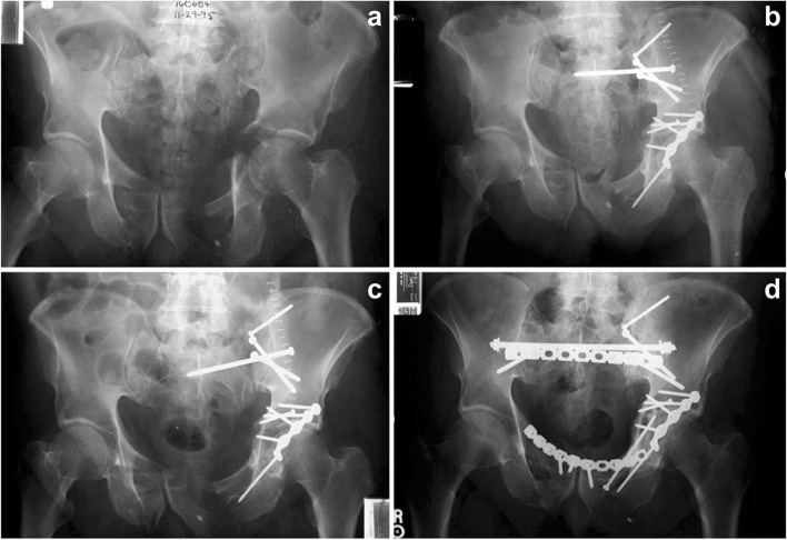
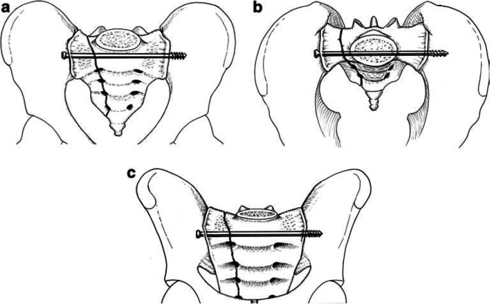
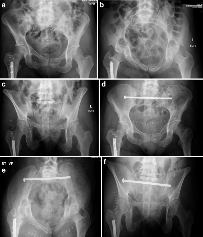
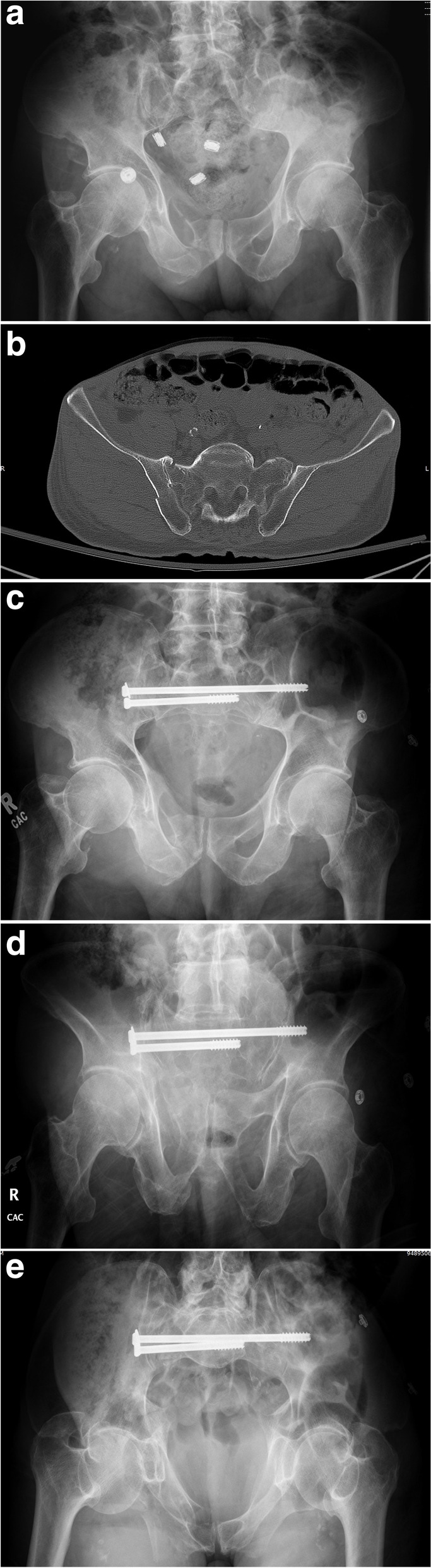
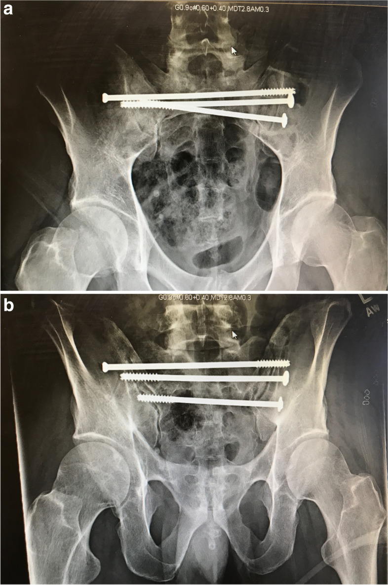
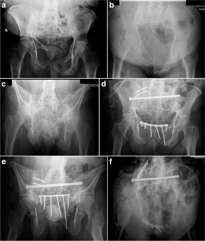
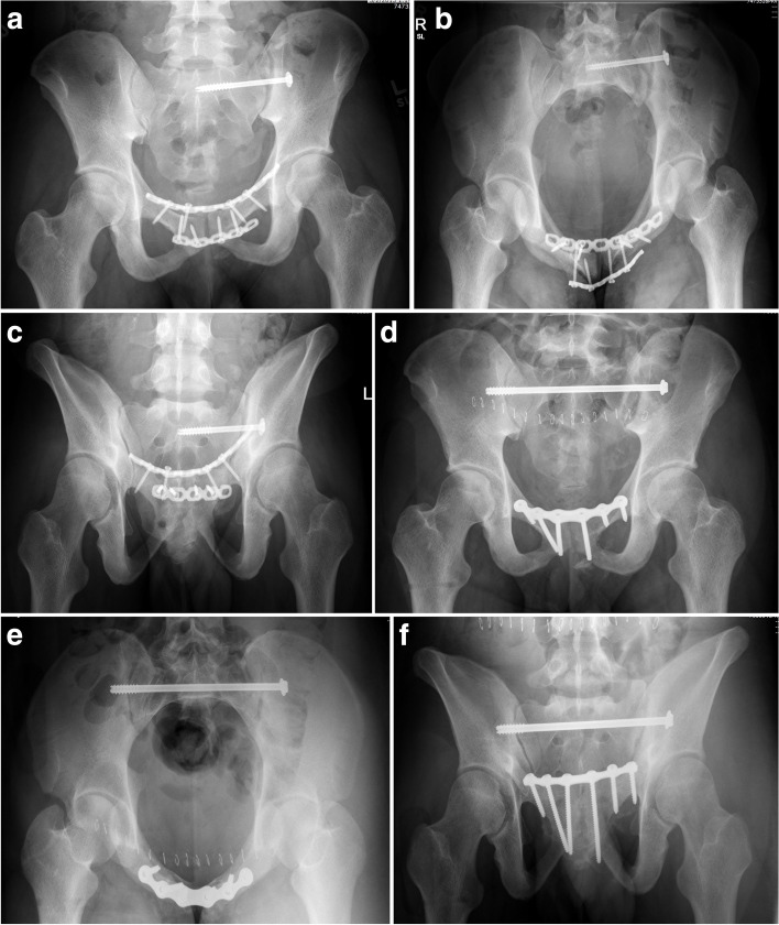
Posterior pelvic ring injuries (i.e., sacro-iliac joint dislocations, fracture-dislocations, sacral fractures, pelvic non-unions/malunions) are challenging injury patterns which require a significant level of surgical training and technical expertise. The modality of surgical management depends on the specific injury patterns, including the specific bony fracture pattern, ilio-sacral joint involvement, and the soft tissue injury pattern. The workhorse for posterior pelvic ring stabilization has been cannulated iliosacral screws, however, trans-sacral screws may impart increased fixation strength. Depending on injury pattern and sacral anatomy, trans-sacral screws can potentially be more beneficial than iliosacral screws. In this article, the authors will briefly review pelvic mechanics and discuss their rationale for ilio-sacral and/or trans-sacral screw fixation.

Keywords: Iliosacral; Injury mechanism; Pelvic fracture; Posterior pelvic ring; Screw; Trans-sacral.


Conflict of interest statement

The authors declare that they have no competing interests.

Figures

Fig. 1
Fig. 1
Illustration demonstration the effect of loading on the pelvis during gait. During weight-bearing the reactive normal force vector, directed cranially, results in nutation of the ipsilateral pelvis/sacrum. The sacral base (promontory) tilts anteriorly and inferiorly and the distal sacrum tilts posteriorly relative to the ilium (nutation, or “to nod”). The ipsilateral hemipelvis shifts slightly in the y-axis through the pubic symphysis. The opposite motion occurs in the contralateral pelvis (counternutation). The motion is exaggerated in the images for demonstration. This alternating motion during gait is reciprocal unilateral motion
Fig. 2
Fig. 2
Illustration demonstrating the color-coded bone mineral density of the posterior pelvic ring. Higher bone density is located near the SI joints, the S1 body, and the superior endplate of S1
Fig. 3
Fig. 3
Illustration of a vertical sacral fracture with both iliosacral screw fixation and trans-sacral screw fixation. During loading on the ipsilateral side, the force vector is directed cranially. This force is distributed along a longer implant length in the trans-sacral screw versus the iliosacral screw. Further, the contralateral iliac cortex has higher bone density than the S1 body
Fig. 4
Fig. 4
Drawing of a right SI fracture-dislocation treated with an iliosacral screw (A). The usual mode of failure for this screw is rotation in the coronal plane with subsequent cranial fracture displacement (B)
Fig. 5
Fig. 5
Radiographic images of a combined left SI fracture-dislocation and T-shaped acetabular fracture (A). After fracture fixation (B), the posterior pelvic fixation failed (C) necessitating revision with a trans-sacral screw, posterior tension band plate, and anterior ring fixation (D)
Fig. 6
Fig. 6
Antero-posterior (AP) pelvis, inlet, and outlet drawings of a right vertical sacral fracture fixed with a right trans-sacral screw
Fig. 7
Fig. 7
Radiographic pelvic images demonstrating a right SI fracture-dislocation (A-C) reduced and fixed with a trans-sacral screw (E-F)
Fig. 8
Fig. 8
Radiographic and axial CT pelvic image of a right SI fracture-dislocation (A-B) treated with a right trans-sacral screw and right ilio-sacral screw to prevent implant failure and fracture re-displacement (C-E)
Fig. 9
Fig. 9
Post-operative pelvic radiographs (A-B) of a posterior pelvic ring injury fixed with three trans-sacral screws in both S1 and S2
Fig. 10
Fig. 10
Pre- (A-C) and post-operative (D-F) radiographic images of a pelvic malunion from a right SI fracture-dislocation and pubic symphyseal dislocation. The malunion correction was performed in stages resulting in both anterior and posterior ring fixation
Fig. 11
Fig. 11
Pre- and post-operative radiographic images of a left SI fracture-dislocation and pubic symphyseal dislocation that was fixed in a mal-reduced position (A-C). The malunion was re-reduced and fixed with a trans-sacral screw and anterior ring fixation (D-F)

References

    1. Ziran NM, Smith WR. Pelvic fractures. Management of musculoskeletal injuries in the trauma patient. New York: Springer; 2014. pp. 143–163.
    1. Tile M, Helfet D. Fractures of the pelvis and acetabulum. 3. Baltimore: Williams & Wilkins; 2003.
    1. Gray’s Anatomy. The anatomical basis of clinical practice. 41st ed. Standring S editor. New York: Elsevier Limited; 2016.
    1. Chamberlain WE. The symphysis pubis in the roentgen examination of the sacro-iliac joint. Am J Roentgenol. 1930;24:621–625.
    1. Chamberlain WE. The X-ray examination of the sacroiliac joint. Delaware Med J. 1932;4:195–201.

LinkOut - more resources