. 2022 Jul 22;23(15):8072.
doi: 10.3390/ijms23158072.

Distinct Patterns of GR Transcriptional Regulation in Liver and Muscle of LPS-Challenged Weaning Piglets

Affiliations

Distinct Patterns of GR Transcriptional Regulation in Liver and Muscle of LPS-Challenged Weaning Piglets

Jie Liu et al. Int J Mol Sci. .

Abstract

Glucocorticoid receptor (GR), which is ubiquitously expressed in nearly all cell types of various organs, mediates the tissue-specific metabolic and immune responses to maintain homeostasis and ensure survival under stressful conditions or pathological challenges. The neonatal period is metabolically demanding, and piglets are subjected to multiple stressors in modern intensive farms, especially around weaning. The liver is more responsive to LPS challenge compared to muscle, which is indicated by significantly increased TLR4 and p-p65, TNF-α, and IL-6 levels in association with GR down-regulation at both mRNA and protein levels. GR binding to the putative nGRE on TNF-α and IL-6 gene promoters decreased in the liver, but not muscle, upon LPS stimulation. The transcriptional regulation of GR also showed striking differences between liver and muscle. GR exon 1 mRNA variants 1-4, 1-5, and 1-6 were down-regulated in both liver and muscle, but a significant up-regulation of GR exon 1-9/10 mRNA variants abolished the change of total GR mRNA in the muscle in response to LPS stimulation. The significant down-regulation of GR in the liver corresponded with significantly decreased binding of p-GR and diminished histone acetylation in GR gene promoters. These results indicate that tissue-specific GR transcriptional regulation is involved in the differential inflammation responses between liver and muscle.

Keywords: GR; liver; muscle; pig; tissue specificity; transcriptional regulation.


Conflict of interest statement

The authors declare no conflict of interest.

Figures
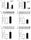
Figure 1
Figure 1
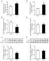
Inflammatory response in liver and muscle when treatment with LPS. (A) Liver cytokine levels of TNF-α and IL-6. (B) Western blot and densitometric analyses of TLR4 and GAPDH proteins in the liver. (C) Western blot and densitometric analyses of p-P65 and GAPDH proteins in the liver. (D) Muscle cytokine level of TNF-α and IL-6. (E) Western blot and densitometric analyses of TLR4 and GAPDH proteins in muscle. (F) Western blot and densitometric analyses of p-P65 and GAPDH proteins in muscle. Values were expressed as means ± SEM, n = 4 in each group, * p < 0.05.
Figure 2
Figure 2
Cortisol content and GR expression in liver and muscle in response to LPS challenge. (A) Liver cortisol level. (B) GR mRNA expression of the liver was evaluated by qRT-PCR. (C) Western blot and densitometric analyses of GR and GAPDH proteins in the liver. (D) Muscle cortisol level. (E) GR mRNA expression of muscle was evaluated by qRT-PCR. (F) Western blot and densitometric analyses of GR and GAPDH proteins in muscle. Values were expressed as means ± SEM, n = 4 in each group, * p < 0.05.
Figure 3
Figure 3
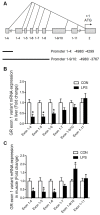
GR exon 1 variants expression in liver and muscle when treatment with LPS. (A) Genomic location of the porcine GR alternative promoters. (B) GR exon 1 variants expression in liver. (C) GR exon 1 variants expression in muscle. Values were expressed as means ± SEM, n = 4 in each group, * p < 0.05.
Figure 4
Figure 4
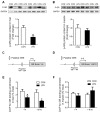
The change of p-GR and GR binding with its promoter in liver and muscle under LPS treatment. (A) Western blot and densitometric analyses of p-GR and GAPDH proteins in the liver. (B) Western blot and densitometric analyses of p-GR and GAPDH proteins in muscle. (C) The putative fragment GRE site on the promoter region of GR exon 1–4. (D) The putative fragment GRE site on the promoter region of GR exon 1–9/10. (E) ChIP analyses GR binding to GRE sites on the promoter of GR exon 1–4 and 1–9/10 in the liver. (F) ChIP analyses p-GR binding to GRE sites on the promoter of GR exon 1–4 and 1–9/10 in muscle. Values were expressed as means ± SEM, n = 4 in each group, * p < 0.05.
Figure 5
Figure 5
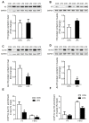
Histone H3 protein content and the enrichment of histone H3 acetylation on GR promoters 1–4 and 1–9/10 under LPS stimulation. (A) Western blot and densitometric analyses of H3 and GAPDH proteins in the liver. (B) Western blot and densitometric analyses of H3 and GAPDH proteins in muscle. (C) Western blot and densitometric analyses of the global histone H3 acetylation level in the liver. (D) Western blot and densitometric analyses of the global histone H3 acetylation level in muscle. (E) ChIP analyses for Ac-H3 enrichment on the promoter of GR exon 1–4 and 1–9/10 in the liver. (F) ChIP analyses Ac-H3 enrichment on the promoter of GR exon 1–4 and 1–9/10 in muscle. Values were expressed as means ± SEM, n = 4 in each group, * p < 0.05.
Figure 6
Figure 6
GR binding to TNF-α and IL-6 gene promoters in liver and muscle. (A) The putative fragment nGRE site on the promoter region of TNF-α. (B) The putative fragment nGRE site on the promoter region of IL-6. (C) ChIP analyses GR binding to nGRE sites on the promoter of TNF-α exon 1–4 and 1–9/10 in the liver. (D) ChIP analyses GR binding to nGRE sites on the promoter of IL-6 exon 1–4 and 1–9/10 in the liver. (E) ChIP analyses GR binding to nGRE sites on the promoter of TNF-α exon 1–4 and 1–9/10 in muscle. (F) ChIP analyses GR binding to nGRE sites on the promoter of IL-6 exon 1–4 and 1–9/10 in muscle. Values were expressed as means ± SEM, n = 4 in each group, * p < 0.05.
Figure 7
Figure 7
Different transcriptional regulation of GR in liver and muscle after LPS challenge. The decrease in GR expression in the liver after LPS stimulation was associated with the decrease in GR exon 1–4/5/6 expression, the binding of GR to GRE, and the acetylation of H3. Decreased GR results in increased expression of inflammatory factors TNF-α and IL-6, leading to an inflammatory response. In muscle, LPS did not affect the expression of GR, so there was no inflammatory response. Arrows for promotion, lines for inhibition, and crosses for no influence.

Similar articles

Cited by

References

    1. Oakley R.H., Cidlowski J.A. The biology of the glucocorticoid receptor: New signaling mechanisms in health and disease. J. Allergy Clin. Immunol. 2013;132:1033–1044. doi: 10.1016/j.jaci.2013.09.007. - DOI - PMC - PubMed
    1. Baschant U., Tuckermann J. The role of the glucocorticoid receptor in inflammation and immunity. J. Steroid Biochem. Mol. Biol. 2010;120:69–75. doi: 10.1016/j.jsbmb.2010.03.058. - DOI - PubMed
    1. Meers G.K., Bohnenberger H., Reichardt H.M., Luhder F., Reichardt S.D. Impaired resolution of DSS-induced colitis in mice lacking the glucocorticoid receptor in myeloid cells. PLoS ONE. 2018;13:e0190846. doi: 10.1371/journal.pone.0190846. - DOI - PMC - PubMed
    1. Carrillo-de Sauvage M.A., Maatouk L., Arnoux I., Pasco M., Sanz Diez A., Delahaye M., Herrero M.T., Newman T.A., Calvo C.F., Audinat E., et al. Potent and multiple regulatory actions of microglial glucocorticoid receptors during CNS inflammation. Cell Death Differ. 2013;20:1546–1557. doi: 10.1038/cdd.2013.108. - DOI - PMC - PubMed
    1. Cain D.W., Cidlowski J.A. Specificity and sensitivity of glucocorticoid signaling in health and disease. Best Pract. Res. Clin. Endocrinol. Metab. 2015;29:545–556. doi: 10.1016/j.beem.2015.04.007. - DOI - PMC - PubMed

MeSH terms