. 2022 Jul 22;23(15):8086.
doi: 10.3390/ijms23158086.

miR-489 Confines Uncontrolled Estrogen Signaling through a Negative Feedback Mechanism and Regulates Tamoxifen Resistance in Breast Cancer

Affiliations

miR-489 Confines Uncontrolled Estrogen Signaling through a Negative Feedback Mechanism and Regulates Tamoxifen Resistance in Breast Cancer

Mithil Soni et al. Int J Mol Sci. .

Abstract

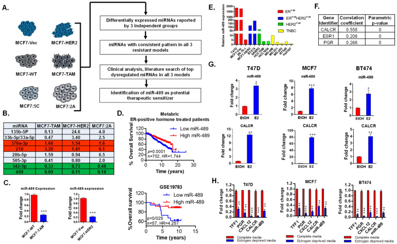
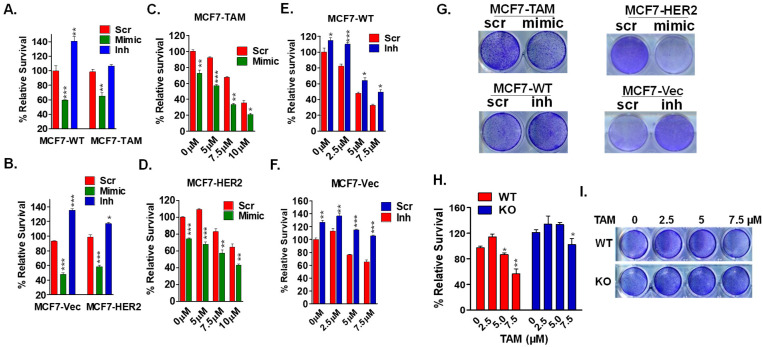
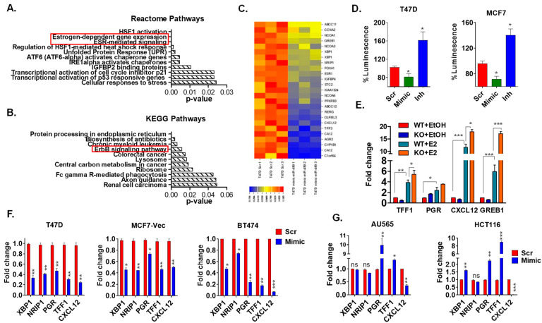
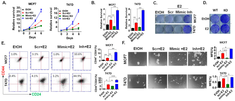
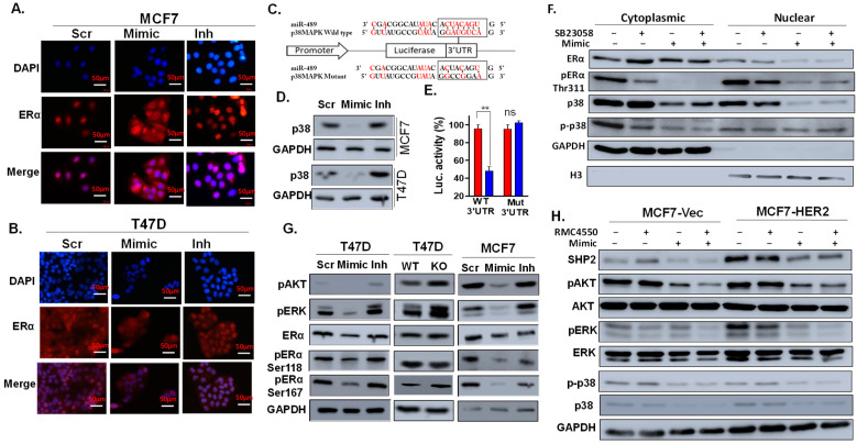
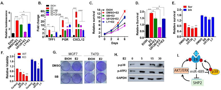
Approximately 75% of diagnosed breast cancer tumors are estrogen-receptor-positive tumors and are associated with a better prognosis due to response to hormonal therapies. However, around 40% of patients relapse after hormonal therapies. Genomic analysis of gene expression profiles in primary breast cancers and tamoxifen-resistant cell lines suggested the potential role of miR-489 in the regulation of estrogen signaling and development of tamoxifen resistance. Our in vitro analysis showed that loss of miR-489 expression promoted tamoxifen resistance, while overexpression of miR-489 in tamoxifen-resistant cells restored tamoxifen sensitivity. Mechanistically, we found that miR-489 is an estrogen-regulated miRNA that negatively regulates estrogen receptor signaling by using at least the following two mechanisms: (i) modulation of the ER phosphorylation status by inhibiting MAPK and AKT kinase activities; (ii) regulation of nuclear-to-cytosol translocation of estrogen receptor α (ERα) by decreasing p38 expression and consequently ER phosphorylation. In addition, miR-489 can break the positive feed-forward loop between the estrogen-Erα axis and p38 MAPK in breast cancer cells, which is necessary for its function as a transcription factor. Overall, our study unveiled the underlying molecular mechanism by which miR-489 regulates an estrogen signaling pathway through a negative feedback loop and uncovered its role in both the development of and overcoming of tamoxifen resistance in breast cancers.

Keywords: CRISPR/Cas9; breast cancer; estrogen receptor; miR-489; tamoxifen resistance.


Conflict of interest statement

The authors declare no conflict of interest.

Figures

Figure 1
Figure 1
miR-489 expression is lost in tamoxifen resistance, predicts breast cancer aggressiveness, and is regulated by the estrogen/ERα axis. (A) Schematic diagram for the identification and validation of miRNAs involved in tamoxifen resistance. miRNA analysis from three independent studies using three independent tamoxifen-resistant model systems. (B) List of top dysregulated miRNAs in all three tamoxifen-resistant cell lines. (C). qRT-PCR validation of miR-489 in MCF7-TAM and MCF7-HER2 cell lines. (D) Clinical analysis of two datasets analyzing miR-489 expression in ER+ breast cancer patients receiving hormone therapy. (E) miR-489 expression in breast cancer cell lines. (F) Correlation of miR-489 expression with expression of ERα and PGR. (G) qRT-PCR analysis of miR-489 expression upon estrogen stimulation in three ER+ breast cancer cell lines. (H) qRT-PCR analysis of miR-489 expression upon estrogen deprivation in three ER+ breast cancer cell lines. * p < 0.05; ** p < 0.01; *** p < 0.001. Data are representative of three independent experiments.
Figure 2
Figure 2
miR-489 restoration overcomes acquired and de novo tamoxifen resistance. (A,B) Effect of miR-489 modulation on proliferation of two pairs of tamoxifen-resistant cell lines. Cells were transfected with 28 nM of Scramble siRNA, miR-489 mimic, and miR-489 inhibitor for 72 h, followed by an MTT-based viability assay. (C,D) miR-489 restoration sensitized MCF7-TAM and MCF7-HER2 cell lines to tamoxifen. Scramble siRNA or the miR-489 mimic were transfected with or without tamoxifen for 72 h, followed by an MTT-based viability assay. (E,F) Depletion of miR-489 promoted tamoxifen resistance in tamoxifen-sensitive cell lines MCF7-TAM and MCF7-HER2. (G) Colony formation assay showing that miR-489 modulated tamoxifen resistance. Cells were treated with indicated microRNA mimics or inhibitors with or without tamoxifen for 72 h, followed by a colony formation assay for 7–10 days. (H,I) miR-489 knockout conferred tamoxifen resistance. WT and KO T47D cells were treated with an indicated concentration of tamoxifen, and viability was examined using MTT (H) and colony formation (I) assays. * p < 0.05; ** p < 0.01; *** p < 0.001. Data are representative of three independent experiments.
Figure 3
Figure 3
Gene expression analysis revealed enrichment of multiple pathways involved in estrogen signaling and tamoxifen resistance. (A,B) Whole-transcriptome analysis of miR-489-transfected and control T47D cells followed by pathway enrichment analysis revealed multiple pathways involved in tamoxifen resistance, including ESR-mediated signaling and ErBB2 pathways. (C) Heatmap demonstrating downregulation of multiple estrogen-responsive genes in miR-489-transfected T47D cells. (D) miR-489 negatively regulated estrogen-induced transcription by transient transfection of cells with the ERE-reporter system. (E) miR-489 WT and knockout T47D cells were treated with ethanol or estrogen for 6 h, and expression of estrogen-responsive genes was examined using qRT-PCR. (F,G) qRT-PCR analysis of estrogen-responsive genes upon miR-489 restoration showed downregulation of estrogen-responsive genes only in ER+ breast cancer cell lines. ns, not significant; * p < 0.05; ** p < 0.01; *** p < 0.001. Data are representative of three independent experiments.
Figure 4
Figure 4
miR-489 acts as an endogenous negative feedback loop to dampen estrogen activities. (A) miR-489 restoration inhibited the proliferation of ER+ breast cancer cell lines, while the inhibition of endogenous miR-489 dramatically enhanced the proliferation of MCF7 and T47D. Scramble siRNA, miR-489 mimic, or miR-489 inhibitor were transfected in the presence or absence of estrogen for 72 h, followed by an MTT-based viability assay. (B) Quantification of viable cells on day 6 post-treatment of MCF7 and T47D. (C) Inhibition of endogenous miR-489 dramatically enhanced estrogen-induced colony formation. MCF7 and T47D cells were transfected with scramble siRNA, miR-489 mimic, and miR-489 inhibitor in the presence or absence of estrogen, followed by a colony formation assay. (D) Hormone-starved miR-489 WT and KO T47D cells were seeded in a 12-well plate and treated with ethanol or E2 for 6 days, and cell viability was measured by crystal violet staining. (E) Breast cancer cell lines were transfected with scr, mimic, or inhibitor in the presence of estrogen for 72 h, followed by flow cytometry to examine CD24 and CD44 surface markers. (F) Breast cell lines were transfected with scr, mimic, or inhibitor in the presence of estrogen for 72 h, followed by a mammosphere assay. Scr = Scramble control; Inh = Inhibitor of miR-489. * p < 0.05; ** p < 0.01; *** p < 0.001. Data are representative of three independent experiments.
Figure 5
Figure 5
miR-489 inhibits estrogen-induced signaling by inhibiting p38 MAPK, PI3K-AKT, and MAPK-ERK pathways. (A,B) MCF7 and T47D cells were transfected with a scramble, mimic, or inhibitor for 72 h in estrogen-deprived media, followed by estrogen stimulation for 15 min. The data represent the localization of estrogen receptors using immunofluorescence. (C) miR-489 binding site in p38 MAPK 3′ UTR. (D) A scramble, mimic, or inhibitor was transfected, and Western blot analysis was performed to examine the total p38 MAPK protein. (E) 3′ UTR transfection assay showing that miR-489 directly bound to 3′UTR of p38 MAPK. Data are means of three replicates ± SEM. ns, not significant; ** p < 0.01. (F) MCF7 cells were transfected with a scramble or mimic in the presence of DMSO or the p38 MAPK inhibitor, SB23508. At 72 h post-transfection, cytoplasmic and nuclear fractionations were prepared for Western blot analysis of expression and phosphorylation statuses of ERα and p38. (G) Cells were transfected with a scramble, mimic, or inhibitor for 72 h, and ERα phosphorylation and responsible kinases were examined using Western blot analysis. (H) MCF7 cells were transfected with a scramble or mimic in the presence of DMSO or SHP2 allosteric inhibitor RMC4550. ERα phosphorylation and responsible kinases were examined using Western blot analysis.
Figure 6
Figure 6
P38 MAPK, PI3K-Akt, and MAPK inhibitors recapitulate the effect of miR-489 in an ER+ breast cancer cell line. (A) MCF7 cells were treated with inhibitors of p38 MAPK, ERK, and PI3k-AKT pathways, and their role on estrogen-induced transcription was examined using a luciferase reporter assay. (B) MCF7 cells were treated with inhibitors for 24 h, and expression of estrogen-responsive genes was examined using qRT-PCR. (C,D) MCF7 cells were treated with inhibitors, and the effects on estrogen-induced proliferation were examined using an MTT assay. (E) MCF7 cells were transfected with a scramble or inhibitors and treated with inhibitors for 72 h in the presence of estrogen, and the effects on proliferation were examined using an MTT assay. (F) miR-489 WT and KO T47D cells were treated with inhibitors in the presence of estrogen, and cell viability was examined using an MTT assay. (G) MCF7 cells were treated with DMSO or a p38 MAPK inhibitor in the presence or absence of estrogen and a colony formation assay was performed. (H) MCF7 cells were treated with estrogen for the indicated time, and activation of p38 MAPK signaling was examined using Western blot. (I) A proposed model for estrogen-induced miR-489 to restrict estrogen signaling via a negative feedback mechanism. * p < 0.05; ** p < 0.01; *** p < 0.001. Data are means of three replicates ± SEM.

Similar articles

Cited by

References

    1. Nass N., Kalinski T. Tamoxifen resistance: From cell culture experiments towards novel biomarkers. Pathol. Res. Pract. 2015;211:189–197. doi: 10.1016/j.prp.2015.01.004. - DOI - PubMed
    1. Clarke R., Tyson J.J., Dixon J.M. Endocrine resistance in breast cancer--An overview and update. Pt 3Mol. Cell. Endocrinol. 2015;418:220–234. doi: 10.1016/j.mce.2015.09.035. - DOI - PMC - PubMed
    1. Jeselsohn R., Buchwalter G., De Angelis C., Brown M., Schiff R. ESR1 mutations-a mechanism for acquired endocrine resistance in breast cancer. Nat. Rev. Clin. Oncol. 2015;12:573–583. doi: 10.1038/nrclinonc.2015.117. - DOI - PMC - PubMed
    1. Osborne C.K., Schiff R. Mechanisms of endocrine resistance in breast cancer. Annu Rev. Med. 2011;62:233–247. doi: 10.1146/annurev-med-070909-182917. - DOI - PMC - PubMed
    1. Fan P., Maximov P.Y., Curpan R.F., Abderrahman B., Jordan V.C. The molecular, cellular and clinical consequences of targeting the estrogen receptor following estrogen deprivation therapy. Pt 3Mol. Cell. Endocrinol. 2015;418:245–263. doi: 10.1016/j.mce.2015.06.004. - DOI - PMC - PubMed

MeSH terms