. 2022 Jul 22;23(15):8104.
doi: 10.3390/ijms23158104.

The LAMMER Kinase MoKns1 Regulates Growth, Conidiation and Pathogenicity in Magnaporthe oryzae

Affiliations

The LAMMER Kinase MoKns1 Regulates Growth, Conidiation and Pathogenicity in Magnaporthe oryzae

Lin Li et al. Int J Mol Sci. .

Abstract

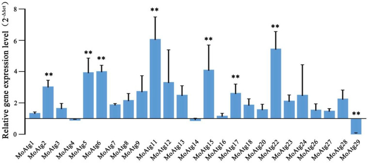
Magnaporthe oryzae is an important pathogen that causes a devastating disease in rice. It has been reported that the dual-specificity LAMMER kinase is conserved from yeast to animal species and has a variety of functions. However, the functions of the LAMMER kinase have not been reported in M. oryzae. In this study, we identified the unique LAMMER kinase MoKns1 and analyzed its function in M. oryzae. We found that in a MoKNS1 deletion mutant, growth and conidiation were primarily decreased, and pathogenicity was almost completely lost. Furthermore, our results found that MoKns1 is involved in autophagy. The ΔMokns1 mutant was sensitive to rapamycin, and MoKns1 interacted with the autophagy-related protein MoAtg18. Compared with the wild-type strain 70-15, autophagy was significantly enhanced in the ΔMokns1 mutant. In addition, we also found that MoKns1 regulated DNA damage stress pathways, and the ΔMokns1 mutant was more sensitive to hydroxyurea (HU) and methyl methanesulfonate (MMS) compared to the wild-type strain 70-15. The expression of genes related to DNA damage stress pathways in the ΔMokns1 mutant was significantly different from that in the wild-type strain. Our results demonstrate that MoKns1 is an important pathogenic factor in M. oryzae involved in regulating autophagy and DNA damage response pathways, thus affecting virulence. This research on M. oryzae pathogenesis lays a foundation for the prevention and control of M. oryzae.

Keywords: LAMMER kinase; Magnaporthe oryzae; conidiation; pathogenicity.


Conflict of interest statement

The authors declare no conflict of interest.

Figures

Figure 1
Figure 1
Sequence alignment of the amino acids of Kns1 in different species. A. The sequence alignment of Kns1 amino acids was made by the DNAMAN 8. The compared sequences were from Magnaporthe oryzae (MoKns1), Colletotrichum orbiculare (Lkh1, TDZ24089), Fusarium graminearum (Kns1, EYB28111.1), Homo sapiens (6FYK_A), Mus musculus (NP_001156904.1), Neurospora crassa (XP_957701.3), Saccharomyces cerevisiae (CAD6636070.1). All these Kns1 proteins have a conserved LAMMER motif (blue framed part).
Figure 2
Figure 2
MoKns1 is important for growth, conidiation and pathogenicity. (A). The wild-type 70−15 strain, the ΔMokns1 mutant and the ΔMokns1N1–C, ΔMokns1N2–C, ΔMokns1N3–C, ΔMokns1N1+N2–C, ΔMokns1N2+N3–C, ΔMokns1N1+N3–C, ΔMokns1–C complemented strains were grown on CM medium for 9 days. (B). Statistical analysis of the diameters of hyphae from the WT, the ΔMokns1 mutant and the ΔMokns1N1–C, ΔMokns1N2–C, ΔMokns1N3–C, ΔMokns1N1+N2–C, ΔMokns1N2+N3–C, ΔMokns1N1+N3–C, ΔMokns1–C complemented strains on CM (Ducan’ test, ** p < 0.01). (C). Statistical analysis of conidia production (Ducan’ test, ** p < 0.01). (D). Map of MoKns1 with three domains.
Figure 3
Figure 3
MoKns1 is involved in the DNA damage stress response. (A,C), sensitivity test for DNA damage stress using hydroxyurea (HU) and methyl methanesulfonate (MMS). The strains were incubated on CM supplemented with 20 mM HU or 0.02% MMS at 28 °C for 8 days. (B,D), the inhibition rate was determined by plotting the percentage of colonies in the presence of various stresses in comparison with the control. Asterisks denote statistical significances (p < 0.01). (E). Relative mRNA levels of DNA damage checkpoint genes (MoMSH1/MGG_00879, MoRAD17/MGG_17052, MoCDS1/MGG_04790, MoCHK1/MGG_03729), mitotic exit genes (MoCDC14/MGG_04637, MoCDC15/MGG_04100, MoDBF2/MGG_02757, MoNUD1/MGG_02395, MoMOB1/MGG_03151, MoCDC5/MGG_09960, MoTEM1/MGG_04896), anaphase-promoting genes (MoAPC3/MGG_17195, MoAPC1/MGG_03314, MoCDC201/MGG_01236) were quantified using qPCR, and the results for the aerial mycelia of the ∆Mokns1 mutant were normalized to α-ACTIN expression and compared to the results for the WT. Error bars represent standard deviation. Significant differences compared with the wild-type strain were estimated using Duncan’s test (** p < 0.01).
Figure 4
Figure 4
MoKns1–N3 interacts with MoAtg18. (A). Yeast two−hybrid analysis. The pair of plasmids pGBKT7−53 and pGADT7−T was used as the positive control. Plates were incubated at 30 °C for 3 days before being photographed. (B). Pull down assays. The recombinant GST–MoKns1–N3 or GST bound to glutathione Sepharose beads was incubated with E. coli cell lysate containing His–MoAtg18. Eluted proteins were analyzed by immunoblot (IB) with the monoclonal anti–His and anti−GST antibodies.
Figure 5
Figure 5
MoKns1–N2 interacts with MoAtg18. (A). Yeast two-hybrid analysis. The pair of plasmids pGBKT7−53 and pGADT7−T was used as the positive control. The plates were incubated at 30 °C for 3 days before being photographed. (B). Pull-down assays. Recombinant GST–MoKns1–N2 or GST bound to glutathione Sepharose beads was incubated with an E. coli cell lysate containing His−MoAtg18. The eluted proteins were analyzed by immunoblotting (IB) with the monoclonal anti−His and anti−GST antibodies.
Figure 6
Figure 6
MoKns1 is involved in autophagy. (A). Sensitivity test to Rapamycin (Duncan’s test, ** p < 0.01). (B). The inhibition rate was determined by comparing the percentage of colonies in the presence of Rapamycin to the percentage of colonies in the control. Asterisks denote statistical significances (p < 0.01). (C). The localization of GFP−MoAtg8 in the 70−15 strain and the ΔMokns1 mutant. The strains were cultured in liquid CM medium for 2 days, then transferred to SD−N medium for 4 h. Bar, 10 μm. (D). Immunoblot analysis of the degradation of GFP−MoAtg8 in SD−N medium for 2 h and 4 h in the WT strain and ΔMokns1 mutant. (E). Immunoblot analysis of MoAtg8/MoAtg8−PE turnover in the wild-type 70−15 strain and ΔMokns1 mutant.
Figure 7
Figure 7
Analysis of the expression levels of core autophagy genes in M. oryzae. The relative mRNA levels of core autophagy genes (MoATG1/MGG_06393, MoATG2/MGG_16734, MoATG3/MGG_17909, MoATG4/MGG_03580, MoATG5/MGG_09262, MoATG6/MGG_03694, MoATG7/MGG_07297, MoATG8/MGG_01062, MoATG9/MGG_09559, MoATG11/MGG_04486, MoATG12/MGG_00598, MoATG13/MGG_454, MoATG14/MGG_03698, MoATG15/MGG_12828, MoATG16/MGG_05255, MoATG17/MGG_07667, MoATG18/MGG_03139, MoATG20/MGG_12832, MoATG22/MGG_09904, MoATG23/MGG_10579, MoATG24/MGG_03638, MoATG26/MGG_03459, MoATG27/MGG_02386, MoATG28/MGG_08061, MoATG29/MGG_02790) were quantified using qPCR, and the results were normalized to α-ACTIN expression and compared to the WT results in the aerial mycelia of ∆Mokns1 mutant. Error bars represent standard deviation. Significant differences compared with the WT were estimated using Duncan’s test (** p < 0.01).

Similar articles

Cited by

References

    1. Ebbole D.J. Magnaporthe as a Model for Understanding Host-Pathogen Interactions. Annu. Rev. Phytopathol. 2007;45:437–456. doi: 10.1146/annurev.phyto.45.062806.094346. - DOI - PubMed
    1. Kong L.A., Li G.T., Liu Y., Liu M.G., Zhang S.J., Yang J., Zhou X.Y., Peng Y.L., Xu J.R. Differences between appressoria formed by germ tubes and appressorium-like structures developed by hyphal tips in Magnaporthe oryzae. Fungal Genet. Biol. 2013;56:33–41. doi: 10.1016/j.fgb.2013.03.006. - DOI - PubMed
    1. Hamer J.E., Talbot N.J. Infection-related development in the rice blast fungus Magnaporthe grisea. Curr. Opin. Microbiol. 1998;1:693–697. doi: 10.1016/S1369-5274(98)80117-3. - DOI - PubMed
    1. Liu X.-H., Lin F.-C. Investigation of the biological roles of autophagy in appressorium morphogenesis in Magnaporthe oryzae. J. Zhejiang Univ. Sci. B. 2008;9:793–796. doi: 10.1631/jzus.B0860013. - DOI - PMC - PubMed
    1. Marroquin-Guzman M., Sun G., Wilson R.A. Glucose-ABL1-TOR Signaling Modulates Cell Cycle Tuning to Control Terminal Appressorial Cell Differentiation. PLoS Genet. 2017;13:e1006557. doi: 10.1371/journal.pgen.1006557. - DOI - PMC - PubMed

Substances

Supplementary concepts

LinkOut - more resources