Review
. 2022 Jul 24;23(15):8141.
doi: 10.3390/ijms23158141.

The Role of HDACs in the Response of Cancer Cells to Cellular Stress and the Potential for Therapeutic Intervention

Affiliations
Review

The Role of HDACs in the Response of Cancer Cells to Cellular Stress and the Potential for Therapeutic Intervention

Rahma K Alseksek et al. Int J Mol Sci. .

Abstract

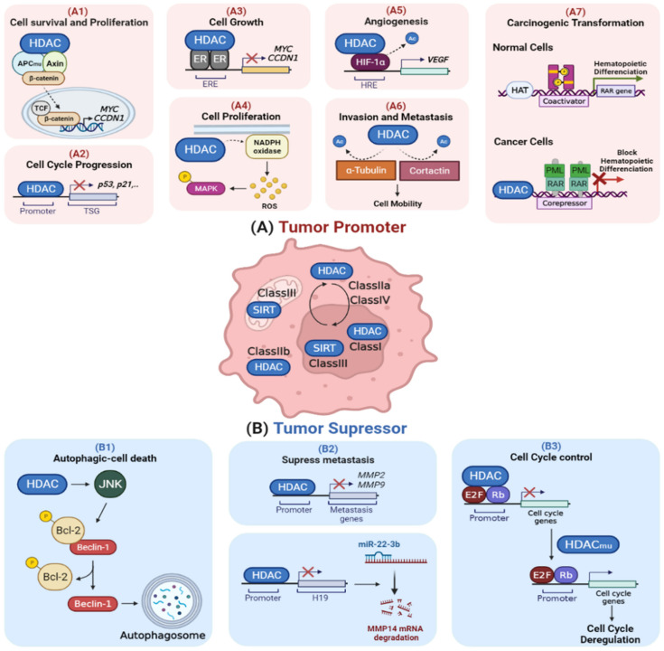
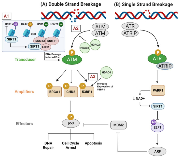
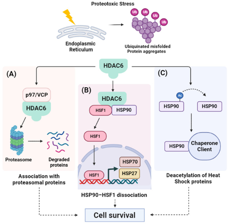
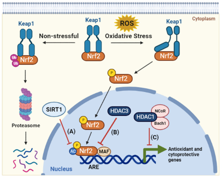
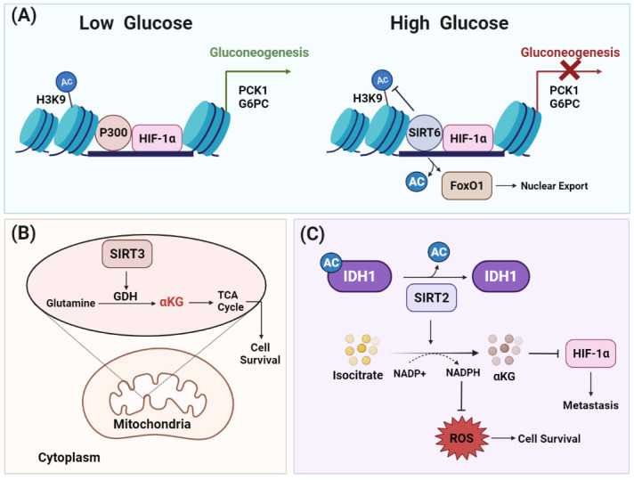
Throughout the process of carcinogenesis, cancer cells develop intricate networks to adapt to a variety of stressful conditions including DNA damage, nutrient deprivation, and hypoxia. These molecular networks encounter genomic instability and mutations coupled with changes in the gene expression programs due to genetic and epigenetic alterations. Histone deacetylases (HDACs) are important modulators of the epigenetic constitution of cancer cells. It has become increasingly known that HDACs have the capacity to regulate various cellular systems through the deacetylation of histone and bounteous nonhistone proteins that are rooted in complex pathways in cancer cells to evade death pathways and immune surveillance. Elucidation of the signaling pathways involved in the adaptive responses to cellular stress and the role of HDACs may lead to the development of novel therapeutic agents. In this article, we overview the dominant stress types including metabolic, oxidative, genotoxic, and proteotoxic stress imposed on cancer cells in the context of HDACs, which guide stress adaptation responses. Next, we expose a closer view on the therapeutic interventions and clinical trials that involve HDACs inhibitors, in addition to highlighting the impact of using HDAC inhibitors in combination with stress-inducing agents for the management of cancer and to overcome the resistance to current cancer therapy.

Keywords: HDAC inhibitors; HDACs; cellular stress; immune modulation.


Conflict of interest statement

The authors declare no conflict of interest.

Figures

Figure 1
Figure 1
HDACs as tumor promoters and suppressors. (A) HDACs are overexpressed in cancer, which promotes cellular proliferation and suppresses apoptosis and cell cycle arrest. (A1) Some HDACs (e.g., HDAC2) stabilize the beta-catenin complex, boosting cell survival and proliferation. (A2) HDACs are involved in the disruption of cell cycle checkpoints by obstructing the expression of tumor suppressor genes such as p53 and p21. (A3) Certain HDACs, such as HDAC1, reduce the transcription activity of estrogen receptor-α (ER-α), resulting in growth promotion. (A4) HDACs (e.g., HDAC6) increase the activation of MAPK through inducing the production of reactive oxygen species (ROS) via NADPH oxidase, thus promoting cell proliferation and survival. (A5) Under hypoxia, HDACs (e.g., HDAC1) stabilize HIF-1α through deacetylation, which, in turn, activates the transcription of genes involved in oxygen delivery, energy metabolism, angiogenesis, and apoptosis. (A6) The deacetylation of cell motility proteins (tubulin and cortactin) by HDACs (e.g., HDAC6) drives the progression of a primary tumor to invasion and metastasis. (A7) In hematological malignancies, PML-RAR oncofusion proteins act as altered transcription factors, which aberrantly recruit HDACs to the promoter site of retinoic acid (RA) genes, thus suppressing myeloid differentiation and causing malignant transformation. (B) Some HDACs have tumor suppressor activities and they are genetically downregulated in cancer. (B1) HDACs suppress tumor growth through activating JNK-mediated Beclin1 dissociation from Bcl-2 to induce caspase-independent autophagy death. (B2) HDACs (e.g., HDAC10) inhibit invasion and metastasis through reducing the histone acetylation level at the promoter sites of the matrix metalloproteinases (MMP2 and MMP9) genes, thereby suppressing their expression. Additionally, HDACs (e.g., HDAC2) suppress cancer metastasis through inhibiting expression of LncRNA H19, a miR-22-3P sponge that upregulates the expression of MMP14, by histone H3K27 deacetylation at its promoter site. (B3) Some HDACs (e.g., HDAC1) are involved in cell cycle regulation by forming a complex with E2F and RB that represses cell cycle progression genes. Mutations in HDAC1 reduce its recruitment and binding to E2F-regulated promoters, thereby reducing their interaction with retinoblastoma protein (Rb). This results in preventing the repression of cell cycle genes by retinoblastoma (Rb). Abbreviations: APC, adenomatous polyposis coli; TSG, tumor suppressor gene; ER, estrogen receptor; ERE, estrogen response element; ROS, reactive oxygen species; MAPK, mitogen-activated protein kinase; HIF-1α, hypoxia-inducible factor-1; HRE, hypoxia-response element; VEGF, vascular endothelial growth factor; RAR, retinoic acid receptor; PML, promyelocytic leukemia; HAT, histone acetyltransferase; JNK, c-Jun N-terminal kinases; Bcl-2, B-cell lymphoma 2; MMP, matrix metalloproteinases; miR, microRNA; Rb, retinoblastoma.
Figure 2
Figure 2
Role of HDACs in the modulation of DNA repair machinery. (A) After DNA double strand breakage, (A1) SIRT1 is directed towards the DNA damage site to induce chromatin reorganization by recruiting epigenetic machinery including DNMT1, DNMT3B, and EZH2. These epigenetic modifiers induce histone repressive modifications, such as hypoacetylation of H4K16 and methylation of H3K9 and H3K27, that help to establish the compaction of chromatin around the damaged site. (A2) HDAC1 and HDAC2 contribute to ATM activation in several tumor types, thereby enhancing the subsequent phosphorylation of BRCA1, CHK2, and p53, which decreases the susceptibility of DNA breakage. (A3) HDAC4 increases the expression of the tumor suppressor gene 53BP1, a protein involved in the early stages of DNA damage signaling. This signaling cascade facilitates the phosphorylation and activation of p53 protein, which promotes cell cycle arrest to allow DNA repair and/or apoptosis. (B) Upon induction of single stand breaks, poly(ADP-ribose) synthesis is catalyzed by PARP1 at unrepaired single break sites, which reduces the activity of NAD+-dependent deacetylase SIRT1 through decreasing the cellular concentration of NAD+. Consequently, the acetylation of E2F1 is maintained, which activates the transcription of ARF, which inhibits MDM2, a negative regulator of p53. This allows p53 to exert its tumor suppressor transcriptional regulation or/and to induce apoptosis. Abbreviations: DNMT, DNA methyltransferase 1; DNMT3B, DNA methyltransferase 3B; EZH2, enhancer of zeste homolog 2; ATM, ataxia-telangiectasia mutated; BRCA1, breast cancer gene 1; CHK2, checkpoint kinase 2; 53BP1, p53-binding protein 1; ATR, ataxia-telangiectasia and Rad3-related; ATRIP, ATR interacting protein; PARP1, poly(ADP-ribose) polymerase 1; NAD+, nicotinamide adenine dinucleotide; E2F1, E2F transcription factor 1; ARF, alternative reading frame; MDM2, mouse double minute 2.
Figure 3
Figure 3
HDAC6 regulates the response to misfolded protein aggregates. Under proteotoxic stress, cancer cells evolve an intricate set of signaling to allow the cell to respond to the presence of misfolded proteins within the endoplasmic reticulum (ER). HDAC6 is considered a master regulator in misfolded protein processing through three different pathways: (A) HDAC6 forms a complex through its ZnF-UBP domain with proteasomal proteins such as p97/VCP that binds to polyubiquitinated proteins, facilitating their proteasomal degradation; (B) In unstressed cells, HDAC6 usually exists in complex with an inactive HSF1 and HSP90. Upon ER stress, this complex is dissociated and the liberated HSF1 activates the expression of heat shock genes (HSP70 and HSP27). These heat shock proteins are essential components of the cell machinery that are required for the proper folding of proteins and the degradation of damaged proteins to protect against the adverse effects of proteotoxic stress; and (C) HDAC6 deacetylates HSP90, enhancing its chaperone activity, and facilitates the recruitment of other chaperones to reduce the level of unfolded proteins. At the end, upgrading the chaperone capacity in processing misfolded proteins by HDAC6 promotes cell survival. Abbreviations: Ub, ubiquitin; VCP, valosin-containing protein; HSF1, heat shock transcription factor 1; HSP90, heat shock protein 90; HSP70, heat shock protein 70; HSP27, heat shock protein 27.
Figure 4
Figure 4
HDACs coordinate the cellular response to oxidative stress. HDACs coordinate the cellular response to oxidative stress. As a defense mechanism against oxidative stressors, Nrf2 acts as a redox-sensitive transcription factor. Under homeostatic conditions, Keap1 sequesters Nrf2 in the cytoplasm, resulting in its degradation by proteasome. When the cells are subjected to oxidative stress, Nrf2 is activated through its dissociation from the Keap1 complex. Then, the Nrf2 protein is transported to the nucleus, where it is acetylated and dimerized with the MAF protein at the antioxidant reactive element (ARE) site in the promoters of antioxidant genes. Through this process, antioxidant genes are activated to scavenge excess reactive oxygen species (ROS) and maintain mitochondrial function. In response to oxidative stress, HDACs act as a negative regulator of the Nrf2 pathway. (A) SIRT1 inhibits Nrf2 acetylation, which reduces its binding to ARE and thereby obstructs Nrf2-mediated gene expression. (B) HDAC3 is recruited to the ARE site and induces a histone hypoacetylation state, hence repressing ARE-dependent gene expression. (C) HDAC1 forms a complex with Bach1, which competes with Nrf2 for the binding to transcription cofactor MAF in oxidative-stress-response genes. Bach1 acts as a functional inhibitor of Nrf2 by forming a complex with nuclear co-repressor NCoR. Abbreviations: Nrf2, NF-E2–related factor 2; Keap1, Kelch-like ECH associated protein 1; ROS, reactive oxygen species; MAF, musculoaponeurotic fibrosarcoma; ARE, antioxidant responsive element; Bach1, BTB domain and CNC homolog 1; NCoR, nuclear receptor corepressor 1.
Figure 5
Figure 5
HDACs elicit metabolic reprogramming to support cell survival under metabolic stress. Different HDACs are involved in reglueing several metabolic pathways. (A) Under low glucose levels, histone acetyltransferase p300 acts as coactivator for HIF-1α to induce transcription of genes involved in gluconeogenesis. Under high glucose levels, SIRT6 inhibits gluconeogenesis by acting as a co-repressor of HIF-1α transcriptional activity by deacetylating H3K9 at the promoter sites of its target glycolytic genes such as PCK1 and G6PC, which are a rate-limiting enzymes in glucose synthesis. SIRT6 deacetylates the transcription factor FoxO1, which results in its export to the cytoplasm. Excluding FoxO1 from the nucleus reduces the expression of PCK1 and G6PCm thereby inhibiting gluconeogenesis. (B) The mitochondrial lysine deacetylase SIRT3 promotes glutamine flux to the TCA cycle via glutamate dehydrogenase, which, in turn, shields the cancer cells from metabolic stress and maintains cell survival. (C) SIRT2 deacetylates IDH1 at lysine 224 and stimulates its metabolic activity. IDH catalyzes the decarboxylation of isocitrate to produce alpha-ketoglutarate (αKG), which is important for the hydroxylation and degradation of HIF-1α. Consequently, SIRT2-dependent IDH1 deacetylation inhibits metastasis as well as decreasing reactive oxygen species (ROS) levels by producing NADPH, an ultimate donor for ROS-detoxifying enzymes. Abbreviations: HIF-1α, hypoxia-inducible factor-1; FoxO1, Forkhead box protein O1; PCK1, phosphoenolpyruvate carboxykinase 1; G6PC, glucose-6-phosphatase catalytic subunit; IDH1, isocitrate dehydrogenase 1; αKG, alpha-ketoglutarate; ROS, reactive oxygen species; GDH, glutamate dehydrogenase; TCA, tricarboxylic acid.

References

    1. Poljšak B., Milisav I. Clinical implications of cellular stress responses. Bosn. J. Basic Med. Sci. 2012;12:122–126. doi: 10.17305/bjbms.2012.2510. - DOI - PMC - PubMed
    1. Raj L., Ide T., Gurkar A.U., Foley M., Schenone M., Li X., Tolliday N.J., Golub T.R., Carr S.A., Shamji A.F., et al. Selective killing of cancer cells by a small molecule targeting the stress response to ROS. Nature. 2011;475:231–234. doi: 10.1038/nature10167. - DOI - PMC - PubMed
    1. Chen M., Xie S. Therapeutic targeting of cellular stress responses in cancer. Thorac. Cancer. 2018;9:1575–1582. doi: 10.1111/1759-7714.12890. - DOI - PMC - PubMed
    1. Hamilton J.P. Epigenetics: Principles and Practice. Dig. Dis. 2011;29:130–135. doi: 10.1159/000323874. - DOI - PMC - PubMed
    1. Nair S.S., Kumar R. Chromatin remodeling in Cancer: A Gateway to regulate gene Transcription. Mol. Oncol. 2012;6:611–619. doi: 10.1016/j.molonc.2012.09.005. - DOI - PMC - PubMed