

Fluorescence Imaging Using Enzyme-Activatable Probes for Detecting Diabetic Kidney Disease and Glomerular Diseases
- PMID: 35897725
- PMCID: PMC9332157
- DOI: 10.3390/ijms23158150
Fluorescence Imaging Using Enzyme-Activatable Probes for Detecting Diabetic Kidney Disease and Glomerular Diseases
Abstract
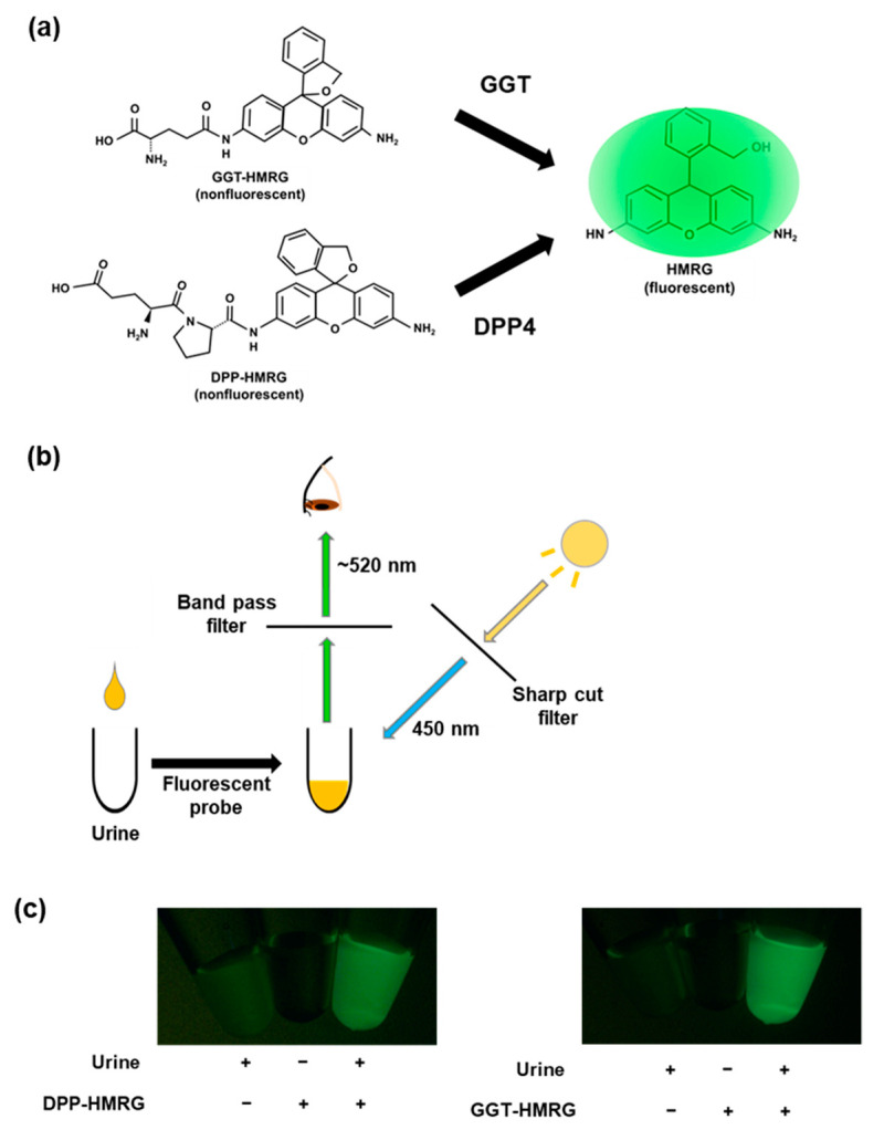
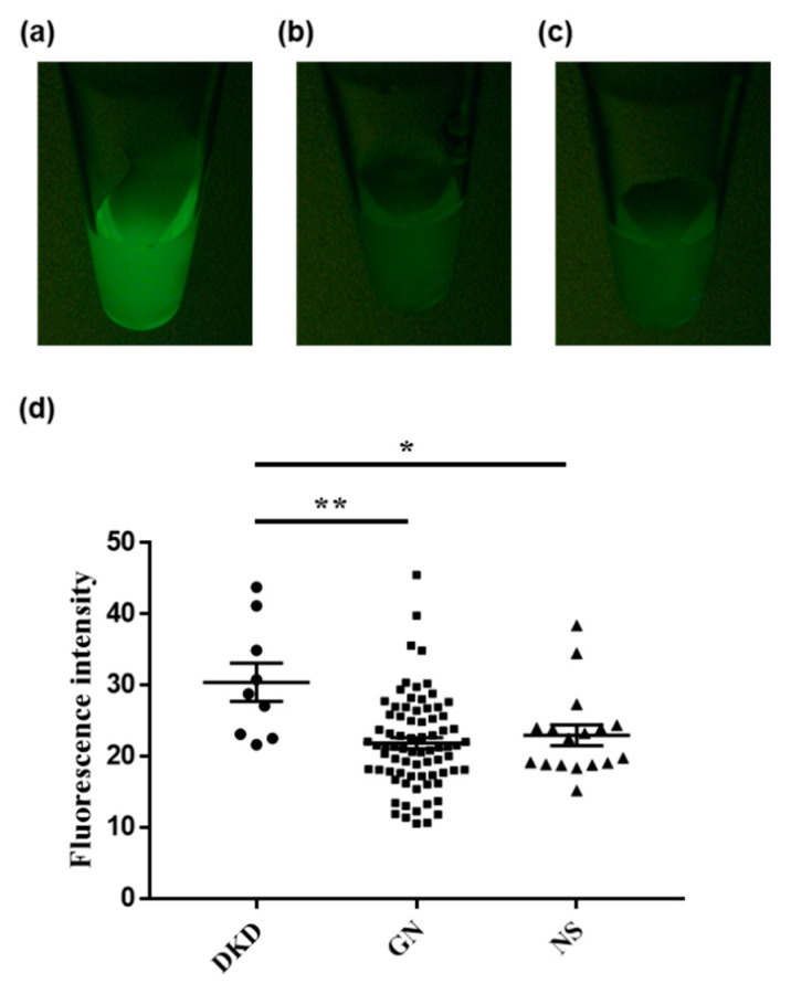
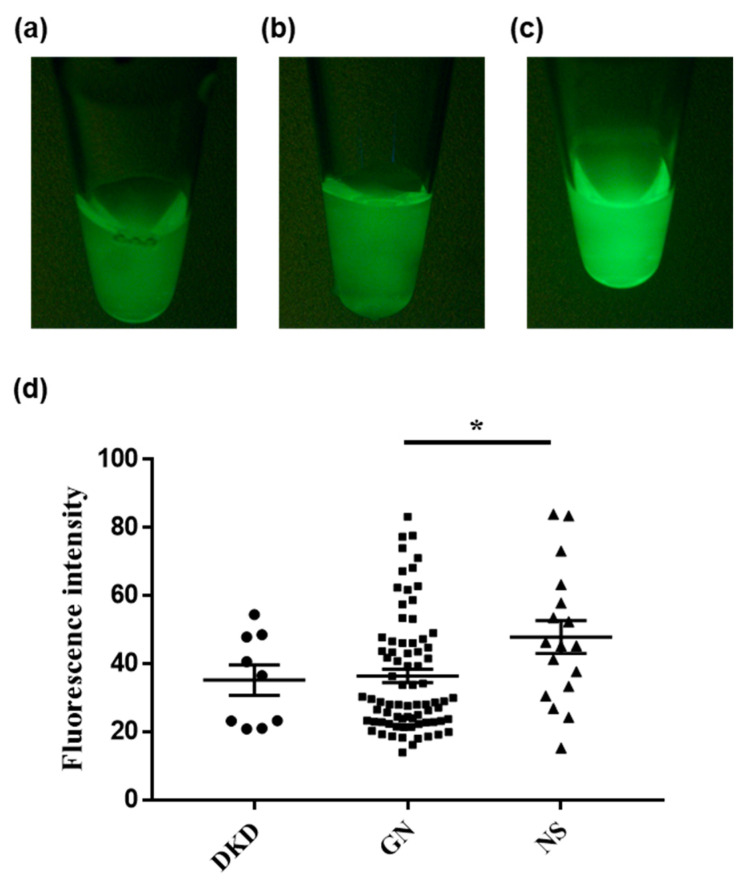
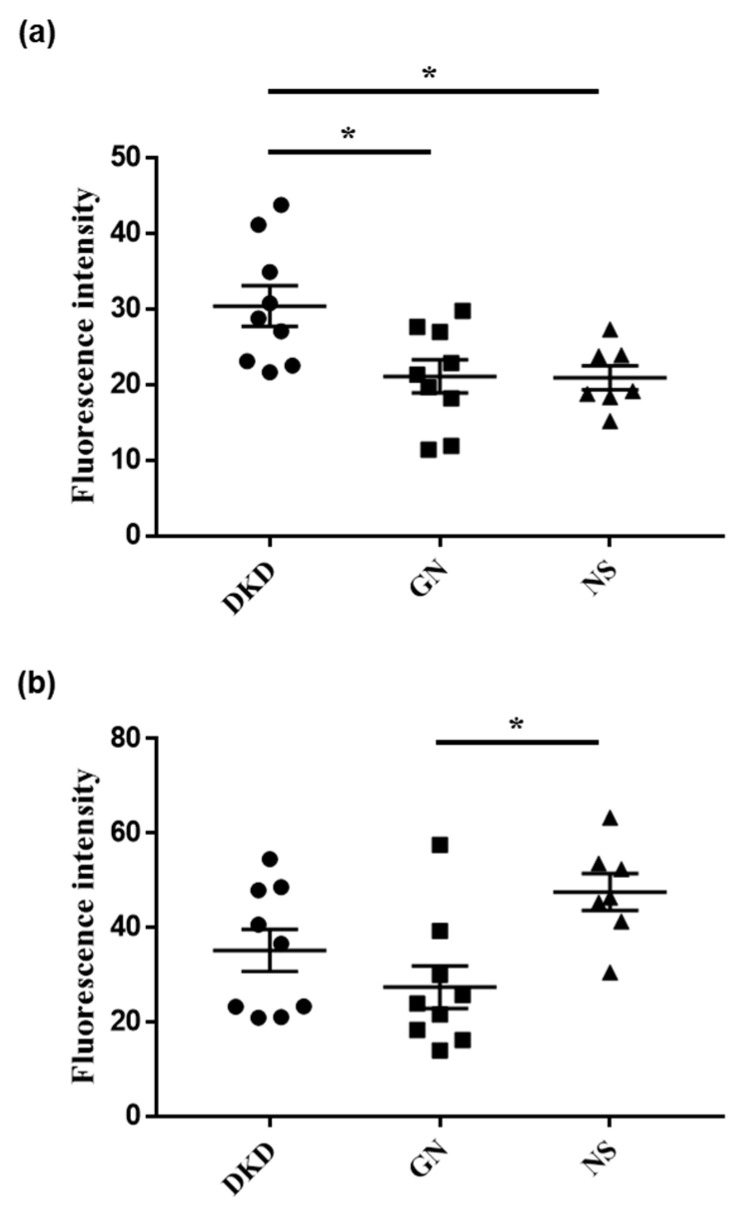
A clear identification of the etiology of glomerular disease is essential in patients with diabetes. Renal biopsy is the gold standard for assessing the underlying nephrotic pathology; however, it has the risk for potential complications. Here, we aimed to investigate the feasibility of urinary fluorescence imaging using an enzyme-activatable probe for differentiating diabetic kidney disease and the other glomerular diseases. Hydroxymethyl rhodamine green (HMRG)-based fluorescent probes targeting gamma-glutamyl transpeptidase (GGT) and dipeptidyl-peptidase (DPP) were used. Urinary fluorescence was compared between groups which were classified by their histopathological diagnoses (diabetic kidney disease, glomerulonephritis, and nephrosclerosis) as obtained by ultrasound-guided renal biopsy. Urinary fluorescence was significantly stronger in patients with diabetic kidney disease compared to those with glomerulonephritis/nephrosclerosis after DPP-HMRG, whereas it was stronger in patients with nephrosclerosis than in patients with glomerulonephritis after GGT-HMRG. Subgroup analyses of the fluorescence performed for patients with diabetes showed consistent results. Urinary fluorescence imaging using enzyme-activatable fluorescence probes thus represents a potential noninvasive assessment technique for kidney diseases in patients with diabetes.
Keywords: diabetic kidney disease; diabetic nephropathy; dipeptidyl-peptidase; enzyme-activatable probe; fluorescence imaging; fluorescent probe; gamma-glutamyl transpeptidase; glomerulonephritis; nephrosclerosis.
Conflict of interest statement
The authors declare no conflict of interest.
Figures
References
-
- Xie Y., Bowe B., Mokdad A.H., Xian H., Yan Y., Li T., Maddukuri G., Tsai C.Y., Floyd T., Al-Aly Z. Analysis of the Global Burden of Disease study highlights the global, regional, and national trends of chronic kidney disease epidemiology from 1990 to 2016. Kidney Int. 2018;94:567–581. doi: 10.1016/j.kint.2018.04.011. - DOI - PubMed
-
- Urano Y., Sakabe M., Kosaka N., Ogawa M., Mitsunaga M., Asanuma D., Kamiya M., Young M.R., Nagano T., Choyke P.L., et al. Rapid Cancer Detection by Topically Spraying a γ-Glutamyltranspeptidase–Activated Fluorescent Probe. Sci. Transl. Med. 2011;3:110ra119. doi: 10.1126/scitranslmed.3002823. - DOI - PMC - PubMed
-
- Onoyama H., Kamiya M., Kuriki Y., Komatsu T., Abe H., Tsuji Y., Yagi K., Yamagata Y., Aikou S., Nishida M., et al. Rapid and sensitive detection of early esophageal squamous cell carcinoma with fluorescence probe targeting dipeptidylpeptidase IV. Sci. Rep. 2016;6:26399. doi: 10.1038/srep26399. - DOI - PMC - PubMed
MeSH terms
Substances
LinkOut - more resources
Full Text Sources
Other Literature Sources
Medical
Miscellaneous
