. 2022 Jul 24;23(15):8161.
doi: 10.3390/ijms23158161.

The Pt(S-pr-thiosal)2 and BCL1 Leukemia Lymphoma: Antitumor Activity In Vitro and In Vivo

Affiliations

The Pt(S-pr-thiosal)2 and BCL1 Leukemia Lymphoma: Antitumor Activity In Vitro and In Vivo

Zana Besser Silconi et al. Int J Mol Sci. .

Abstract

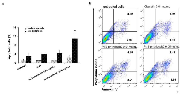
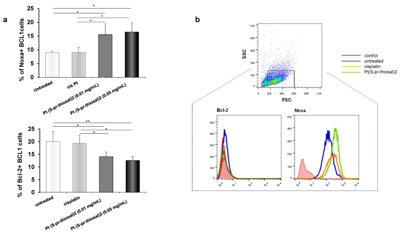
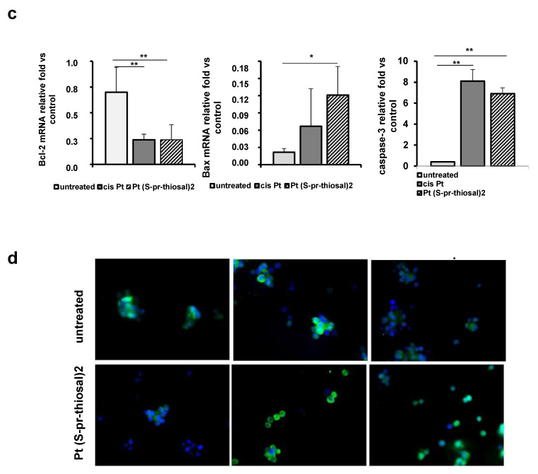
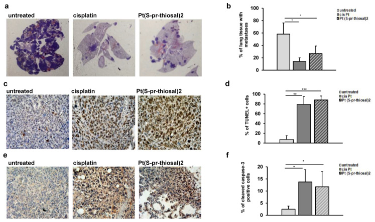
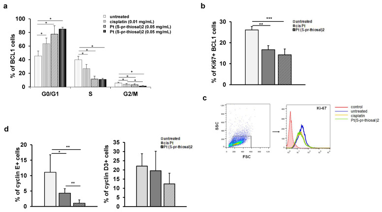
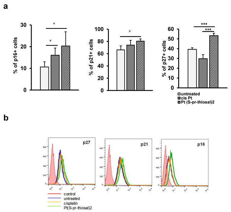
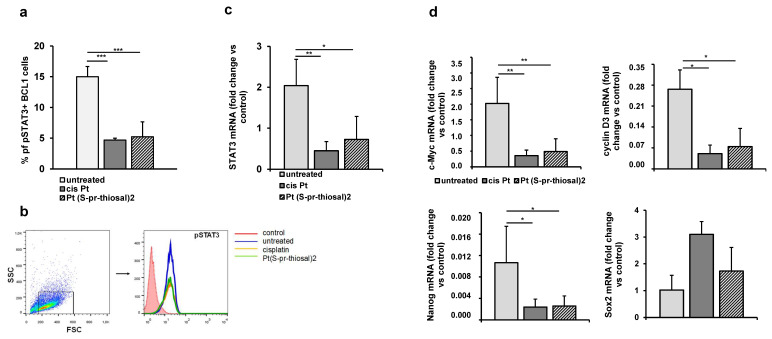
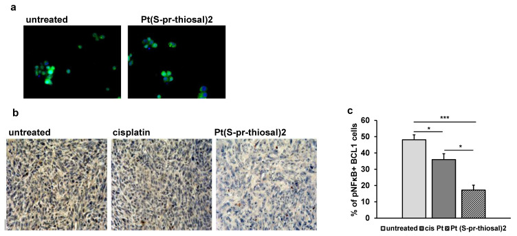
B cell malignancies are, despite the development of targeted therapy in a certain percentage of the patients still a chronic disease with relapses, requiring multiple lines of therapy. Regimens that include platinum-based drugs provide high response rates in different B cell lymphomas, high-risk chronic lymphocytic leukemia (CLL), and devastating complication of CLL, Richter's syndrome. The aim of this study was to explore the potential antitumor activity of previously synthetized platinum(IV) complex with alkyl derivatives of thyosalicilc acid, PtCl2(S-pr-thiosal)2, toward murine BCL1 cells and to delineate possible mechanisms of action. The PtCl2(S-pr-thiosal)2 reduced the viability of BCL1 cells in vitro but also reduced the growth of metastases in the leukemia lymphoma model in BALB/c mice. PtCl2(S-pr-thiosal)2 induced apoptosis, inhibited proliferation of BCL1 cells, and induced cell cycle disturbance. Treatment of BCL1 cells with PtCl2(S-pr-thiosal)2 inhibited expression of cyclin D3 and cyclin E and enhanced expression of cyclin-dependent kinase inhibitors p16, p21, and p27 resulting in cell cycle arrest in the G1 phase, reduced the percentage of BCL1 cells in the S phase, and decreased expression of Ki-67. PtCl2(S-pr-thiosal)2 treatment reduced expression of phosphorylated STAT3 and downstream-regulated molecules associated with cancer stemness and proliferation, NANOG, cyclin D3, and c-Myc, and expression of phosphorylated NFκB in vitro and in vivo. In conclusion, PtCl2(S-pr-thiosal)2 reduces STAT3 and NFκB phosphorylation resulting in inhibition of BCL1 cell proliferation and the triggering of apoptotic cell death.

Keywords: BCL1; Bcl-2; Pt(S-pr-thiosal)2 complex; STAT3; antitumor activity.


Conflict of interest statement

The authors declare no conflict of interest.

Figures

Figure 1
Figure 1
Pt(S-pr-thiosal)2 induces apoptotic death of BCL1 cells. (a) Results for the percentage of cells in early (annexin V+ propidium iodide-) and late (annexin V+ propidium iodide+) apoptosis detected by flow cytometry are shown as mean + SD for 3 independent experiments. The statistical significance of the difference in the percentage of apoptotic cells was determined by Student’s t test; * p < 0.05, ** p < 0.01 and indicates the difference in the percentage of BCL1 cells treated with platinum complex at 0.05 mg/mL and all three other groups of BCL1 cells (untreated; treated with cisplatin; and treated with platinum complex at 0.01 mg/mL). (b) Representative dot plots show the population of BCL1 cells in early and late apoptosis.
Figure 2
Figure 2
Pt(S-pr-thiosal)2 enhances expression of proapoptotic molecules, while reducing expression of antiapoptotic molecules in BCL1 cells. (a) The graphs show the mean for the percentage of Noxa and Bcl2 positive BCL1 cells. The statistical significance of the difference in the percentage of apoptotic cells was determined by Student’s t test; * p < 0.05, ** p < 0.01 and indicates the difference in the percentage of BCL1 cells treated with platinum complex at 0.05 mg/mL and all three other groups of BCL1 cells (untreated; treated with cisplatin; and treated with platinum complex at 0.01 mg/mL). (b) Representative histograms show the expression of Bcl-2 and Noxa in untreated, cisplatin-treated, and Pt(S-pr-thiosal)2 treated BCL1 cells; (c) Mean values +SD for expression of mRNA for Bcl2, Bax and caspase-3 detected in BCL1 cells by real-time qRT-PCR for 5 samples per group are shown. The statistical significance was determined by Student’s t test; * p < 0.05, ** p < 0.01. (d) Immunofluorescence staining for (green stained proteins Bcl-2 (left), Bax (middle), and active form of caspase 3 (right); DNA stained blue) (DAPI)) in BCL1 cells untreated and treated with platinum complex for 12 h at a dose of 0.05 mg/mL, magnification 200×.
Figure 2
Figure 2
Pt(S-pr-thiosal)2 enhances expression of proapoptotic molecules, while reducing expression of antiapoptotic molecules in BCL1 cells. (a) The graphs show the mean for the percentage of Noxa and Bcl2 positive BCL1 cells. The statistical significance of the difference in the percentage of apoptotic cells was determined by Student’s t test; * p < 0.05, ** p < 0.01 and indicates the difference in the percentage of BCL1 cells treated with platinum complex at 0.05 mg/mL and all three other groups of BCL1 cells (untreated; treated with cisplatin; and treated with platinum complex at 0.01 mg/mL). (b) Representative histograms show the expression of Bcl-2 and Noxa in untreated, cisplatin-treated, and Pt(S-pr-thiosal)2 treated BCL1 cells; (c) Mean values +SD for expression of mRNA for Bcl2, Bax and caspase-3 detected in BCL1 cells by real-time qRT-PCR for 5 samples per group are shown. The statistical significance was determined by Student’s t test; * p < 0.05, ** p < 0.01. (d) Immunofluorescence staining for (green stained proteins Bcl-2 (left), Bax (middle), and active form of caspase 3 (right); DNA stained blue) (DAPI)) in BCL1 cells untreated and treated with platinum complex for 12 h at a dose of 0.05 mg/mL, magnification 200×.
Figure 3
Figure 3
Pt(S-pr-thiosal)2 induces significant apoptotic effect in vivo. (a) Representative sections of lung tissue obtained from mice injected intravenously with BCL1 cells and treated with platinum complex, cisplatin, or saline, stained with hematoxylin and eosin. (b) The mean value ± SD of the percentage of lung tissue affected by metastases (n = 10 for each of the groups) calculated by analyzing lung tissue preparations stained with hematoxylin and eosin in Image J. (c) Representative lung sections obtained four weeks after application of BCL1 cells stained by the TUNEL method. (d) The percentage of TUNEL-positive nuclei (brown) presented as mean values + SD for 5 mice per group. (e) Representative immunohistochemistry of active caspase-3 expression in tumor tissue in the lungs of untreated mice and mice treated with cisplatin and complex. (f) The percentage of active caspase-3 positive tumor cells (brown color) in relation to the total number of cells in the visual field presented as mean values + SD. * p < 0.05, ** p < 0.01, *** p < 0.001.
Figure 4
Figure 4
Pt(S-pr-thiosal)2 has antiproliferative effect on BCL1 cells. (a) The graph shows the mean percentage of BCL1 cells by phases of the cell cycle + SD; (b) Percentage of Ki67+ BCL1 cells + SD; (c) Representative histograms show Ki-67 expression in untreated, cisplatin-treated and Pt(S-pr-thiosal)2 treated BCL1 cells. (d) Percentage of cyclin E+ and cyclin D3+ BCL1 cells + SD from three independent experiments, for 3 replicates. Statistical significance was determined by Student’s t test, * p < 0.05, ** p < 0.01, *** p < 0.001. (d) representative histograms show Ki-67 expression in untreated, cisplatin-treated and Pt(S-pr-thiosal)2 treated BCL1 cells.
Figure 5
Figure 5
Pt(S-pr-thiosal)2 increases the percentage of BCL1 cells expressing cyclin-dependent kinase inhibitors p16, p21, and p27. After treatment with Pt(S-pr-thiosal)2 complex (dose 0.05 mg/mL) and cisplatin for 12 h, BCL1 cells were stained with anti-p16, anti-p21, and anti-p27 antibodies, and the percentage of positive cells was detected by flow cytometry. (a) The graph shows the mean values of the percentage of positive BCL1 cells +SD for 3 replicates. Statistical significance was determined by Student’s t test; * p < 0.05; *** p < 0.001. (b) Representative histograms show the expression of p27, p21, and p16 in untreated, cisplatin-treated, and Pt(S-pr-thiosal)2 treated BCL1 cells.
Figure 6
Figure 6
Pt(S-pr-thiosal)2 decreases the expression of phosphorylated STAT3 and STAT3 regulated genes in BCL1 cells. (a) The percentage of (phospho Y705) STAT3 positive BCL1 cells was detected by flow cytometry, the relative expression of mRNA for (c) STAT3 and (d) c-Myc, cyclin D3, Nanog, and Sox2, normalized to β-actin, was analyzed by real-time qRT-PCR and presented as mean values + SD for 5 samples per group. The statistical significance was determined by Student’s t test; * p < 0.05, ** p < 0.01, *** p < 0.001. (b) Representative histograms show the expression of phosphorylated STAT3 in untreated, cisplatin-treated, and Pt(S-pr-thiosal)2 treated BCL1 cells.
Figure 7
Figure 7
The expression of the phosphorylated form of NF-κB is decreased in BCL1 cells after exposure to Pt(S-pr-thiosal)2. (a) Immunofluorescence (green stained phosphorylated NF-κB and DNA stained blue) (DAPI)) BCL1 cells untreated and treated with platinum complex for 12 h, magnification 200×. (b) Representative images of phosphorylated NF-κB expression in tumor tissue (detected by immunohistochemistry) in the lungs of untreated mice and mice treated with cisplatin and Pt(S-pr-thiosal)2, four weeks after intravenous administration of BCL1 cells. (c) The percentage of positive cells (expressing NF-κB) in relation to the total number of tumor cells in the field of view presented as the mean values + SD for 5 mice per group. Statistical significance was determined by Student’s t test; * p < 0.05; *** p < 0.001.

Similar articles

Cited by

  • Metal-Based Complexes in Cancer.
    Riccardi C, Piccolo M. Riccardi C, et al. Int J Mol Sci. 2023 Apr 14;24(8):7289. doi: 10.3390/ijms24087289. Int J Mol Sci. 2023. PMID: 37108457 Free PMC article.

References

    1. Fisher S.G., Fisher R.I. The epidemiology of non-Hodgkin’s lymphoma. Oncogene. 2004;23:6524–6534. doi: 10.1038/sj.onc.1207843. - DOI - PubMed
    1. Yao Y., Lin X., Li F., Jin J., Wang H. The global burden and attributable risk factors of chronic lymphocytic leukemia in 204 countries and territories from 1990 to 2019: Analysis based on the global burden of disease study 2019. Biomed. Eng. Online. 2022;21:4. doi: 10.1186/s12938-021-00973-6. - DOI - PMC - PubMed
    1. Klanova M., Klener P. BCL-2 Proteins in Pathogenesis and Therapy of B-Cell Non-Hodgkin Lymphomas. Cancers. 2020;12:938. doi: 10.3390/cancers12040938. - DOI - PMC - PubMed
    1. Tsujimoto Y., Finger L.R., Yunis J., Nowell P.C., Croce C.M. Cloning of the chromosome breakpoint of neoplastic B cells with the t(14;18) chromosome translocation. Science. 1984;226:1097–1099. doi: 10.1126/science.6093263. - DOI - PubMed
    1. Cho-Vega J.H., Rassidakis G.Z., Admirand J.H., Oyarzo M., Ramalingam P., Paraguya A., McDonnell T.J., Amin H.M., Medeiros L.J. MCL-1 expression in B-cell non-Hodgkin’s lymphomas. Hum. Pathol. 2004;35:1095–1100. doi: 10.1016/j.humpath.2004.04.018. - DOI - PubMed