. 2022 Jul 26;23(15):8205.
doi: 10.3390/ijms23158205.

The Effect of N-Acetylation on the Anti-Inflammatory Activity of Chitooligosaccharides and Its Potential for Relieving Endotoxemia

Affiliations

The Effect of N-Acetylation on the Anti-Inflammatory Activity of Chitooligosaccharides and Its Potential for Relieving Endotoxemia

Wentong Hao et al. Int J Mol Sci. .

Abstract

Endotoxemia is mainly caused by a massive burst of inflammatory cytokines as a result of lipopolysaccharide (LPS) invasion. Chitooligosaccharides (COS) is expected to be a potential drug for relieving endotoxemia due to its anti-inflammatory properties. However, the structural parameters of COS are often ambiguous, and the effect of degree of acetylation (DA) of COS on its anti-inflammatory remains unknown. In this study, four COSs with different DAs (0%, 12%, 50% and 85%) and the same oligomers distribution were successfully obtained. Their structures were confirmed by 1H NMR and MS analysis. Then, the effect of DA on the anti-inflammatory activity and relieving endotoxemia potential of COS was researched. The results revealed that COS with a DA of 12% had better anti-inflammatory activity than COSs with other DAs, mainly in inhibiting LPS-induced inflammatory cytokines burst, down-regulating its mRNA expression and reducing phosphorylation of IκBα. Furthermore, this COS showed an obviously protective effect on endotoxemia mice, such as inhibiting the increase in inflammatory cytokines and transaminases, alleviating the injury of liver and intestinal tissue. This study explored the effect of DA on the anti-inflammatory activity of COS for the first time and lays the foundation for the development of COS as an anti-inflammatory drug against endotoxemia.

Keywords: anti-inflammation; chitooligosaccharides; degree of acetylation; endotoxemia.


Conflict of interest statement

The authors declare no conflict of interest.

Figures

Figure 1
Figure 1
1H NMR spectra of COSs with different DAs. (AD) represent 0% COS, 12% COS, 50% COS and 85% COS, respectively. The peak at 2.8–3.2 ppm is the hydrogen on the second carbon of GlcN, and the peak at 1.9 ppm is the hydrogen on the acetyl group in GlcNAc. The peak intensity at 1.9 ppm increases linearly with DA increasing.
Figure 2
Figure 2
Mass spectrum analysis of the four prepared COSs with different DAs. (A) The positive-ion mode ESI-MS spectrum of COS with DA = 0%, (B) The positive-ion mode ESI-MS spectrum of COS with DA = 12%, (C) The positive-ion mode ESI-MS spectrum of COS with DA = 50%, (D) The positive-ion mode ESI-MS spectrum of COS with DA = 85%. The numbers in the figure only represent the number of monosaccharide units.
Figure 3
Figure 3
Effects of different concentrations of COSs on macrophage cell viability. At the concentrations used in this study, these COSs had no cytotoxic effects on cells. The data are presented as means ± SD of three replicates. * There is a significant difference between 0% COS with 85% COS in 800 μg/mL, p ≤ 0.05.
Figure 4
Figure 4
COSs with different DAs inhibited LPS-induced secretion of NO, IL-6 and TNF-α. 0%, 12%, 50%, 85% represent COS with DA of 0%, 12%, 50%, 85%, respectively. The data are presented as means ± SD of three replicates. From “a” to “e”, the mean values decrease in turn. There is no significant difference between groups with the same letter. Different letters indicate individual groups for multiple comparisons with significant differences (one-way ANOVA, Duncan, p < 0.05).
Figure 5
Figure 5
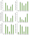
COS can reduce the up-regulation of several inflammation-related genes caused by LPS. 0%, 12%, 50%, 85% represent COS with DA of 0%, 12%, 50%, 85%, respectively. The data are presented as means ± SD of three replicates. From “a” to “e”, the mean values decrease in turn. There is no significant difference between groups with the same letter. Different letters indicate individual groups for multiple comparisons with significant differences (one-way ANOVA, Duncan, p < 0.05).
Figure 6
Figure 6
Western blotting of phosphorylated-IκBα and IκBα. After the cells were lysed, the total protein was extracted for WB experiments. 0%, 12%, 50%, 85% represent COS with DA of 0%, 12%, 50%, 85%, respectively. The data are presented as means ± SD of three replicates. From “a” to “d”, the mean values decrease in turn. There is no significant difference between groups with the same letter. Different letters indicate individual groups for multiple comparisons with significant differences (one-way ANOVA, Duncan, p < 0.05).
Figure 7
Figure 7
The immunofluorescent staining assay was used to analyze the nuclear translocation of NF-κB in RAW264.7 cells. NF-κB was stained with rabbit anti-NF-κB p65 IgG, with FITC-conjugated goat anti-rabbit IgG as secondary antibody (green). Nuclei were stained with DAPI (blue). Measuring bar 10 μm.
Figure 8
Figure 8
Changes in pro-inflammatory markers and transaminases with 12% COS treatment. M represents the model group; Dex represents the positive control group. The data are presented as means ± SD of three replicates. From “a” to “e”, the mean values decrease in turn. There is no significant difference between groups with the same letter. Different letters indicate individual groups for multiple comparisons with significant differences (one-way ANOVA, Duncan, p < 0.05).
Figure 9
Figure 9
H&E staining and TEM of liver tissue. (A) represents the H&E staining of liver tissue; (B) represents the nuclei of hepatocytes under the same magnification. The arrow indicates chromatin condensation; (C) represents the hepatocyte mitochondria under the same magnification. Arrows indicate mitochondrial inner cristae.
Figure 10
Figure 10
H&E staining of small intestinal villi. Compared with the model group, COS pretreatment increased villi height and the microvilli in the NC group and COS group can be seen under high magnification, while the microvilli in the model group disappear.

Similar articles

Cited by

References

    1. Raetz C.R., Whitfield C. Lipopolysaccharide endotoxins. Annu. Rev. Biochem. 2002;71:635–700. doi: 10.1146/annurev.biochem.71.110601.135414. - DOI - PMC - PubMed
    1. Mohammad S., Thiemermann C. Role of Metabolic Endotoxemia in Systemic Inflammation and Potential Interventions. Front. Immunol. 2020;11:594150. doi: 10.3389/fimmu.2020.594150. - DOI - PMC - PubMed
    1. Rhee S.H. Lipopolysaccharide: Basic biochemistry, intracellular signaling, and physiological impacts in the gut. Intest. Res. 2014;12:90–95. doi: 10.5217/ir.2014.12.2.90. - DOI - PMC - PubMed
    1. Liu P.Y., Shen H.H., Kung C.W., Chen S.Y., Lu C.H., Lee Y.M. The Role of HSP70 in the Protective Effects of NVP-AUY922 on Multiple Organ Dysfunction Syndrome in Endotoxemic Rats. Front. Pharmacol. 2021;12:724515. doi: 10.3389/fphar.2021.724515. - DOI - PMC - PubMed
    1. Wu T.J., Hsieh Y.J., Lu C.W., Lee C.J., Hsu B.G. Linagliptin Protects against Endotoxin-Induced Acute Kidney Injury in Rats by Decreasing Inflammatory Cytokines and Reactive Oxygen Species. Int. J. Mol. Sci. 2021;22:11190. doi: 10.3390/ijms222011190. - DOI - PMC - PubMed

LinkOut - more resources