. 2022 Jul 26;23(15):8246.
doi: 10.3390/ijms23158246.

Myeloperoxidase as a Marker to Differentiate Mouse Monocyte/Macrophage Subsets

Affiliations

Myeloperoxidase as a Marker to Differentiate Mouse Monocyte/Macrophage Subsets

Cody J Gurski et al. Int J Mol Sci. .

Abstract

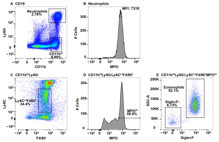
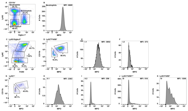
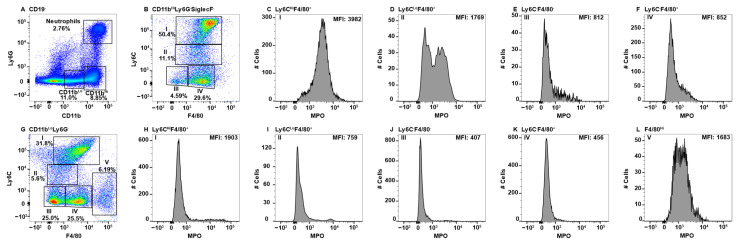
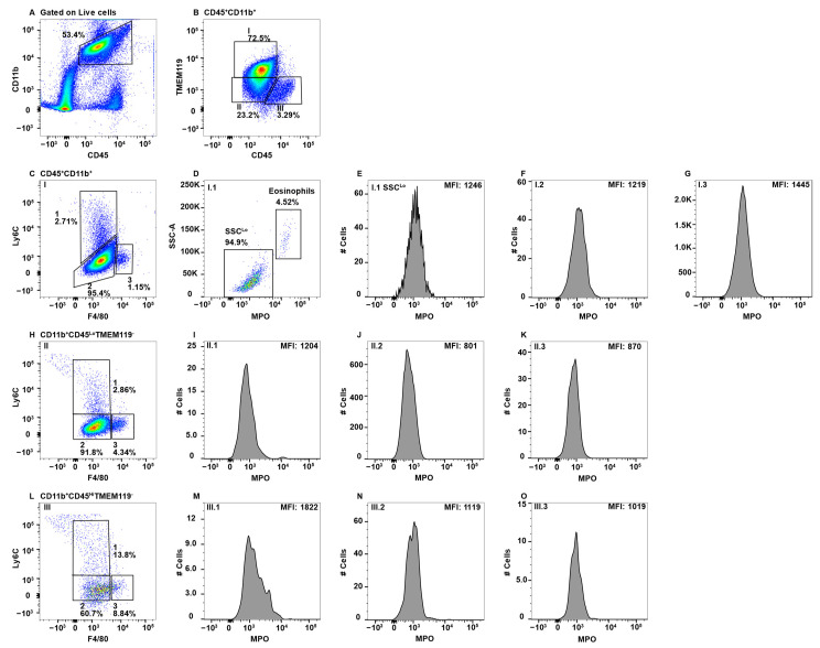
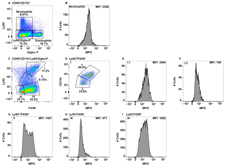
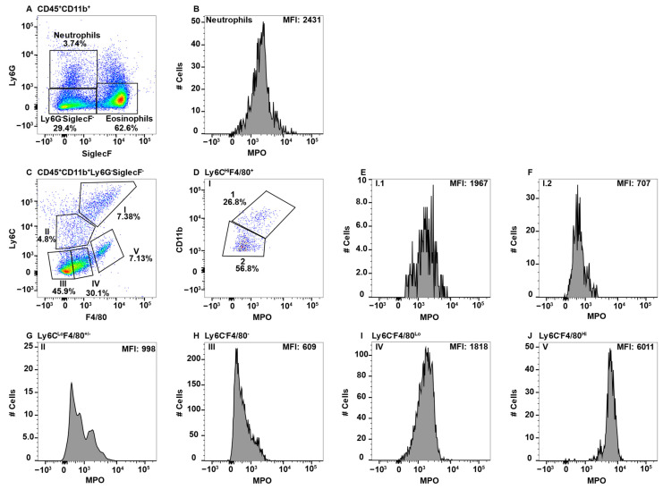
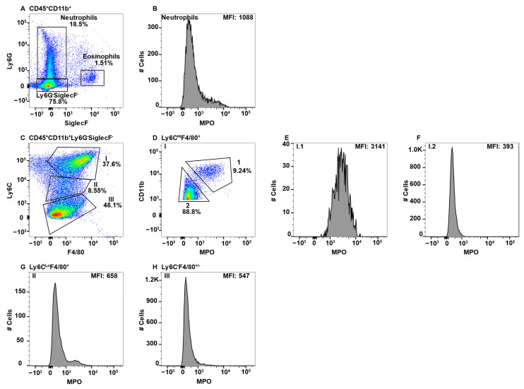
Macrophages are present in every tissue in the body and play essential roles in homeostasis and host defense against microorganisms. Some tissue macrophages derive from the yolk sac/fetal liver that populate tissues for life. Other tissue macrophages derive from monocytes that differentiate in the bone marrow and circulate through tissues via the blood and lymphatics. Circulating monocytes are very plastic and differentiate into macrophages with specialized functions upon entering tissues. Specialized monocyte/macrophage subsets have been difficult to differentiate based on cell surface markers. Here, using a combination of "pan" monocyte/macrophage markers and flow cytometry, we asked whether myeloperoxidase (MPO) could be used as a marker of pro-inflammatory monocyte/macrophage subsets. MPO is of interest because of its potent microbicidal activity. In wild-type SPF housed mice, we found that MPO+ monocytes/macrophages were present in peripheral blood, spleen, small and large intestines, and mesenteric lymph nodes, but not the central nervous system. Only monocytes/macrophages that expressed cell surface F4/80 and/or Ly6C co-expressed MPO with the highest expression in F4/80HiLy6CHi subsets regardless of tissue. These cumulative data indicate that MPO expression can be used as an additional marker to differentiate between monocyte/macrophage subsets with pro-inflammatory and microbicidal activity in a variety of tissues.

Keywords: macrophage; monocyte; myeloperoxidase.


Conflict of interest statement

The authors declare no conflict of interest.

Figures

Figure 1
Figure 1
Using neutrophils as a positive control for MPO expression by flow cytometry, eosinophils were found to give a false positive signal. Splenocytes were stained with antibodies specific for CD19, CD11b, Ly6G, F4/80, Ly6C, MPO, and SiglecF and using flow cytometry, the CD19 cells were gated out. CD11b+Ly6G+ neutrophils (A) were analyzed for MPO expression (B). CD11bHiLy6G cells were analyzed for F4/80 and Ly6C expression (C) and F4/80+Ly6CLo MPO expression is shown (D). MPOHi cells were further analyzed for SSCHi and SiglecF expression to identify eosinophils (E). The percent positive cells in dot plot gates and the MFI of histograms is provided. Data shown are representative of three mice.
Figure 2
Figure 2
Blood monocyte/macrophage subsets differentially express MPO. PBMC were stained with antibodies specific for CD45, CD11b, Ly6C, Ly6G, F4/80, MPO, and SiglecF and using flow cytometry, CD45+ cells were gated on CD11b. Neutrophils and eosinophils were identified by expression of Ly6G and SiglecF, respectively (A). Neutrophil MPO positive staining is shown (B). CD11b+Ly6GSiglecF cells were analyzed for F4/80 and Ly6C expression and four Subpop (I–IV) were identified (C). Subpop I F4/80+Ly6CHi cells were separated into CD11bHi (1) and CD11bLo (2) populations (D), and MPO expression is shown (E and F, respectively). Subpop II Ly6CLo cells were separated into CD11bHi (1) and CD11bLo (2) populations (G), and MPO expression is shown (H and I, respectively). MPO expression in Subpop III F4/80Ly6C cells (J) and Subpop IV F4/80+Ly6C cells (K) is shown. The percent positive cells in dot plot gates and the MFI of histograms is provided. Data shown are representative of three mice.
Figure 3
Figure 3
Splenic monocyte/macrophage subsets differentially express MPO. Splenocytes were stained with antibodies specific for CD19, CD11b, Ly6C, Ly6G, F4/80, MPO, and SiglecF and using flow cytometry, CD19 cells were analyzed for Ly6G and CD11b to identify double positive neutrophils and Ly6GCD11bHi and Ly6GCD11bLo cells (A). CD11bHiLy6GSiglecF cells were analyzed for F4/80 and Ly6C expression and four Subpop (I–IV) were identified (B). MPO expression in Subpop I F4/80+Ly6CHi (C), II Ly6CLoF4/80+ (D), III Ly6CF4/80 (E), I and IV Ly6CF4/80+ (F) is shown. CD11bLoLy6GSiglecF cells were analyzed for F4/80 and Ly6C expression and five Subpop (I–V) were identified (G). MPO expression in Subpop I F4/80+Ly6CHi (H), II Ly6CLoF4/80+ (I), III Ly6CF4/80 (J), IV Ly6CF4/80+ (K), and V Ly6CF4/80Hi (L) is shown. The percent positive cells in dot plot gates and the MFI of histograms is provided. Data shown are representative of three mice.
Figure 4
Figure 4
CNS microglial cells and perivascular macrophages do not express MPO. Mononuclear cells from brains and spinal cords were stained with antibodies specific for CD45, CD11b, Ly6C, F4/80, MPO, and TMEM119 and using flow cytometry cells were gated on CD45+CD11b+ (A), which were analyzed for TMEM119 expression identifying three Subpop (I-III) (B). Subpop I CD45LoCD11b+TMEM119+ cells were analyzed for Ly6C and F4/80 expression, which identified three subsets (I.1, I.2, and I.3) (C). SSCHI eosinophils were eliminated in Subset I.1 (D) prior to analysis of MPO expression (E). Subsets I.2 and I.3 did not require eosinophil exclusion before MPO analysis (F and G, respectively). Subpop II CD45LoCD11b+TMEM119 cells were analyzed for Ly6C and F4/80 expression (H), which identified three subsets (II.1, II.2, and II.3), which were further analyzed for MPO expression (I,J,K, respectively). Subpop III CD45HiCD11b+TMEM119 cells analyzed for Ly6C and F4/80 expression identified three subsets (III.1, III.2, and III.3) (L), which were further analyzed for MPO expression (M,N,O, respectively). The percent positive cells in dot plot gates and the MFI of histograms is provided. Data shown are representative of three mice.
Figure 5
Figure 5
Large intestinal macrophage subsets differentially express MPO. Large intestine mononuclear cells were stained with antibodies specific for CD45, CD11b, Ly6C, Ly6G, F4/80, MPO, and SiglecF and using flow cytometry, CD45+CD11b+ cells were analyzed for Ly6G and SiglecF expression to identify Ly6G+SiglecF neutrophils and Ly6GSiglecF+ eosinophils (A). Neutrophil MPO-positive staining is shown (B). CD45+CD11b+Ly6GSiglecF cells analyzed for F4/80 and Ly6C identified four Subpop (I–IV) (C). Subpop I F4/80+Ly6CHi cells were separated into CD11bHi (1) and CD11bLo (2) populations (D), and MPO expression is shown (E and F, respectively). MPO expression in Subpop II Ly6CLoF4/80+ (G), III Ly6CF4/80 (H) and IV Ly6CF4/80+ (I) is shown. The percent positive cells in dot plot gates and the MFI of histograms is provided. Data shown are representative of three mice.
Figure 6
Figure 6
Small intestinal macrophage subsets differentially express MPO. Small intestine mononuclear cells were stained with antibodies specific for CD45, CD11b, Ly6C, Ly6G, F4/80, MPO, and SiglecF and using flow cytometry, CD45+CD11b+ cells were analyzed for Ly6G and SiglecF expression to identify Ly6G+SiglecF neutrophils and Ly6GSiglecF+ eosinophils (A). Neutrophil MPO-positive staining is shown (B). CD11bHiLy6GSiglecF cells were analyzed for F4/80 and Ly6C expression and five Subpop (I–V) were identified (C). Subpop I F4/80+Ly6CHi cells were separated into CD11bHi (1) and CD11bLo (2) populations (D), and MPO expression is shown (E and F, respectively). MPO expression in Subpop II Ly6CLoF4/80+ (G), III Ly6CF4/80 (H), IV Ly6CF4/80+ (I), and Ly6CF4/80Hi V (J) is shown. The percent positive cells in dot plot gates and the MFI of histograms is provided. Data shown are representative of three mice.
Figure 7
Figure 7
MLN macrophage subsets differentially express MPO. MLN cells were stained with antibodies that were specific for CD45, CD11b, Ly6C, Ly6G, F4/80, MPO, and SiglecF and using flow cytometry CD45+CD11b+ cells were analyzed for Ly6G and SiglecF expression to identify Ly6G+SiglecF neutrophils and Ly6GSiglecF+ eosinophils (A). Neutrophil expression of MPO is shown (B). CD45+CD11bLy6GSiglecF cells were analyzed for F4/80 and Ly6C expression and three Subpop (III) were identified (C). Subpop I F4/80+Ly6CHi cells were separated into CD11bHi (1) and CD11bLo (2) populations (D), and MPO expression is shown (E and F, respectively). MPO expression in Subpop II Ly6CLoF4/80+ (G) and III Ly6CF4/80 (H) is shown. The percent positive cells in dot plot gates and the MFI of histograms is provided. Data shown are representative of three mice.

References

    1. Aratani Y. Myeloperoxidase: Its role for host defense, inflammation, and neutrophil function. Arch. Biochem. Biophys. 2018;640:47–52. doi: 10.1016/j.abb.2018.01.004. - DOI - PubMed
    1. El-Benna J., Hurtado-Nedelec M., Marzaioli V., Marie J.C., Gougerot-Pocidalo M.A., Dang P.M. Priming of the neutrophil respiratory burst: Role in host defense and inflammation. Immunol. Rev. 2016;273:180–193. doi: 10.1111/imr.12447. - DOI - PubMed
    1. Weiss S.J., Klein R., Slivka A., Wei M. Chlorination of taurine by human neutrophils. Evidence for hypochlorous acid generation. J. Clin. Investig. 1982;70:598–607. doi: 10.1172/JCI110652. - DOI - PMC - PubMed
    1. Nauseef W.M. Myeloperoxidase in human neutrophil host defence. Cell. Microbiol. 2014;16:1146–1155. doi: 10.1111/cmi.12312. - DOI - PMC - PubMed
    1. Klebanoff S.J. Myeloperoxidase: Friend and foe. J. Leukoc. Biol. 2005;77:598–625. doi: 10.1189/jlb.1204697. - DOI - PubMed