. 2022 Jul 27;23(15):8267.
doi: 10.3390/ijms23158267.

COX-2/sEH Dual Inhibitor Alleviates Hepatocyte Senescence in NAFLD Mice by Restoring Autophagy through Sirt1/PI3K/AKT/mTOR

Affiliations

COX-2/sEH Dual Inhibitor Alleviates Hepatocyte Senescence in NAFLD Mice by Restoring Autophagy through Sirt1/PI3K/AKT/mTOR

Chen-Yu Zhang et al. Int J Mol Sci. .

Abstract

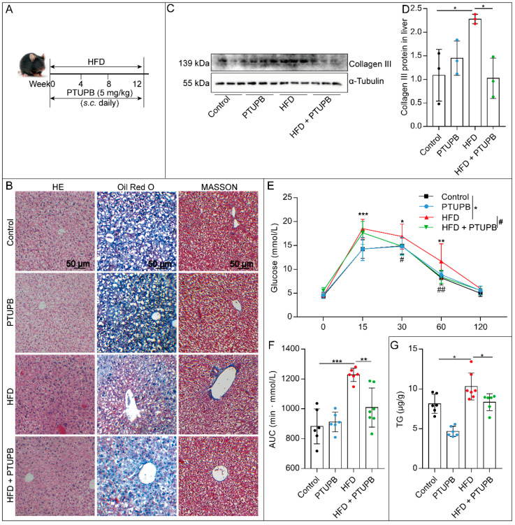
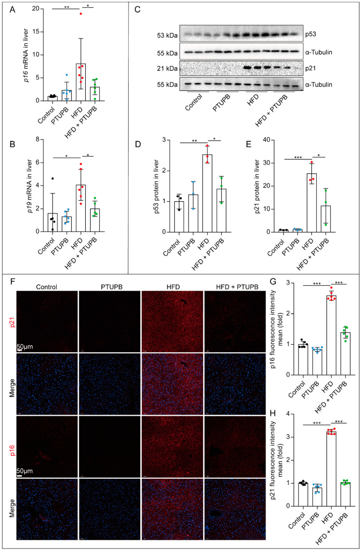
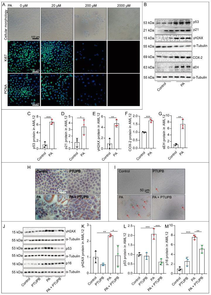
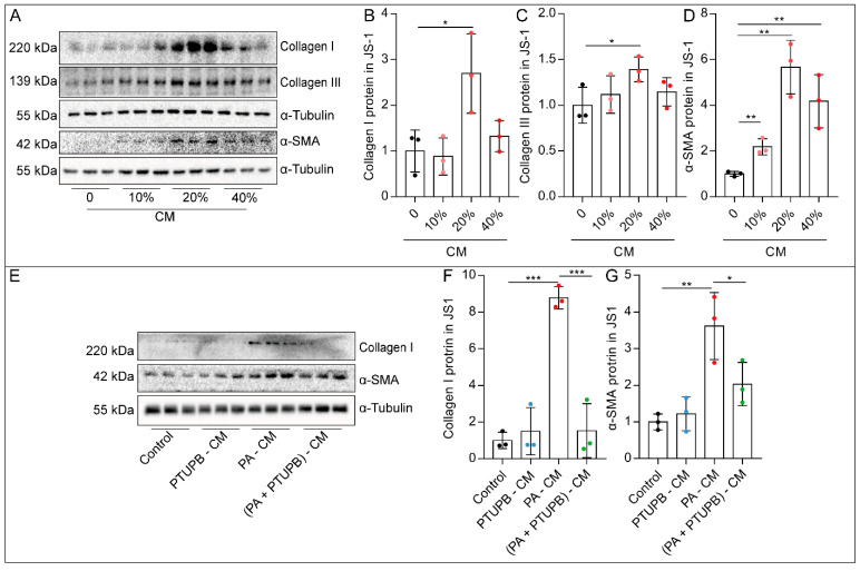
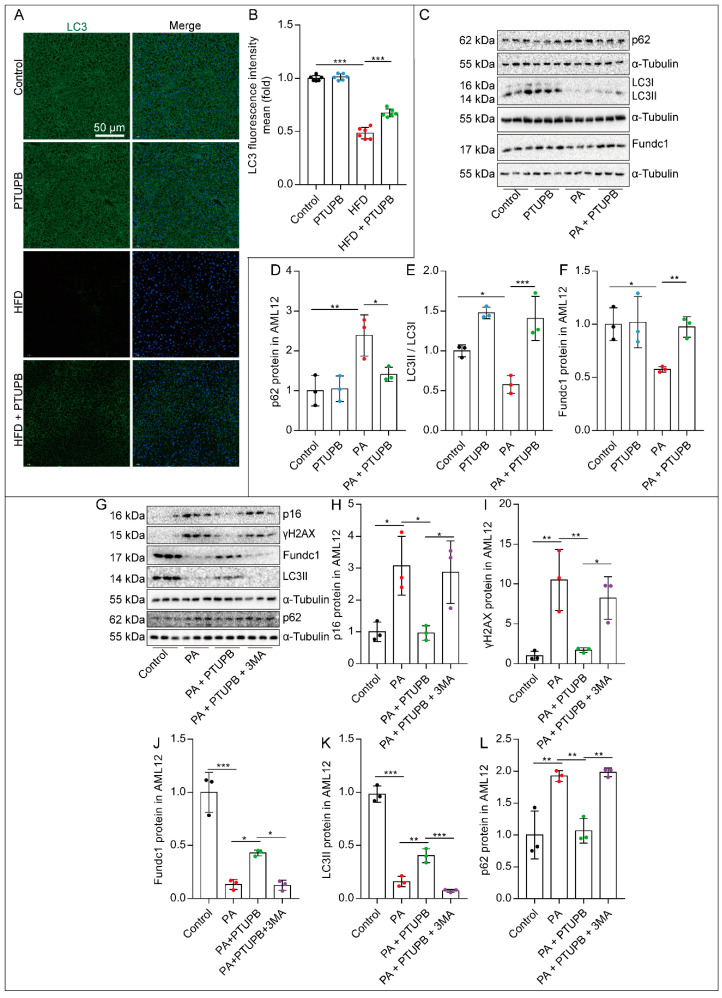
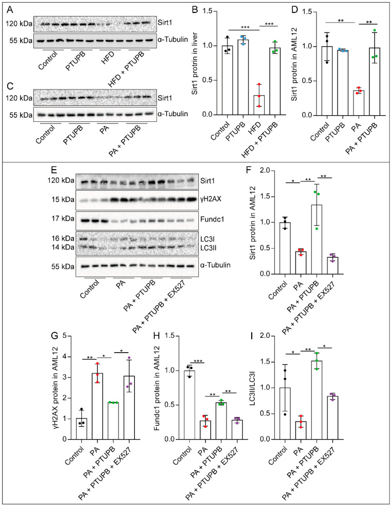
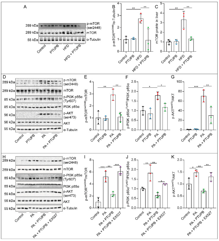
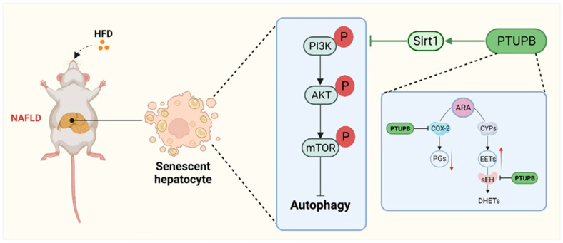
We previously found that the disorder of soluble epoxide hydrolase (sEH)/cyclooxygenase-2 (COX-2)-mediated arachidonic acid (ARA) metabolism contributes to the pathogenesis of the non-alcoholic fatty liver disease (NAFLD) in mice. However, the exact mechanism has not been elucidated. Accumulating evidence points to the essential role of cellular senescence in NAFLD. Herein, we investigated whether restoring the balance of sEH/COX-2-mediated ARA metabolism attenuated NAFLD via hepatocyte senescence. A promised dual inhibitor of sEH and COX-2, PTUPB, was used in our study to restore the balance of sEH/COX-2-mediated ARA metabolism. In vivo, NAFLD was induced by a high-fat diet (HFD) using C57BL/6J mice. In vitro, mouse hepatocytes (AML12) and mouse hepatic astrocytes (JS1) were used to investigate the effects of PTUPB on palmitic acid (PA)-induced hepatocyte senescence and its mechanism. PTUPB alleviated liver injury, decreased collagen and lipid accumulation, restored glucose tolerance, and reduced hepatic triglyceride levels in HFD-induced NAFLD mice. Importantly, PTUPB significantly reduced the expression of liver senescence-related molecules p16, p53, and p21 in HFD mice. In vitro, the protein levels of γH2AX, p53, p21, COX-2, and sEH were increased in AML12 hepatocytes treated with PA, while Ki67 and PCNA were significantly decreased. PTUPB decreased the lipid content, the number of β-gal positive cells, and the expression of p53, p21, and γH2AX proteins in AML12 cells. Meanwhile, PTUPB reduced the activation of hepatic astrocytes JS1 by slowing the senescence of AML12 cells in a co-culture system. It was further observed that PTUPB enhanced the ratio of autophagy-related protein LC3II/I in AML12 cells, up-regulated the expression of Fundc1 protein, reduced p62 protein, and suppressed hepatocyte senescence. In addition, PTUPB enhanced hepatocyte autophagy by inhibiting the PI3K/AKT/mTOR pathway through Sirt1, contributing to the suppression of senescence. PTUPB inhibits the PI3K/AKT/mTOR pathway through Sirt1, improves autophagy, slows down the senescence of hepatocytes, and alleviates NAFLD.

Keywords: COX-2/sEH dual inhibitor; autophagy; non-alcoholic fatty liver disease; senescence.


Conflict of interest statement

The authors declared no conflict of interests.

Figures

Figure 1
Figure 1
PTUPB reduces HFD-induced NAFLD in mice. PTUPB (5 mg/kg/day, s.c.) was administered daily (A). HE-stained histological images of liver sections from mice (B), left column, scale bars = 50 μm, n = 6. Oil Red O staining of liver sections from mice (B), middle column, scale bars = 50 μm, n = 6. Masson staining of liver sections from mice (B), right column, scale bars = 50 μm, n = 6. Western blot was used to detect collagen III protein expressions and quantitative analysis ((C, D), n = 6–7). Measurement of blood glucose and AUC during IGTT of mice fed with HFD or normal chow diet for 12 weeks ((E,F), n = 6–7). Content of TG in liver tissue of mice ((G), n = 6–7). Data are expressed as the mean ± SD. Differences among multiple groups were performed using ANOVA. Tukey’s test was used as a post hoc test for pairwise comparisons. # or * p < 0.05, ** p < 0.01, and *** p < 0.001.
Figure 2
Figure 2
PTUPB inhibits hepatocyte senescence induced by HFD in mice. p16 and p19 mRNA expressions in the liver were detected using real-time PCR ((A,B), n = 5–6). Western blot was used to detect p53 and p21 protein expressions and quantitative analysis ((CE), n = 6). The fluorescence intensity of p21 and p16 were detected by immunofluorescence ((F), scale bars = 50 μm, n = 6). Image J was used to analyze the fluorescence intensity of p21 and p16 ((G,H), n = 6). Data are expressed as the mean ± SD. Differences among multiple groups were performed using ANOVA. Tukey’s test was used as a post hoc test for pairwise comparisons. * p < 0.05, ** p < 0.01, and *** p < 0.001.
Figure 3
Figure 3
PTUPB attenuates PA-induced hepatocyte senescence in vitro. AML12 cells were treated with a series of concentrations of PA (0, 20, 200, and 2000 μM) for 48 h, and then the changes in cell morphology and number were photographed under a microscope ((A), scale bars = 100 μm, n = 3). The fluorescence intensity of Ki67 and PCNA was detected by immunofluorescence ((A), scale bars = 100 μm, n = 3). AML12 cells were treated with PTUPB (1 μM) for 1 h before treatment with PA (200 µM). Forty-eight hours after the PA administration, the protein expressions of p53, p21, γH2AX, COX-2, and sEH in AML12 cells were measured by Western blot and quantitatively analyzed ((BG), n = 3). Representative images of Oil Red O staining in AML-12 hepatocytes treated with PA with/without PTUPB for 48 h ((H), bar = 50 µm). Senescence was confirmed by SA-β-gal staining ((I), bar = 50 µm). The protein expressions of γH2AX, p53, and p16 in AML12 cells were measured by Western blot and quantitatively analyzed after PA stimulation with/without PTUPB for 48 h ((JM), n = 3). Data are expressed as the mean ± SD. Differences among multiple groups were performed using ANOVA. Tukey’s test was used as a post hoc test for pairwise comparisons. Comparisons between the two-group were made with an unpaired t-test. * p < 0.05, ** p < 0.01, and *** p < 0.001.
Figure 4
Figure 4
PTUPB reduces hepatic astrocyte activation by inhibiting hepatocyte senescence. AML12 cells were treated with PA (200 µM) for 48 h, and cell culture supernatant was collected as CM. The protein expressions of collagen I, collagen III, and α-SMA in JS1 cells treated with different concentrations of PA-CM stimulation for 48 h were measured by Western blot and quantitatively analyzed ((AD), n = 3). The protein expressions of Collagen I and α-SMA in JS1 cells were measured by Western blot and quantitatively analyzed after different CM stimulation for 48 h ((EG), n = 3). Data are expressed as the mean ± SD. Differences among multiple groups were performed using ANOVA. Tukey’s test was used as a post hoc test for pairwise comparisons. * p < 0.05, ** p < 0.01, and *** p < 0.001.
Figure 5
Figure 5
PTUPB attenuates hepatocyte senescence by enhancing autophagy in AML12 cells. The fluorescence intensity of liver LC3 was detected by immunofluorescence ((A), scale bars = 50 μm, n = 6). Image J was used to analyze the fluorescence intensity of LC3 ((B), n = 6). Cells were treated with PTUPB (1 μM) for 1 h before treatment with PA (200 µM). The protein expressions of p62, LC3I/II, and Fundc1 were measured by Western blot and quantitatively analyzed in AML12 cells treated with PA for 48 h ((CF), n = 3). Cells were treated with PTUPB (1 μM) and 3-MA (5 mM) for 1 h before treatment with PA (200 µM). The protein expressions of p16, γH2AX, Fundc1, LC3II, and p62 were measured by Western blot and quantitatively analyzed in AML12 cells ((GL), n = 3). Data are expressed as the mean ± SD. Differences among multiple groups were performed using ANOVA. Tukey’s test was used as a post hoc test for pairwise comparisons. * p < 0.05, ** p < 0.01, and *** p < 0.001.
Figure 6
Figure 6
PTUPB enhances hepatocyte autophagy by promoting Sirt1 expression. The protein expressions of Sirt1 in the liver ((A,B), n = 6) and AML12 cells ((C,D), n = 3) were measured by Western blot and quantitatively analyzed. Cells were treated with PTUPB (1 μM) or EX527 (10 μM) for 1 h before treatment with PA (200 µM) for 48 h. The protein expressions of Sirt1, γH2AX, Fundc1, and LC3II/I in AML12 cells were measured by Western blot and quantitatively analyzed ((EI), n = 3). Data are expressed as the mean ± SD. Differences among multiple groups were performed using ANOVA. Tukey’s test was used as a post hoc test for pairwise comparisons. * p < 0.05, ** p < 0.01, and *** p < 0.001.
Figure 7
Figure 7
PTUPB inhibits the PI3K/AKT/mTOR signaling pathway by promoting Sirt1 expression. The mTOR protein and its phosphorylation level in the liver were measured by Western blot and quantitatively analyzed ((AC), n = 6). Cells were treated with PTUPB (1 μM) for 1 h before treatment with PA (200 µM). The levels of p-mTOR, p-AKT, and p-PI3K in AML12 cells were measured by Western blot and quantitatively analyzed after PA stimulation for 30 min ((DG), n = 3). Cells were treated with PTUPB (1 μM) or EX527 (10 μM) for 1 h before treatment with PA (200 µM). The levels of p-mTOR, p-AKT, and p-PI3K in AML12 cells were measured by Western blot and quantitatively analyzed after PA stimulation for 30 min ((HK), n = 3). Data are expressed as the mean ± SD. Differences among multiple groups were performed using ANOVA. Tukey’s test was used as a post hoc test for pairwise comparisons. * p < 0.05, ** p < 0.01, and *** p < 0.001.
Figure 8
Figure 8
Schematic illustration. PTUPB alleviates HFD-induced NAFLD by reducing hepatocyte senescence through autophagy in mice.

References

    1. Francque S., Szabo G., Abdelmalek M.F., Byrne C.D., Cusi K., Dufour J.-F., Roden M., Sacks F., Tacke F. Nonalcoholic steatohepatitis: The role of peroxisome proliferator-activated receptors. Nat. Rev. Gastroenterol. Hepatol. 2021;18:24–39. doi: 10.1038/s41575-020-00366-5. - DOI - PubMed
    1. Abdelmalek M.F. Nonalcoholic fatty liver disease: Another leap forward. Nat. Rev. Gastroenterol. Hepatol. 2021;18:85–86. doi: 10.1038/s41575-020-00406-0. - DOI - PMC - PubMed
    1. Tanwar S., Rhodes F., Srivastava A., Trembling P.M., Rosenberg W. Inflammation and fibrosis in chronic liver diseases including non-alcoholic fatty liver disease and hepatitis C. World J. Gastroenterol. 2020;26:109–133. doi: 10.3748/wjg.v26.i2.109. - DOI - PMC - PubMed
    1. Liu T., Luo X., Li Z.-H., Wu J.-C., Luo S.-Z., Xu M.-Y. Zinc-α2-glycoprotein 1 attenuates non-alcoholic fatty liver disease by negatively regulating tumour necrosis factor-α. World J. Gastroenterol. 2019;25:5451–5468. doi: 10.3748/wjg.v25.i36.5451. - DOI - PMC - PubMed
    1. Funk C.D. Prostaglandins and Leukotrienes: Advances in Eicosanoid Biology. Science. 2001;294:1871–1875. doi: 10.1126/science.294.5548.1871. - DOI - PubMed

MeSH terms