Review
. 2022 Jul 26;27(15):4787.
doi: 10.3390/molecules27154787.

Conformational Variability of Amyloid-β and the Morphological Diversity of Its Aggregates

Affiliations
Review

Conformational Variability of Amyloid-β and the Morphological Diversity of Its Aggregates

Maho Yagi-Utsumi et al. Molecules. .

Abstract

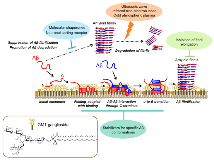
Protein folding is the most fundamental and universal example of biomolecular self-organization and is characterized as an intramolecular process. In contrast, amyloidogenic proteins can interact with one another, leading to protein aggregation. The energy landscape of amyloid fibril formation is characterized by many minima for different competing low-energy structures and, therefore, is much more enigmatic than that of multiple folding pathways. Thus, to understand the entire energy landscape of protein aggregation, it is important to elucidate the full picture of conformational changes and polymorphisms of amyloidogenic proteins. This review provides an overview of the conformational diversity of amyloid-β (Aβ) characterized from experimental and theoretical approaches. Aβ exhibits a high degree of conformational variability upon transiently interacting with various binding molecules in an unstructured conformation in a solution, forming an α-helical intermediate conformation on the membrane and undergoing a structural transition to the β-conformation of amyloid fibrils. This review also outlines the structural polymorphism of Aβ amyloid fibrils depending on environmental factors. A comprehensive understanding of the energy landscape of amyloid formation considering various environmental factors will promote drug discovery and therapeutic strategies by controlling the fibril formation pathway and targeting the consequent morphology of aggregated structures.

Keywords: NMR spectroscopy; aggregation; amyloid-β; cryo-electron microscopy; fibril; ganglioside; molecular chaperone.


Conflict of interest statement

The authors declare no conflict of interest.

Figures

Figure 1
Figure 1
Transient interaction of Aβ with binding molecules. Each binding site on Aβ with the spherical complex displaying GM1 glycans (green), SorLA Vsp10 domain (orange), and apical domain of GroEL (blue) is represented with the primary structure of Aβ. The molecular graphics of GroEL and SorLA Vps10 domain with Aβ are based on PDB: 1KP8 and 3WSZ, respectively. The molecular graphics of the spherical complex displaying GM1 glycans are adopted with permission from reference [21]. 2015, WILEY–VCH Verlag GmbH & Co. KGaA, Weinheim, Germany.
Figure 2
Figure 2
Schematic representation of the structural basis of the conformational transition and molecular assembly of Aβ promoted on GM1 ganglioside clusters on the neuronal cell membrane and the structure-based therapeutic strategies. After the initial encounter, the GM1 cluster captures Aβ at the hydrophobic/hydrophilic interface, which facilitates α-helix formation, thereby restricting the spatial rearrangements of Aβ molecules. Consequently, a specific intermolecular interaction between Aβ molecules is enhanced on the GM1 cluster, leading to their α-to-β conformational transition, resulting in amyloid fibril formation. Several proteins, including molecular chaperones, capture Aβ and thereby suppress its fibrillization. Irradiation with ultrasonic waves, an infrared free-electron laser, and cold atmospheric plasma can break down amyloid fibrils. Adapted with permission from reference [12]. 2019, The Pharmaceutical Society of Japan.
Figure 3
Figure 3
Aβ fibril structures solved by solid-state NMR and cryo-EM. The variety of fibril structures of Aβ(1–40) (blue, PDB: 2LMN, 2LMP) and Aβ(1–42) (magenta, PDB: 2MXU, 5KK3, 5AEF, 2NAO, 5OQV) fibrils prepared in vitro. Ex vivo, Aβ(1–40) seeded fibrils, which were formed by seed aggregation with recombinant Aβ(1–40) and ex vivo fibrils (green, PDB: 6W0O, 6SHS, 2M4J). The Aβ(1–42) fibrils were extracted from human AD brains (orange, PDB: 7Q4B, 7Q4M).

Similar articles

Cited by

References

    1. Dobson C.M. Protein folding and misfolding. Nature. 2003;426:884–890. doi: 10.1038/nature02261. - DOI - PubMed
    1. Chiti F., Dobson C.M. Protein Misfolding, Amyloid Formation, and Human Disease: A Summary of Progress over the Last Decade. Annu. Rev. Biochem. 2017;86:27–68. doi: 10.1146/annurev-biochem-061516-045115. - DOI - PubMed
    1. Chiti F., Dobson C.M. Protein misfolding, functional amyloid, and human disease. Annu. Rev. Biochem. 2006;75:333–366. doi: 10.1146/annurev.biochem.75.101304.123901. - DOI - PubMed
    1. Knowles T.P., Vendruscolo M., Dobson C.M. The amyloid state and its association with protein misfolding diseases. Nat. Rev. Mol. Cell Biol. 2014;15:384–396. doi: 10.1038/nrm3810. - DOI - PubMed
    1. Hartl F.U., Hayer-Hartl M. Converging concepts of protein folding in vitro and in vivo. Nat. Struct. Mol. Biol. 2009;16:574–581. doi: 10.1038/nsmb.1591. - DOI - PubMed

LinkOut - more resources