. 2022 Jul 11:9:926591.
doi: 10.3389/fmolb.2022.926591. eCollection 2022.

Cations Do Not Alter the Membrane Structure of POPC-A Lipid With an Intermediate Area

Affiliations

Cations Do Not Alter the Membrane Structure of POPC-A Lipid With an Intermediate Area

Sergei Kurakin et al. Front Mol Biosci. .

Abstract

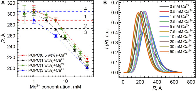
Combining small-angle neutron scattering (SANS), small-angle X-ray scattering (SAXS), and densitometric measurements, we have studied the interactions of the divalent cations Ca2+ and Mg2+ with the lipid vesicles prepared of a mixed-chain palmitoyl-oleoyl-phosphatidylcholine (POPC) at 25°C. The structural parameters of the POPC bilayer, such as the bilayer thickness, lateral area, and volume per lipid, displayed no changes upon the ion addition at concentrations up to 30 mM and minor changes at > 30 mM Ca2+ and Mg2+, while some decrease in the vesicle radius was observed over the entire concentration range studied. This examination allows us to validate the concept of lipid-ion interactions governed by the area per lipid suggested previously and to propose the mixed mode of those interactions that emerge in the POPC vesicles. We speculate that the average area per POPC lipid that corresponds to the cutoff length of lipid-ion interactions generates an equal but opposite impact on ion bridges and separate lipid-ion pairs. As a result of the dynamic equilibrium, the overall structural properties of bilayers are not affected. As the molecular mechanism proposed is affected by the structural properties of a particular lipid, it might help us to understand the fundamentals of processes occurring in complex multicomponent membrane systems.

Keywords: SANS; SAXS; cations; densitometry; lipid bilayer; structure.


Conflict of interest statement

The authors declare that the research was conducted in the absence of any commercial or financial relationships that could be construed as a potential conflict of interest.

Figures

FIGURE 1
FIGURE 1
Volume per lipid V L in POPC vesicles for different Ca2+ concentrations as a function of temperature. The error bars show systematic errors calculated based on sample preparation protocol (uncertainties in lipid weighing, and buffer volume added to the lipid powder, etc.).
FIGURE 2
FIGURE 2
Dependence of volume per lipid V L on the concentration of Ca2+ and Mg2+ in POPC vesicles at T = 25°C. The dashed line represents V L of neat POPC MLVs with the wide gray band depicting its standard deviation.
FIGURE 3
FIGURE 3
(A) SANS experimental curves measured at T = 25°C for POPC samples (points) and the best-fit results of their model analysis by spherical vesicles (lines). (B) SAXS curves measured at T = 25°C for POPC (points) and their best-fit results of the Gaussian model (lines). SANS and SAXS curves are presented for 0 mM (1), 1 mM (2), 5 mM (3), 10 mM (4), and 50 mM (5) of Ca2+ concentration.
FIGURE 4
FIGURE 4
(A) Inner vesicle radii R extracted from the SANS experiments for the POPC bilayers (lipid concentrations of 0.5 wt%, 1.0 wt%, and 3.0 wt%) loaded with various Ca2+ and Mg2+ concentrations. Horizontal dashed lines represent R of POPC ULVs without Ca2+/Mg2+ obtained with the same model, where numbers (1–4) represent different POPC concentrations in the sample: 0.5 wt% (1), 1 wt% (2–3), and 3 wt% (4). (B) Distribution functions of POPC vesicle radii upon Ca2+ additions extracted from the fitting of SANS data. The functions are presented in accordance with the Schultz distribution described in the Supplementary Material.
FIGURE 5
FIGURE 5
The changes of POPC bilayer thickness (d L and d HH ) as a function of Ca2+ and Mg2+ concentrations at 25°C according to (A) SANS and (B) SAXS measurements. Dashed lines represent d L and d HH of neat POPC ULVs with the wide gray bands depicting their standard deviations.
FIGURE 6
FIGURE 6
Area per lipid A L of the POPC bilayer as a function of Ca2+ and Mg2+ concentrations. The dashed line represents A L of neat POPC with the wide gray band depicting its standard deviation.

References

    1. Akashi K.-i., Miyata H., Itoh H., Kinosita K. (1998). Formation of Giant Liposomes Promoted by Divalent Cations: Critical Role of Electrostatic Repulsion. Biophysical J. 74 (6), 2973–2982. 10.1016/S0006-3495(98)78004-X - DOI - PMC - PubMed
    1. Akutsu H., Seelig J. (1981). Interaction of Metal Ions with Phosphatidylcholine Bilayer Membranes. Biochemistry 20 (26), 7366–7373. 10.1021/bi00529a007 - DOI - PubMed
    1. Allolio C., Harries D. (2021). Calcium Ions Promote Membrane Fusion by Forming Negative-Curvature Inducing Clusters on Specific Anionic Lipids. ACS Nano 15 (8), 12880–12887. 10.1021/acsnano.0c08614 - DOI - PubMed
    1. Alsop R. J., Maria Schober R., Rheinstädter M. C. (2016). Swelling of Phospholipid Membranes by Divalent Metal Ions Depends on the Location of the Ions in the Bilayers. Soft Matter 12 (32), 6737–6748. 10.1039/C6SM00695G - DOI - PubMed
    1. Altenbach C., Seelig J. (1984). Calcium Binding to Phosphatidylcholine Bilayers as Studied by Deuterium Magnetic Resonance. Evidence for the Formation of a Calcium Complex with Two Phospholipid Molecules. Biochemistry 23 (17), 3913–3920. 10.1021/bi00312a019 - DOI - PubMed