Case Report: Disorder of Sexual Development in a Chinese Crested Dog With XX/XY Leukocyte Chimerism and Mixed Cell Testicular Tumors
- PMID: 35898552
- PMCID: PMC9309221
- DOI: 10.3389/fvets.2022.937991
Case Report: Disorder of Sexual Development in a Chinese Crested Dog With XX/XY Leukocyte Chimerism and Mixed Cell Testicular Tumors
Abstract
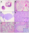
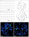
A 10-year-old intact female Chinese Crested dog was presented for evaluation and further diagnostics due to persistent symptoms of vulvar swelling, vaginal discharge, and an 8-year history of acyclicity. At presentation, generalized hyperpigmentation and truncal alopecia were identified, with no aberrations of the female phenotype. Vaginal cytology confirmed the influence of estrogen at multiple veterinary visits, and hormonal screening of progesterone and anti-Mullerian hormone indicated gonadal presence. Based on findings from abdominal laparotomy and gonadectomy, the tissue was submitted for histopathology. Histopathologic evaluation identified the gonads to be abnormal testes containing multiple Sertoli and interstitial (Leydig) cell tumors. The histopathologic diagnosis of testes and concurrent normal external female phenotype in the patient lead to a diagnosis of a disorder of sexual development (DSD). Karyotype evaluation by conventional and molecular analysis revealed a two cell line chimeric pattern of 78,XX (80%) and 78,XY (20%) among blood leukocytes, as well as a positive PCR test for the Y-linked SRY gene. Cytogenetic analysis of skin fibroblasts revealed the presence of 78,XX cells exclusively, and PCR tests for the Y-linked SRY gene were negative in the hair and skin samples. These results are consistent with an XX/XY blood chimerism. This is one of the few case reports of a canine with the diagnosis of leukocyte chimerism with normal female phenotypic external genitalia. This case illustrates a distinct presentation for hormonally active Sertoli cell tumorigenesis and demonstrates surgery as a curative treatment option for clinically affected patients.
Keywords: Leydig cell tumor; Sertoli cell tumor; canine; chimerism; disorder of sexual development (DSD).
Copyright © 2022 Schwartz, Sugai, Eden, Castaneda, Jevit, Raudsepp and Cecere.
Conflict of interest statement
The authors declare that the research was conducted in the absence of any commercial or financial relationships that could be construed as a potential conflict of interest.
Figures


Similar articles
-
XX/XY Chimerism in Internal Genitalia of a Virilized Heifer.Animals (Basel). 2022 Oct 26;12(21):2932. doi: 10.3390/ani12212932. Animals (Basel). 2022. PMID: 36359056 Free PMC article.
-
A case of leucocyte chimerism (78,XX/78,XY) in a dog with a disorder of sexual development.Reprod Domest Anim. 2014 Jun;49(3):e31-4. doi: 10.1111/rda.12318. Epub 2014 Apr 16. Reprod Domest Anim. 2014. PMID: 24735223
-
A case report of a normal fertile woman with 46,XX/46,XY somatic chimerism reveals a critical role for germ cells in sex determination.Hum Reprod. 2024 Apr 3;39(4):849-855. doi: 10.1093/humrep/deae026. Hum Reprod. 2024. PMID: 38420683
-
Disorders of Sex Development Are an Emerging Problem in French Bulldogs: A Description of Six New Cases and a Review of the Literature.Sex Dev. 2019;13(4):205-211. doi: 10.1159/000506582. Epub 2020 Mar 24. Sex Dev. 2019. PMID: 32203972 Review.
-
Abnormalities of gonadal differentiation.Baillieres Clin Endocrinol Metab. 1998 Apr;12(1):133-42. doi: 10.1016/s0950-351x(98)80512-0. Baillieres Clin Endocrinol Metab. 1998. PMID: 9890065 Review.
Cited by
-
An Unusual Case of Collision Testicular Tumor in a Female DSD Dog.Vet Sci. 2023 Mar 27;10(4):251. doi: 10.3390/vetsci10040251. Vet Sci. 2023. PMID: 37104406 Free PMC article.
-
Sex cord-stromal (granulosa cell) tumor in an ovotestis from a cow.Can Vet J. 2024 Nov;65(11):1141-1148. Can Vet J. 2024. PMID: 39494182 Free PMC article.
References
Publication types
LinkOut - more resources
Full Text Sources

