. 2022 Jul 18:2022:9376338.
doi: 10.1155/2022/9376338. eCollection 2022.

Small Extracellular Vesicles from Inflamed Adipose Derived Stromal Cells Enhance the NF- κ B-Dependent Inflammatory/Catabolic Environment of Osteoarthritis

Affiliations

Small Extracellular Vesicles from Inflamed Adipose Derived Stromal Cells Enhance the NF- κ B-Dependent Inflammatory/Catabolic Environment of Osteoarthritis

Carola Cavallo et al. Stem Cells Int. .

Abstract

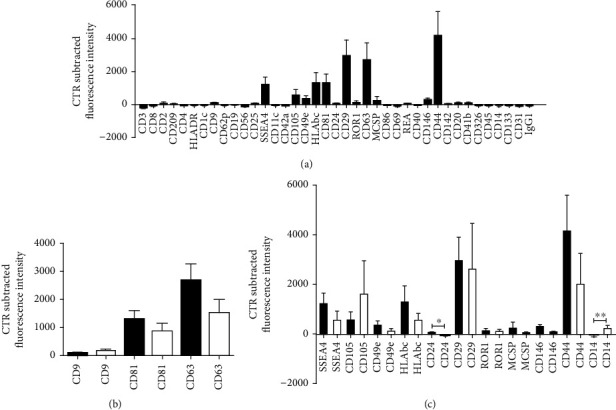
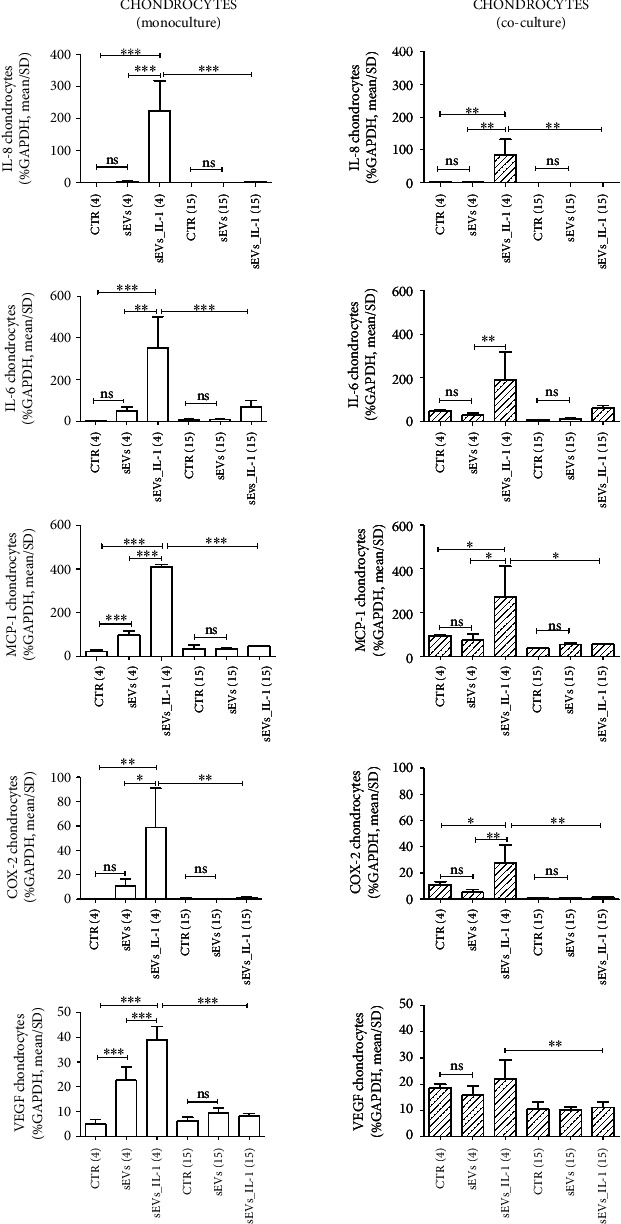
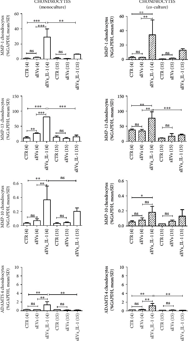
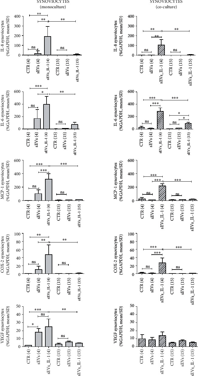
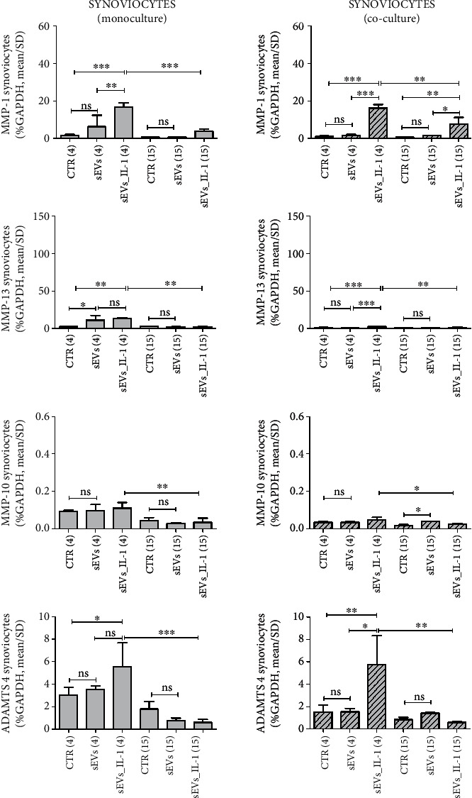
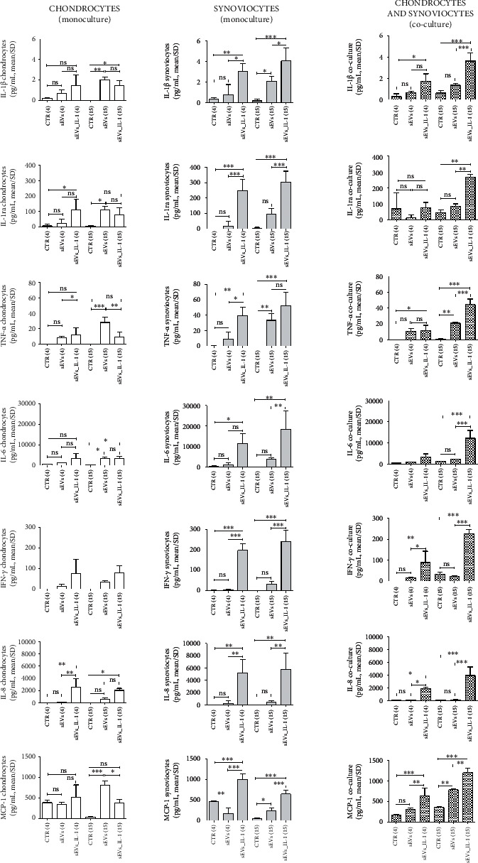
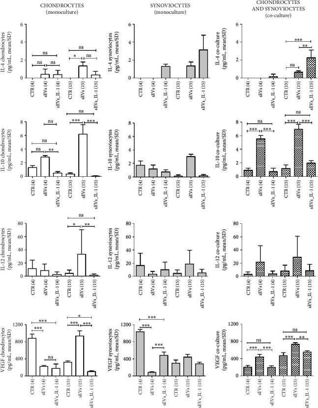
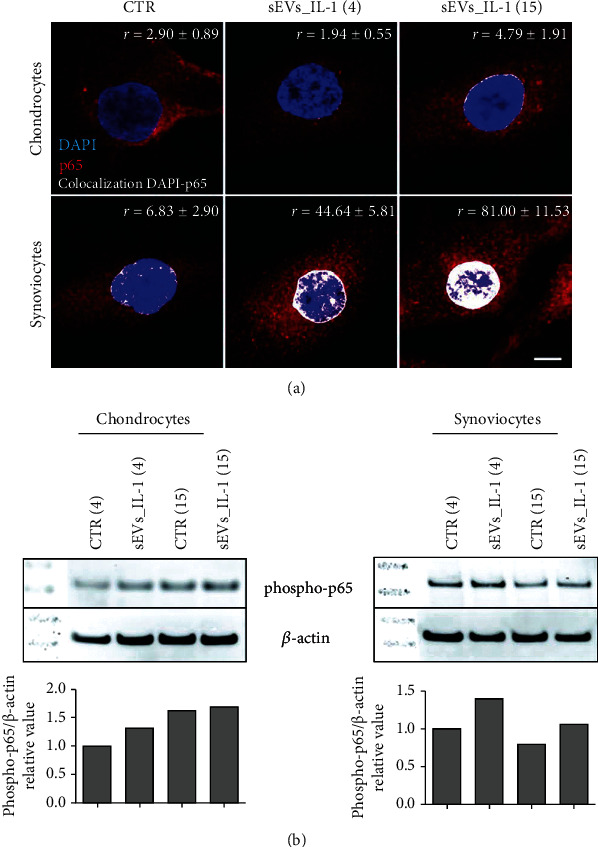
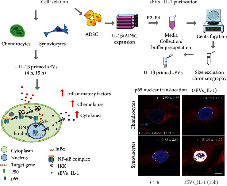
The last decade has seen exponentially growing efforts to exploit the effects of adipose derived stromal cells (ADSC) in the treatment of a wide range of chronic degenerative diseases, including osteoarthritis (OA), the most prevalent joint disorder. In the perspective of developing a cell-free advanced therapy medicinal product, a focus has been recently addressed to the ADSC secretome that lends itself to an allogeneic use and can be further dissected for the selective purification of small extracellular vesicles (sEVs). sEVs can act as "biological drug carriers" to transfer information that mirror the pathophysiology of the providing cells. This is important in the clinical perspective where many OA patients are also affected by the metabolic syndrome (MetS). ADSC from MetS OA patients are dysfunctional and "inflammatory" primed within the adipose tissue. To mimic this condition, we exposed ADSC to IL-1β, and then we investigated the effects of the isolated sEVs on chondrocytes and synoviocytes, either cultured separately or in co-culture, to tease out the effects of these "IL-1β primed sEVs" on gene and protein expression of major inflammatory and catabolic OA markers. In comparison with sEVs isolated from unstimulated ADSC, the IL-1β primed sEVs were able to propagate NF-κB activation in bystander joint cells. The effects were more prominent on synoviocytes, possibly because of a higher expression of binding molecules such as CD44. These findings call upon a careful characterization of the "inflammatory fingerprint" of ADSC to avoid the transfer of an unwanted message as well as the development of in vitro "preconditioning" strategies able to rescue the antiinflammatory/anticatabolic potential of ADSC-derived sEVs.


Conflict of interest statement

The authors declare that there is no conflict of interest regarding the publication of this paper.

Figures

Figure 1
Figure 1
sEVs_IL-1 Surface Epitope characterization. (a) Whole MACSPlex Exosome Kit surface assessment of 37 surface markers and 2 isotype controls of different purified sEVs_IL-1 preparations (n = 3) from IL-1β treated ADSC (black graphs). Data are expressed as net fluorescence intensity (control subtracted fluorescence intensity) and displayed as mean ± SD. (b) Side-by-side comparison of the expression of tetraspanins (CD9, CD81, and CD63) in sEVs obtained from IL-1β treated ADSC (sEVs_IL-1, n = 3, black graphs) or untreated ADSC (sEVs, n = 3, white graphs). (c) Side-by-side comparison of the expression of other 11 detectable surface markers in sEVs obtained from IL-1β treated ADSC (sEVs_IL-1, n = 3, black graphs) or untreated ADSC (sEVs, n = 3, white graphs). The latter correspond to data presented in [5]. Since the ADSC cultures were the same, comparisons were undertaken by mean of the Student's t-test for paired samples and statistically significant differences evidenced as ∗p < 0.05 and ∗∗p < 0.01.
Figure 2
Figure 2
sEVs_IL-1 effects on gene expression of major inflammatory genes in chondrocytes kept in control conditions. The figure reports the effects on gene expression of major inflammatory molecules (IL-8, IL-6, and MCP-1) and molecules involved in angiogenesis (VEGF) and pain (COX-2) in chondrocytes grown in both monoculture (white pattern, left graphs) and co-cultured with synoviocytes (white dashed pattern, right graphs) in CTR, sEVs, or sEVs_IL-1 conditions. Data were normalized to GAPDH. Each graph reports data collected from 3 independent sEVs samples and expressed as means ± SD. The means of the groups were compared by ANOVA, followed by Tukey's post hoc test, with ∗p < 0.05, ∗∗p < 0.01, and ∗∗∗p < 0.001.
Figure 3
Figure 3
sEVs_IL-1 effects on gene expression of catabolic genes in chondrocytes kept in control conditions. The figure reports the effects on gene expression of major catabolic enzymes (MMP-1, MMP-13, MMP-10, and ADAMTS 4) in chondrocytes grown in both monoculture (white pattern, left graphs) and co-cultured with synoviocytes (white dashed pattern, right graphs) in CTR, sEVs, or sEVs_IL-1 conditions. Data were normalized to GAPDH. Each graph reports data collected from 3 independent sEVs samples and expressed as mean ± SD. The means of the groups were compared by ANOVA, followed by Tukey's post hoc test, with ∗p < 0.05, ∗∗p < 0.01, and ∗∗∗p < 0.001.
Figure 4
Figure 4
sEVs_IL-1 effects on gene expression of major inflammatory genes in synoviocytes kept in control conditions. The figure reports the effects on gene expression of major inflammatory molecules (IL-8, IL-6, and MCP-1) and molecules involved in angiogenesis (VEGF) and pain (COX-2) in synoviocytes grown in both monoculture (grey pattern, left graphs) and co-cultured with chondrocytes (grey dashed pattern, right graphs) in CTR, sEVs, or sEVs_IL-1 conditions. Data were normalized to GAPDH. Each graph reports data collected from 3 independent sEVs samples and expressed as means ± SD. The means of the groups were compared by ANOVA, followed by Tukey's post hoc test, with ∗p < 0.05, ∗∗p < 0.01, and ∗∗∗p < 0.001.
Figure 5
Figure 5
sEVs_IL-1 effects on gene expression of catabolic genes in synoviocytes kept in control conditions. The figure reports the effects on gene expression of major catabolic enzymes (MMP-1, MMP-13, MMP-10, and ADAMTS 4) in synoviocytes grown in both monoculture (grey pattern, left graphs) and co-cultured with chondrocytes (grey dashed pattern, right graphs) in CTR, sEVs, or sEVs_IL-1 conditions. Data were normalized to GAPDH. Each graph reports data collected from 3 independent sEVs samples and expressed as mean ± SD. The means of the groups were compared by ANOVA, followed by Tukey's post hoc test, with ∗p < 0.05, ∗∗p < 0.01, and ∗∗∗p < 0.001.
Figure 6
Figure 6
sEVs_IL-1 effects on the protein release of major inflammatory molecules (IL-1β, IL-1ra, TNF-α, IL-6, IFN-γ, IL-8, and MCP-1) as assessed by Bioplex. Flanked results obtained from chondrocytes (white pattern), synoviocytes (grey pattern), and co-culture of these cells (pixelated pattern). For each cell type, results are shown obtained in CTR, sEVs, or sEVs_IL-1 conditions. Each graph reports data collected from 3 independent sEVs samples and expressed as means ± SD. Data were compared by ANOVA, followed by Tukey's post hoc test, with ∗p < 0.05, ∗∗p < 0.01, and ∗∗∗p < 0.001.
Figure 7
Figure 7
sEVs_IL-1 effects on the protein release of another subset of cytokines with reported antiinflammatory effects (IL-4, IL-10, IL-12, and VEGF) as assessed by Bioplex. Flanked results obtained from chondrocytes (white pattern), synoviocytes (grey pattern), and co-culture of these cells (pixelated pattern). For each cell type, results are shown obtained in CTR, sEVs, or sEVs_IL-1 conditions. Each graph reports data collected from 3 independent sEVs samples and expressed as means ± SD. Data were compared by ANOVA, followed by Tukey's post hoc test, with ∗p < 0.05, ∗∗p < 0.01, and ∗∗∗p < 0.001.
Figure 8
Figure 8
sEVs_IL-1 effects on NF-κB activation. (a) p65 immunofluorescence on chondrocytes (upper row) and synoviocytes (lower row) in control conditions (CTR) and at 4 and 15 h after exposure to sEVs_IL-1. Scale bar: 10 μm. Each image reports the level of the colocalization of the red (p65) to the blue (DNA) signal. The colocalization of the fluorochromes (shown in white) was quantified using Pearson's colocalization coefficient (r), derived from 15 analyzed optical sections and expressed as percentage ± SD. 5 cells were considered for each condition. (b) Western blot analysis of samples of chondrocytes (left) and synoviocytes (right) in CTR or sEVs_IL-1 condition at both 4 hr and 15 hr post-delivery. These samples were dedicated to western blot analysis of phosphorylation of p65 and β-actin as loading control. Lower graphs: relative quantification of phospho-p65 signal normalized to that of β-actin.
Figure 9
Figure 9
Experimental outline of the research work undertaken in the paper: sEVs_IL-1 collection from IL-1 treated ADSC; purification of sEVs_IL-1 through a combined precipitation and size exclusion chromatography method; addition of sEVs_IL-1 to primary cultures of chondrocytes and synoviocytes; analysis of the effects at the gene and protein level and molecular hypothesis underlying the observed effects.

References

    1. Filardo G., Tschon M., Perdisa F., et al. Micro-fragmentation is a valid alternative to cell expansion and enzymatic digestion of adipose tissue for the treatment of knee osteoarthritis: a comparative preclinical study. Knee Surgery, Sports Traumatology, Arthroscopy . 2021;30(3) - PubMed
    1. Perdisa F., Gostynska N., Roffi A., Filardo G., Marcacci M., Kon E. Adipose-derived mesenchymal stem cells for the treatment of articular cartilage: a systematic review on preclinical and clinical evidence. Stem Cells International . 2015;2015:13. doi: 10.1155/2015/597652.597652 - DOI - PMC - PubMed
    1. Mazini L., Rochette L., Amine M., Malka G. Regenerative capacity of adipose derived stem cells (ADSCs), comparison with mesenchymal stem cells (MSCs) International Journal of Molecular Sciences . 2019;20(10):p. 2523. doi: 10.3390/ijms20102523. - DOI - PMC - PubMed
    1. D'Arrigo D., Roffi A., Cucchiarini M., Moretti M., Candrian C., Filardo G. Secretome and extracellular vesicles as new biological therapies for knee osteoarthritis: a systematic review. Journal of Clinical Medicine . 2019;8(11):p. 1867. doi: 10.3390/jcm8111867. - DOI - PMC - PubMed
    1. Cavallo C., Merli G., Borzi R. M., et al. Small extracellular vesicles from adipose derived stromal cells significantly attenuate in vitro the NF-kappaB dependent inflammatory/catabolic environment of osteoarthritis. Scientific Reports . 2021;11(1):p. 1053. doi: 10.1038/s41598-020-80032-7. - DOI - PMC - PubMed

LinkOut - more resources