. 2022 Jul 11:13:901442.
doi: 10.3389/fmicb.2022.901442. eCollection 2022.

In vitro and in vivo Characterization of Host-Pathogen Interactions of the L3881 Candida albicans Clinical Isolate

Affiliations

In vitro and in vivo Characterization of Host-Pathogen Interactions of the L3881 Candida albicans Clinical Isolate

Pedro H F Sucupira et al. Front Microbiol. .

Abstract

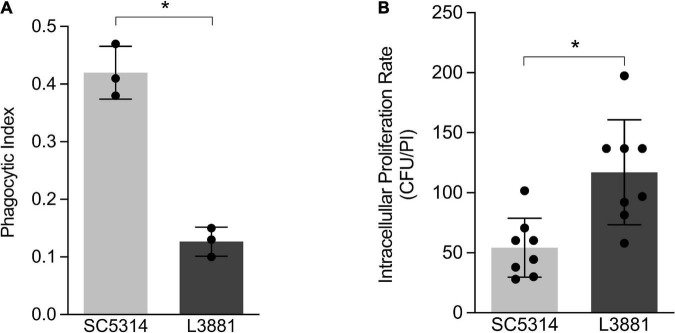
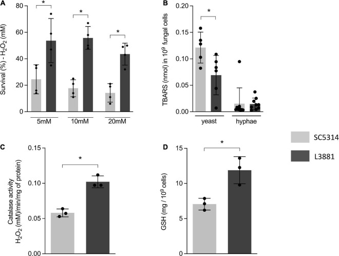
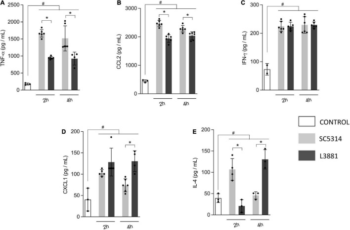
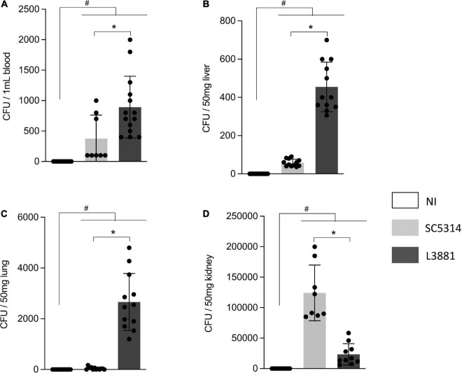
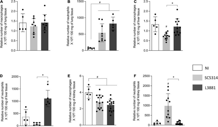
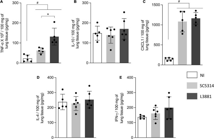
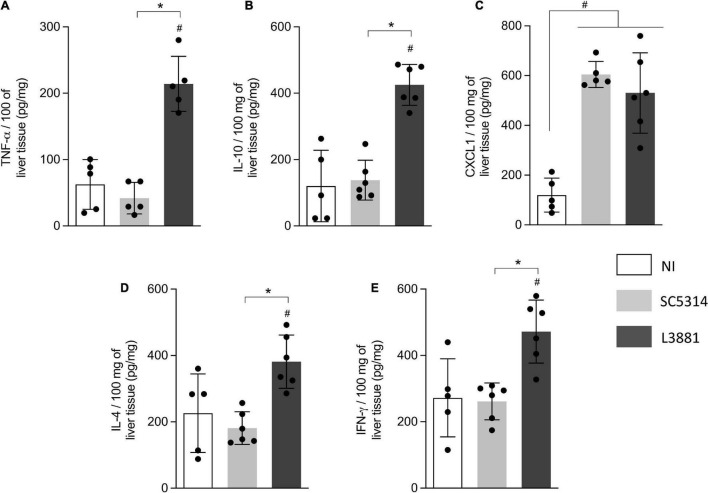
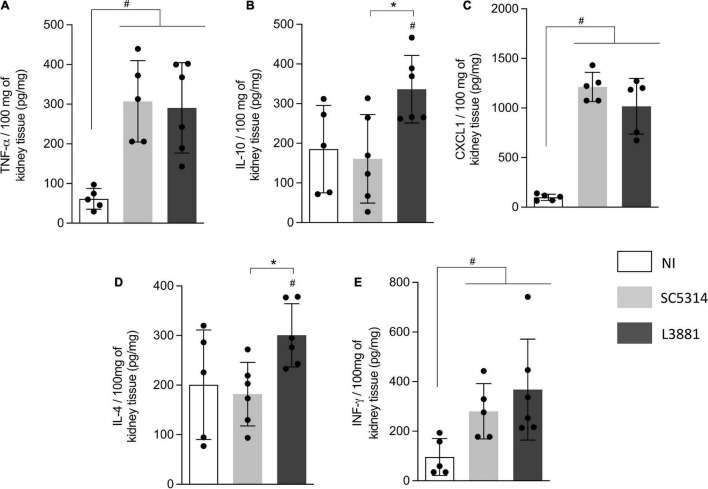
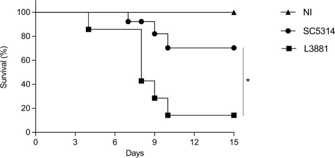
Candida albicans is a human commensal fungus and the etiologic agent of nosocomial infections in immunocompromised individuals. Candida spp. is the most studied human fungal pathogen, and the mechanisms by which this fungus can evade the immune system affecting immunosuppressed individuals have been extensively studied. Most of these studies focus on different species of Candida, and there is much to be understood in virulence variability among lineages, specifically different C. albicans clinical isolates. To better understand the main mechanisms of its virulence variability modulated in C. albicans clinical isolates, we characterized L3881 lineage, which has been previously classified as hypovirulent, and SC5314 lineage, a virulent wild-type control, by using both in vitro and in vivo assays. Our findings demonstrated that L3881 presented higher capacity to avoid macrophage phagocytosis and higher resistance to oxidative stress than the wild type. These characteristics prevented higher mortality rates for L3881 in the animal model of candidiasis. Conversely, L3881 has been able to induce an upregulation of pro-inflammatory mediators both in vitro and in vivo. These results indicated that in vitro and in vivo functional characterizations are necessary for determination of virulence in different clinical isolates due to its modulation in the host-pathogen interactions.

Keywords: Candida albicans infection; clinical isolates; fungal infection; innate immunity; virulence.


Conflict of interest statement

The authors declare that the research was conducted in the absence of any commercial or financial relationships that could be construed as a potential conflict of interest.

Figures

FIGURE 1
FIGURE 1
In vitro characterization of Candida albicans SC5314 and L3881 lineages. (A) Phagocytosis was assessed by cytospin preparations of murine macrophage RAW 264.7 cell line co-cultured with SC5314 and L3881. (B) Killing assay was evaluated by cell lysis with water, the diluted samples were plated in the fungal medium, and colony-forming units (CFUs) were determined 16 h post-incubation (PI). Each dot represents an experimental replica. Data are presented as mean ± standard deviation (SD). Asterisk (*) represents significant differences with p < 0.05.
FIGURE 2
FIGURE 2
Antioxidant activity of SC5314 and L3881 lineages. (A) C. albicans yeast cells of SC5314 and L3881 lineages were incubated with three different concentrations of H2O2 (5, 10, and 20 mM). (B) Lipid peroxidation was measured by the formation of products of oxidation of thiobarbituric acid. (C) Catalase activity was measured by following the absorbance decrease in H2O2. (D) Glutathione (GSH) was quantified using the enzymatic recycling procedure, reducing NAD+ to NADPH in the presence of glutathione reductase. Each dot represents an experimental replica. Data are presented as mean ± SD. Asterisk (*) represents significant differences with p < 0.05.
FIGURE 3
FIGURE 3
In vitro cytokine and chemokine profile of L3881 and SC5314. Murine macrophage RAW 264.7 cell line was infected with L3881 and SC5314 yeast lineages. Supernatants were collected at 2 and 4 h post-incubation and used for ELISA for quantifications of (A) TNF-α, (B) CCL2, (C) IFN-γ, (D) CXCL1, and (E) IL-4 levels. Each dot represents an experimental replica. Data are presented as mean ± SD. Hashtag (#) and asterisk (*) represent significant differences with p < 0.05.
FIGURE 4
FIGURE 4
CFU quantification in mice infected with C. albicans lineages. BALB/c male mice were infected with SC5314 or L3881 intravenously through the tail vein. Mice in the non-infected group (NI) received saline solution. At 2 days post-inoculation, the blood, liver, lungs, and kidney were collected. CFUs were determined for (A) blood and homogenates from the (B) liver, (C) lung, and (D) kidney. Each dot represents tissue from an individual mouse. Data are presented as mean ± SD. Hashtag (#) and asterisk (*) represent significant differences with p < 0.05.
FIGURE 5
FIGURE 5
Relative number of macrophages and neutrophils in the lung, liver, and kidney from mice infected with C. albicans lineages BALB/c male mice were infected with SC5314 or L3881 intravenously through the tail vein. Mice in the non-infected group (NI) received saline solution. At 2 days post-inoculation, the liver, lungs, and kidney were collected for NAG (relative number of macrophages) and MPO (relative number of neutrophils) quantification. (A) NAG in the lung. (B) MPO in the lung. (C) NAG in the liver. (D) MPO in the liver. (E) NAG in the kidney. (F) MPO in the kidney. Each dot represents tissue from an individual mouse. Data are presented as mean ± SD. Hashtag (#) and asterisk (*) represent significant differences with p < 0.05.
FIGURE 6
FIGURE 6
Cytokine and chemokine profiles of lung tissue from mice infected with C. albicans lineages. BALB/c male mice were infected with SC5314 or L3881 intravenously through the tail vein. Mice in the non-infected group (NI) received saline solution. At 2 days post-inoculation, lungs were collected for (A) TNF-α, (B) IL-10, (C) CXCL1, (D) IL-4, and (E) INF-γ by ELISA. Each dot represents tissue from an individual mouse. Data are presented as mean ± SD. Hashtag (#) and asterisk (*) represent significant differences with p < 0.05.
FIGURE 7
FIGURE 7
Cytokine and chemokine profiles of liver tissue from mice infected with C. albicans lineages. BALB/c male mice were infected with SC5314 or L3881 intravenously through the tail vein. Mice in the non-infected group (NI) received saline solution. At 2 days post-inoculation, the liver as collected for (A) TNF-α, (B) IL10, (C) CXCL1, (D) IL4, and (E) INF-γ by ELISA. Each dot represents tissue from an individual mouse. Statistical analyses were performed using the unpaired t-test. Data are presented as mean ± SD. Hashtag (#) and asterisk (*) represent significant differences with p < 0.05.
FIGURE 8
FIGURE 8
Cytokine and chemokine profiles of kidney tissue from mice infected with C. albicans lineages. BALB/c male mice were infected with SC5314 or L3881 intravenously through the tail vein. Mice in the non-infected group (NI) received saline solution. At 2 days post-inoculation, kidneys were collected for (A) TNF-α, (B) IL10, (C) CXCL1, (D) IL4, and (E) INF-γ by ELISA. Each dot represents tissue from an individual mouse. Statistical analyses were performed using the unpaired t-test. Data are presented as mean ± SD. Hashtag (#) and asterisk (*) represent significant differences with p < 0.05.
FIGURE 9
FIGURE 9
Mortality rate of mice infected with C. albicans lineages. BALB/c male mice were infected with SC5314 or L3881 intravenously through the tail vein, and survival was monitored for 15 days. Mice in the non-infected group (NI) received saline solution. Comparative lethality curves of NI and infected groups were performed. Data are presented as mean ± SD. Asterisk (*) represents significant differences with p < 0.05.

Similar articles

Cited by

References

    1. Aebi H. (1984). Catalase in vitro. Methods Enzymol. 105 121–126. 10.1016/s0076-6879(84)05016-3 - DOI - PubMed
    1. Arendrup M. C. (2010). Epidemiology of invasive candidiasis. Curr. Opin. Crit. Care. 16 445–452. 10.1097/MCC.0b013e32833e84d2 - DOI - PubMed
    1. Ashman R. B. (2008). Protective and pathologic immune responses against Candida albicans infection. Front. Biosci. 13:3334–3351. 10.2741/2929 - DOI - PubMed
    1. Bahia D., Satoskar A. R., Dussurget O. (2018). Editorial: cell signaling in host-pathogen interactions: the host point of view. Front. Immunol. 9:221. 10.3389/fimmu.2018.00221 - DOI - PMC - PubMed
    1. Bradford M. M. (1976). A rapid and sensitive method for the quantitation of microgram quantities of protein utilizing the principle of protein-dye binding. Anal. Biochem. 72 248–254. 10.1006/abio.1976.9999 - DOI - PubMed