Review
. 2022 Jul 11:13:928608.
doi: 10.3389/fneur.2022.928608. eCollection 2022.

Clinical Importance of the Persistent Primitive Trigeminal Artery in Vascular Lesions and Its Role in Endovascular Treatment

Affiliations
Review

Clinical Importance of the Persistent Primitive Trigeminal Artery in Vascular Lesions and Its Role in Endovascular Treatment

Yiheng Wang et al. Front Neurol. .

Abstract

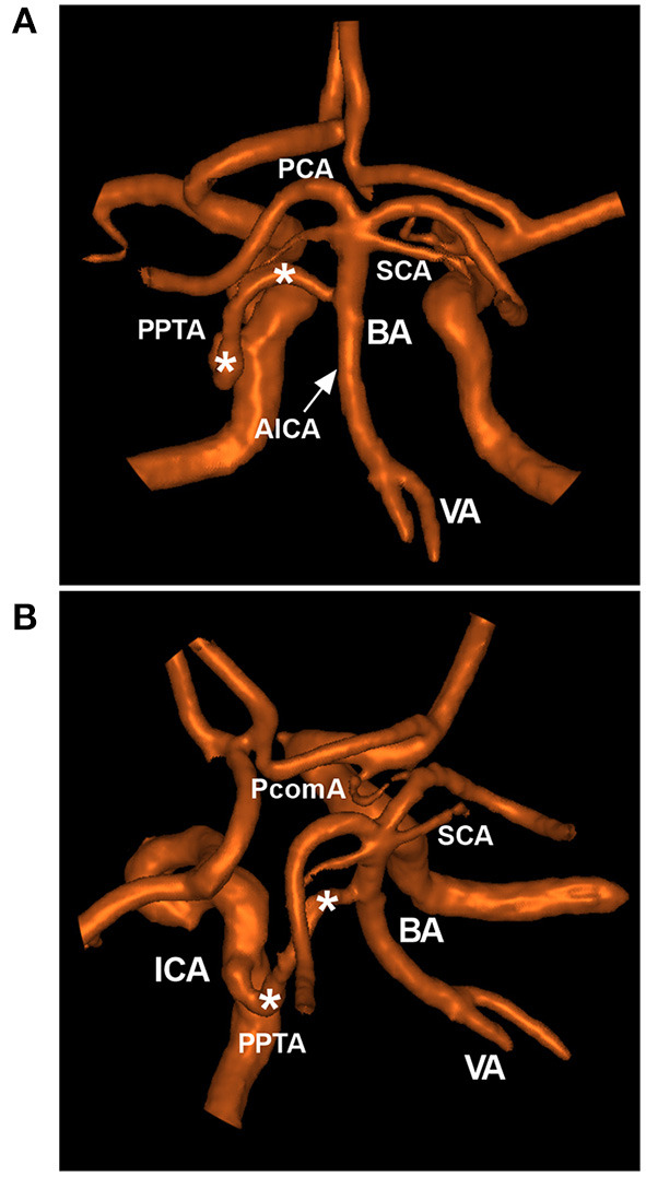
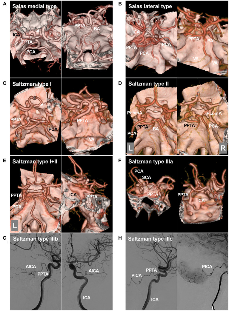
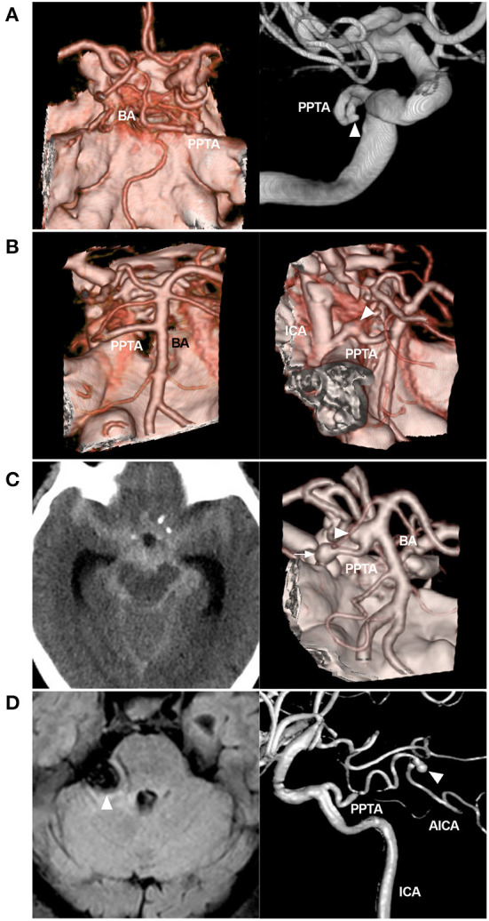
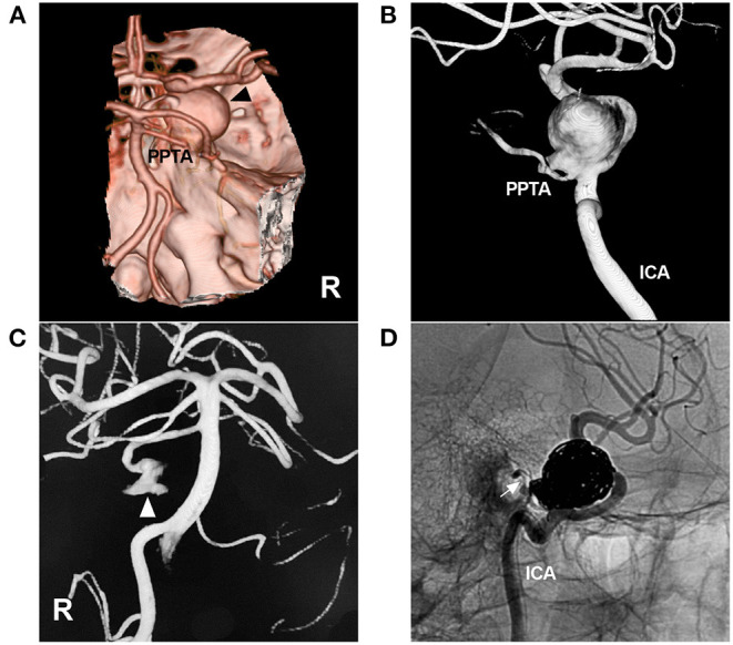
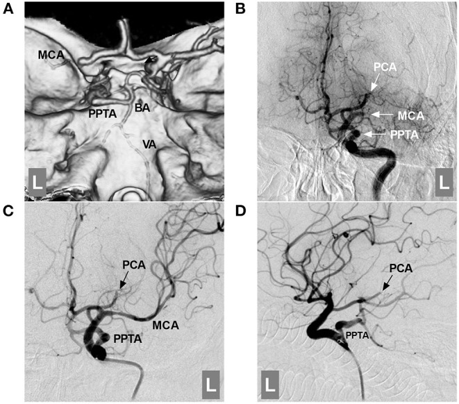
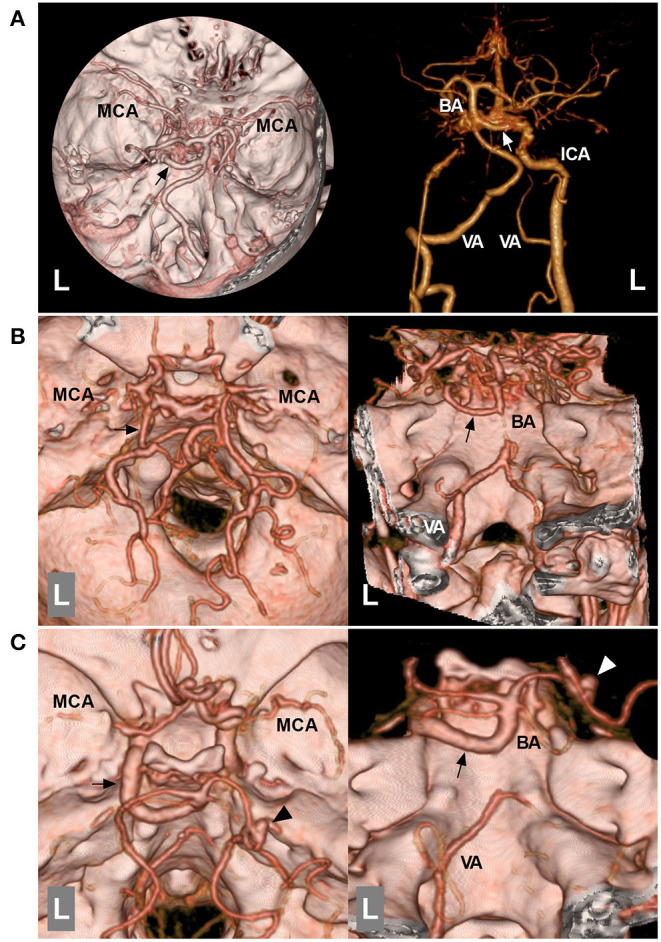
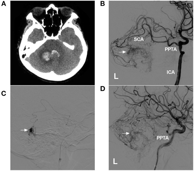
The persistent primitive trigeminal artery (PPTA) extends from the internal carotid artery to the basilar artery between the origins of the anterior inferior cerebellar artery and superior cerebellar artery. PPTAs have complex anatomical characteristics. Salas and Saltzman classifications are most often used in PPTAs. The PPTA can play many roles in vascular lesions, including intracranial aneurysms, brain arteriovenous malformations, trigeminal artery-cavernous fistulas, Moyamoya disease, and large vessel occlusion. For these lesions, surgical treatment is difficult due to the deep location and complex anatomy of the PPTA, but endovascular treatment (EVT) has emerged as a good alternative. Currently, a complete review of the clinical importance of the PPTA in terms of its role in the development and EVT of vascular lesions is lacking. Therefore, we conducted a PubMed search, performed a review of the relevant extracted literature and cataloged our experience with PPTAs. By review, we found that a thorough understanding of the anatomical and angiographic features of this PPTA is of utmost importance when making therapeutic decisions for any of these pathological conditions.

Keywords: clinical importance; endovascular treatment; persistent primitive trigeminal artery; review; vascular lesion.


Conflict of interest statement

The authors declare that the research was conducted in the absence of any commercial or financial relationships that could be construed as a potential conflict of interest.

Figures

Figure 1
Figure 1
Typical PPTA in MRA. (A,B): Posterior anterior (A) and superior oblique MRA views (B) showing a typical PPTA (asterisks) from the cavernous ICA to the BA between the SCA and AICA. The arrow (A) indicates the origin of the AICA. AICA, anterior inferior cerebellar artery; BA, basilar artery; ICA, internal carotid artery; MRA, magnetic resonance angiography; PCA, posterior cerebral artery; PcomA, posterior communicating artery; PPTA, persistent primitive trigeminal artery; SCA, superior cerebellar artery; VA, vertebral artery.
Figure 2
Figure 2
Salas and Saltzman classifications. (A): Salas medial (sphenoidal) type. Left, CTA showing the PPTA arising from the ICA, running medially into the sella (arrowhead), and then crossing the dorsum sellae. Right: CTA showing the PPTA connecting to the BA. (B): Salas lateral (petrosal) type. Left, CTA showing the PPTA arising from the ICA, running laterally, and then crossing the cavernous sinus. Right: CTA showing the PPTA connecting with the BA. (C): Saltzman type I. Left, CTA showing the PPTA arising from the ICA. Right: CTA showing the PPTA connecting with the BA. In this case, the PPTA is strong and hyperplastic, and the PcomAs and vertebrobasilar artery are hypoplastic. (D): Saltzman type II. Left, CTA showing the left PPTA and the left fetal-type PCA; Right, CTA showing the right PCA receiving its blood supply from a patent PcomA. The proximal BA is well developed. (E): Saltzman combined type (I+II). Left, CTA showing that the left PCA is fetal-type and not connected with the BA, the right PCA originates from the BA, and no PcomA can be seen; Right: CTA showing that the PPTA arises from the left ICA. The vertebrobasilar arteries are normally developed. (F): CTA showing a type IIIa PPTA arising from the ICA and then extending to the opposite SCA. (G): DSA of the ICA showing a type IIIb PPTA arising from the ICA and then extending into the AICA. (H): DSA of the ICA showing a type IIIc PPTA arising from the ICA and then extending into the PICA. AICA, anterior inferior cerebellar artery; BA, basilar artery; CTA, computed tomography angiography; DSA, digital subtraction angiography; ICA, internal carotid artery; L, left; PCA, posterior cerebral artery; PcomA, posterior communicating artery; PICA, posterior inferior cerebellar artery; PPTA, persistent primitive trigeminal artery; R, right; SCA, superior cerebellar artery; VA, vertebral artery.
Figure 3
Figure 3
Variant aneurysms of the PPTA. (A): Left: CTA showing the PPTA; Right: DSA of the ICA showing a small aneurysm (arrowhead) on the middle PPTA trunk. (B): Left: CTA showing the PPTA; Right: CTA showing a fusiform aneurysm (arrowhead) on the PPTA trunk near the origin. (C): Left: CT showing a subarachnoid hemorrhage; Right: CTA showing two aneurysms, one (arrow) located on the PPTA trunk and the other (arrowhead) at the junction of the PPTA and BA. (D): Left: MRI showing a mass (arrowhead) in the cerebellopontine angle region, compressing the brainstem; Right: Three-dimensional DSA showing a Saltzman type IIIb PPTA, which continues to the AICA; a distal aneurysm of the AICA can be seen (arrowhead). AICA, anterior inferior cerebellar artery; BA, basilar artery; CT, computed tomography; CTA, computed tomography angiography; DSA, digital subtraction angiography; ICA, internal carotid artery; MRI, magnetic resonance imaging; PPTA, persistent primitive trigeminal artery.
Figure 4
Figure 4
Coiling of an aneurysm of the PPTA-ICA junction. (A): CTA showing a large right parasellar aneurysm (arrowhead); a PPTA can be seen. (B): Three-dimensional DSA of the ICA showing the aneurysm located at the junction of the PPTA and ICA. (C): Three-dimensional DSA of the right VA showing part of the PPTA aneurysm (arrowhead). (D): Unsubtracted DSA showing the aneurysm coiled via the ICA, preserving the PPTA (arrow). CTA, computed tomography angiography; DSA, digital subtraction angiography; ICA, internal carotid artery; PPTA, persistent primitive trigeminal artery; R, right; VA, vertebral artery.
Figure 5
Figure 5
Role of the PPTA in acute large vessel occlusion. (A): CTA showing an occluded left MCA and a PPTA connected with the BA; the proximal BA and VA were hypoplastic. (B): DSA of the ICA showing an occluded left MCA and PCA (arrows with MCA and PCA); the PPTA (arrow with PPTA) is unaffected. (C,D): DSA of the ICA showing that the MCA was revascularized after mechanical thrombectomy; the PCA was fetal-type and occluded distally (arrows with PCA). BA, basilar artery; CTA, computed tomography angiography; DSA, digital subtraction angiography; ICA, internal carotid artery; L, left; MCA, middle cerebral artery; PCA, posterior cerebral artery; PPTA, persistent primitive trigeminal artery; VA, vertebral artery.
Figure 6
Figure 6
Role of the PPTA in MMD. (A): Left: CTA of the superior inferior view showing that the bilateral MCAs have partially disappeared, and the left PPTA (arrow) is indicated. Right: CTA with bone removal showing that the distal ICA termination is occluded, and the left ICA is connected with the PPTA (arrow). (B): Left: CTA of superior inferior view showing that the bilateral MCAs are obscured, and the left PPTA (arrow) is indicated; Right: CTA of posterior anterior view showing that the proximal BA was hypoplastic, and the left PPTA (arrow) is indicated. (C): Left: CTA of superior inferior view showing that the bilateral MCAs have partially disappeared, and a left PPTA (arrow) and an aneurysm (arrowhead) on the right PCA can be seen; Right: CTA of posterior anterior view showing a hypoplastic proximal BA and an aneurysm (arrowhead) on the right PCA, and the left PPTA (arrow) is indicated. BA, basilar artery; CTA, computed tomography angiography; ICA, internal carotid artery; L, left; MCA, middle cerebral artery; MMD, Moyamoya disease; PCA, posterior cerebral artery; PPTA, persistent primitive trigeminal artery; VA, vertebral artery.
Figure 7
Figure 7
Role of the PPTA as a transarterial path for a BAVM. (A): CTA showing hemorrhage of the cerebellar vermis and fourth ventricle. (B): DSA of the left ICA showing a BAVM supplied by a PPTA via the SCA. An intranidal aneurysm is indicated (arrow). (C): Superselective angiogram of the microcatheter showing the aneurysm (arrow). (D): Postoperative DSA of the left ICA showing that the intranidal aneurysm had been embolized (arrow). BAVM, brain arteriovenous malformation; CT, computed tomography; CTA, computed tomography angiography; DSA, digital subtraction angiography; ICA, internal carotid artery; L, left; SCA, superior cerebellar artery; PPTA, persistent primitive trigeminal artery.

Similar articles

Cited by

References

    1. Padget DH. The development of the cranial arteries in the human embryo. Contrib Embryol. (1948) 32:205–62.
    1. Zenteno M, Lee A, Moscote-Salazar LR. Rupture of persistent primitive trigeminal artery-basilar artery aneurysm managed with stent-assisted coiling. Asian J Neurosurg. (2018) 13:817–21. 10.4103/ajns.AJNS_53_16 - DOI - PMC - PubMed
    1. Hiramatsu R, Ohnishi H, Kawabata S, Miyachi S, Kuroiwa T. Successful recanalization for internal carotid artery occlusion with persistent primitive trigeminal artery manifesting only as ischemia of the posterior circulation. BMC Neurol. (2016) 16:41. 10.1186/s12883-016-0559-6 - DOI - PMC - PubMed
    1. O'Uchi E, O'Uchi T. Persistent primitive trigeminal arteries (PTA) and its variant (PTAV): analysis of 103 cases detected in 16,415 cases of MRA over 3 years. Neuroradiology. (2010) 52:1111–9. 10.1007/s00234-010-0669-6 - DOI - PubMed
    1. Meckel S, Spittau B, McAuliffe W. The persistent trigeminal artery: development, imaging anatomy, variants, and associated vascular pathologies. Neuroradiology. (2013) 55:5–16. 10.1007/s00234-011-0995-3 - DOI - PubMed

LinkOut - more resources