

Detergent exposure induces epithelial barrier dysfunction and eosinophilic inflammation in the esophagus
- PMID: 35899466
- PMCID: PMC9797443
- DOI: 10.1111/all.15457
Detergent exposure induces epithelial barrier dysfunction and eosinophilic inflammation in the esophagus
Abstract
Background: Eosinophilic esophagitis (EoE) is a chronic allergic disease associated with type 2 inflammation and epithelial barrier dysfunction. The etiology is unknown, however, genetic heritability studies suggest environmental factors play a key role in pathogenesis. Detergents, such as sodium dodecyl sulfate (SDS), are common ingredients in household products such as dish soap and toothpaste. We hypothesized detergent exposure decreases epithelial barrier function and induces esophageal inflammation.
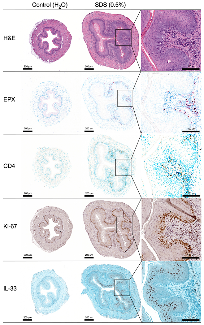
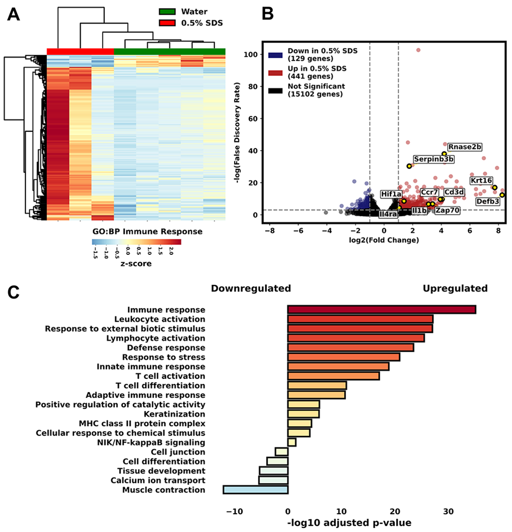
Methods: Immortalized esophageal epithelial cells (EPC2) were cultured in air-liquid interface (ALI) and exposed to SDS. Barrier function/activity was assessed by transepithelial electrical resistance (TEER), FITC-dextran flux, and RT-PCR. Additionally, SDS-treated mouse esophageal organoids were evaluated for morphology. To investigate the effects of SDS in vivo, mice were treated with 0.5% SDS in drinking water for 14 days. Esophagi were assessed by gross morphology, histopathology, protein expression, and bulk RNA sequencing.
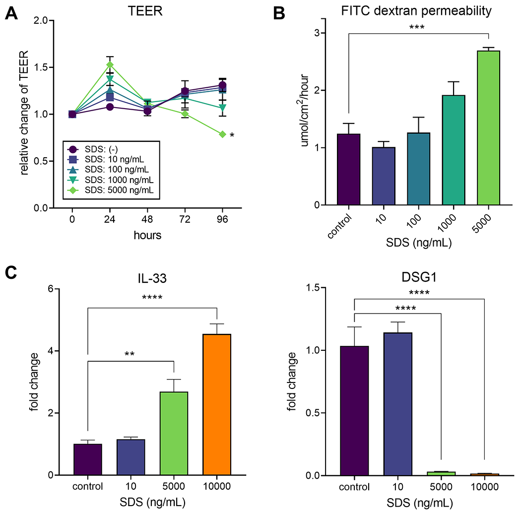
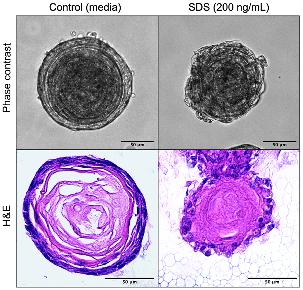
Results: When EPC2 cells were exposed to SDS (5 μg/ml) for 96 h, TEER decreased (p = 0.03), and FITC-dextran flux increased (p = 0.0002). mRNA expression of IL-33 increased 4.5-fold (p = 0.02) at 6 h and DSG1 decreased (p < 0.0001) by 72 h. Disrupted epithelial integrity was noted in SDS-treated esophageal organoids. When mice were exposed to SDS, they showed increased esophageal width, chemokine, and metalloprotease levels. Mice treated with SDS also showed increased IL-33 protein expression, basal zone hyperplasia, CD4+ cell infiltration, and esophageal eosinophilia. RNA sequencing revealed upregulation of immune response pathway genes.
Conclusion: Exposure to SDS decreases esophageal barrier integrity, stimulates IL-33 production, and promotes epithelial hyperplasia and tissue eosinophilia. Detergents may be a key environmental trigger in EoE pathogenesis.
Keywords: IL-33; detergent; eosinophilic esophagitis; epithelium.
© 2022 European Academy of Allergy and Clinical Immunology and John Wiley & Sons Ltd.
Conflict of interest statement
Figures
Comment in
-
Detergent-induced eosinophilic inflammation in the esophagus: A key evidence for the epithelial barrier theory.Allergy. 2023 Jun;78(6):1422-1424. doi: 10.1111/all.15646. Epub 2023 Mar 30. Allergy. 2023. PMID: 36645729 No abstract available.
Similar articles
-
Epithelial overexpression of IL-33 induces eosinophilic esophagitis dependent on IL-13.J Allergy Clin Immunol. 2024 May;153(5):1355-1368. doi: 10.1016/j.jaci.2024.01.017. Epub 2024 Feb 3. J Allergy Clin Immunol. 2024. PMID: 38310974 Free PMC article.
-
TGF-β1 alters esophageal epithelial barrier function by attenuation of claudin-7 in eosinophilic esophagitis.Mucosal Immunol. 2018 Mar;11(2):415-426. doi: 10.1038/mi.2017.72. Epub 2017 Aug 23. Mucosal Immunol. 2018. PMID: 28832026 Free PMC article.
-
Filaggrin and tight junction proteins are crucial for IL-13-mediated esophageal barrier dysfunction.Am J Physiol Gastrointest Liver Physiol. 2018 Sep 1;315(3):G341-G350. doi: 10.1152/ajpgi.00404.2017. Epub 2018 May 10. Am J Physiol Gastrointest Liver Physiol. 2018. PMID: 29746170
-
Epithelial origin of eosinophilic esophagitis.J Allergy Clin Immunol. 2018 Jul;142(1):10-23. doi: 10.1016/j.jaci.2018.05.008. J Allergy Clin Immunol. 2018. PMID: 29980278 Free PMC article. Review.
-
Barrier Dysfunction in Eosinophilic Esophagitis.Curr Gastroenterol Rep. 2023 Dec;25(12):380-389. doi: 10.1007/s11894-023-00904-6. Epub 2023 Nov 11. Curr Gastroenterol Rep. 2023. PMID: 37950816 Review.
Cited by
-
Comparative evaluation of six commercial adult toothpaste formulations reveals cytotoxicity and altered functions in a human oral melanocyte model: an in vitro study.Odontology. 2025 Jan;113(1):163-179. doi: 10.1007/s10266-024-00957-7. Epub 2024 Jun 1. Odontology. 2025. PMID: 38822982
-
Epithelial overexpression of IL-33 induces eosinophilic esophagitis dependent on IL-13.J Allergy Clin Immunol. 2024 May;153(5):1355-1368. doi: 10.1016/j.jaci.2024.01.017. Epub 2024 Feb 3. J Allergy Clin Immunol. 2024. PMID: 38310974 Free PMC article.
-
Immunologic and pathologic characterization of a novel swine biomedical research model for eosinophilic esophagitis.Front Allergy. 2022 Nov 14;3:1029184. doi: 10.3389/falgy.2022.1029184. eCollection 2022. Front Allergy. 2022. PMID: 36452260 Free PMC article.
-
Mammalian esophageal stratified tissue homeostasis is maintained distinctively by the epithelial pluripotent p63+Sox2+ and p63-Sox2+ cell populations.Cell Mol Life Sci. 2023 Sep 26;80(10):305. doi: 10.1007/s00018-023-04952-z. Cell Mol Life Sci. 2023. PMID: 37752383 Free PMC article.
-
Epithelial barrier dysfunction and associated diseases in companion animals: Differences and similarities between humans and animals and research needs.Allergy. 2024 Dec;79(12):3238-3268. doi: 10.1111/all.16343. Epub 2024 Oct 17. Allergy. 2024. PMID: 39417247 Free PMC article. Review.
References
Publication types
MeSH terms
Substances
Supplementary concepts
Grants and funding
LinkOut - more resources
Full Text Sources
Medical
Research Materials
