. 2022 Aug 19;131(5):424-441.
doi: 10.1161/CIRCRESAHA.122.321187. Epub 2022 Jul 28.

Endothelial UCP2 Is a Mechanosensitive Suppressor of Atherosclerosis

Affiliations

Endothelial UCP2 Is a Mechanosensitive Suppressor of Atherosclerosis

Jiang-Yun Luo et al. Circ Res. .

Abstract

Background: Inflamed endothelial cells (ECs) trigger atherogenesis, especially at arterial regions experiencing disturbed blood flow. UCP2 (Uncoupling protein 2), a key mitochondrial antioxidant protein, improves endothelium-dependent relaxation in obese mice. However, whether UCP2 can be regulated by shear flow is unknown, and the role of endothelial UCP2 in regulating inflammation and atherosclerosis remains unclear. This study aims to investigate the mechanoregulation of UCP2 expression in ECs and the effect of UCP2 on endothelial inflammation and atherogenesis.

Methods: In vitro shear stress simulation system was used to investigate the regulation of UCP2 expression by shear flow. EC-specific Ucp2 knockout mice were used to investigate the role of UCP2 in flow-associated atherosclerosis.

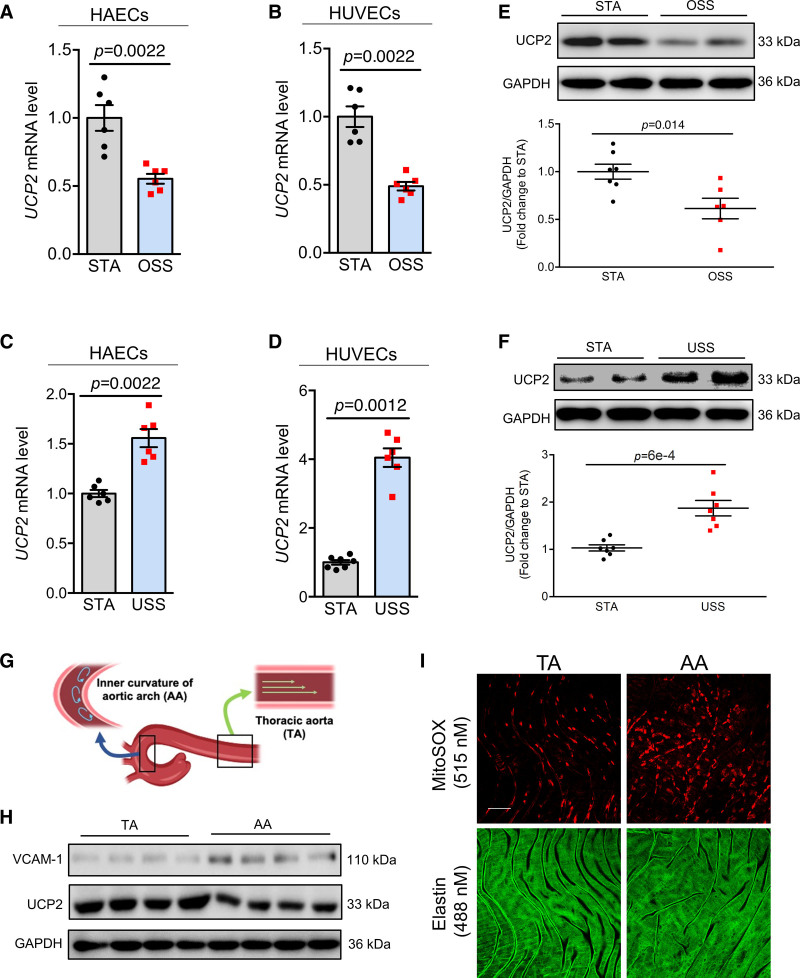
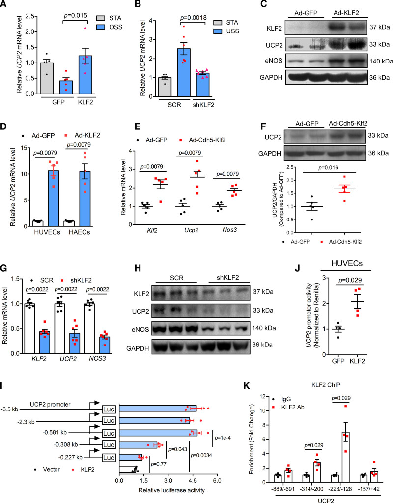
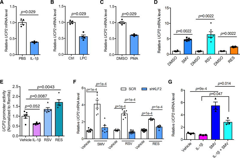
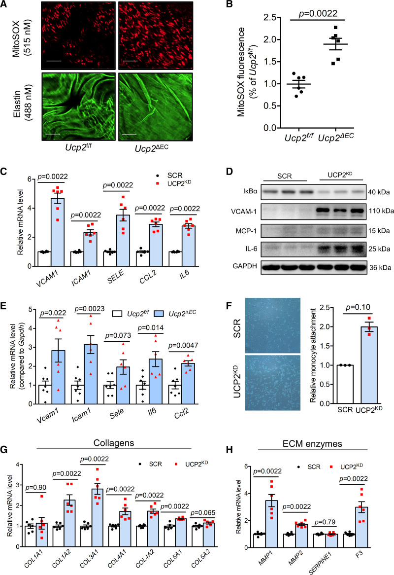
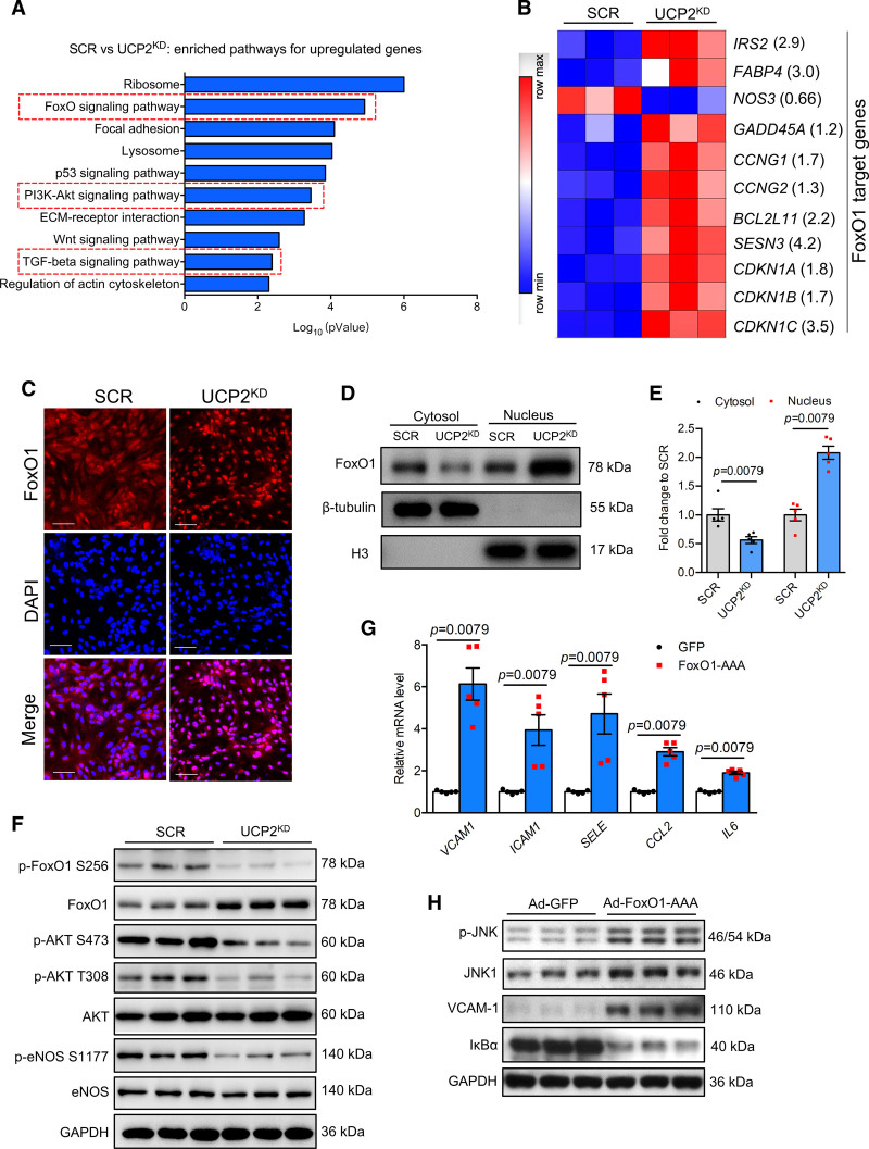
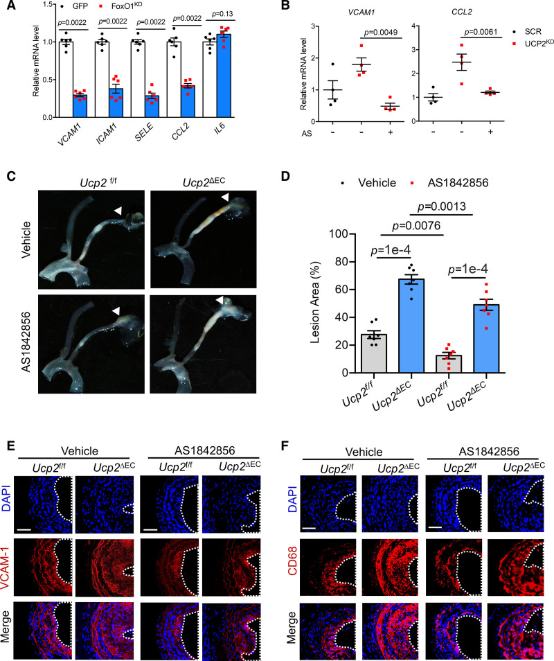
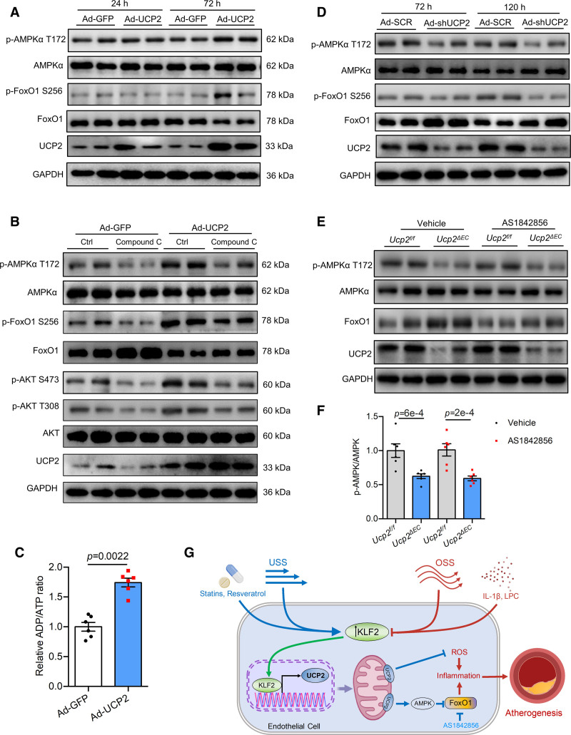
Results: Shear stress experiments showed that KLF2 (Krüppel-like factor 2) mediates fluid shear stress-dependent regulation of UCP2 expression in human aortic and human umbilical vein ECs. Unidirectional shear stress, statins, and resveratrol upregulate whereas oscillatory shear stress and proinflammatory stimuli inhibit UCP2 expression through altered KLF2 expression. KLF2 directly binds to UCP2 promoter to upregulate its transcription in human umbilical vein ECs. UCP2 knockdown induced expression of genes involved in proinflammatory and profibrotic signaling, resulting in a proatherogenic endothelial phenotype. EC-specific Ucp2 deletion promotes atherogenesis and collagen production. Additionally, we found endothelial Ucp2 deficiency aggravates whereas adeno-associated virus-mediated EC-Ucp2 overexpression inhibits carotid atherosclerotic plaque formation in disturbed flow-enhanced atherosclerosis mouse model. RNA-sequencing analysis revealed FoxO1 (forkhead box protein O1) as the major proinflammatory transcriptional regulator activated by UCP2 knockdown, and FoxO1 inhibition reduced vascular inflammation and disturbed flow-enhanced atherosclerosis. We showed further that UCP2 level is critical for phosphorylation of AMPK (AMP-activated protein kinase), which is required for UCP2-induced inhibition of FoxO1.

Conclusions: Altogether, our studies uncover that UCP2 is novel mechanosensitive gene under the control of fluid shear stress and KLF2 in ECs. UCP2 expression is critical for endothelial proinflammatory response and atherogenesis. Therapeutic strategies enhancing UCP2 level may have therapeutic potential against atherosclerosis.

Keywords: antioxidants; atherosclerosis; endothelial cell; inflammation; resveratrol.


Figures

Figure 1.
Figure 1.
Differential regulation of UCP2 (uncoupling protein 2) by distinct flow patterns. Oscillatory shear stress (OSS) for 12 h downregulated UCP2 mRNA level in human aortic endothelial cells (HAECs; A) and human umbilical vein ECs (HUVECs; B), n=6, nonparametric Mann-Whitney test. Unidirectional shear stress (USS) for 24 h upregulates UCP2 mRNA level in HAECs (C) and HUVECs (D), n=6, nonparametric Mann-Whitney test. E, Representative and summarized Western blotting data showing inhibited UCP2 protein expression in HUVECs exposed to OSS for 24 h. n=6, nonparametric Mann-Whitney test. F, Representative and summarized Western blotting data showing increased expression of UCP2 protein level in HUVECs exposed to USS for 24 h. n=6, nonparametric Mann-Whitney test. G, Diagram showing the location of thoracic aorta (TA) and aortic arch (AA) in the mouse aortas. H, Western blotting data showing reduced UCP2 protein level in AA compared with TA, VCAM-1 level was known to be increased in AA and used as a positive control here, n=4. I, En face MitoSOX staining indicative of mitochondrial ROS in the en face endothelium of aortas shows that AA has higher level of mitochondrial ROS compared with TA. Scale bar: 50 μm. Representative images were selected as they best represent images obtained as well as the mean values of each condition. STA indicates static.
Figure 2.
Figure 2.
UCP2 (Uncoupling protein 2) is a target of KLF2 (Krüppel-like factor 2). A, Real-time quantitative polymerase chain reaction (qPCR) data showing KLF2 overexpression by adenovirus KLF2 (Ad-KLF2) reversed oscillatory shear stress (OSS)-induced downregulation of UCP2 mRNA in human umbilical vein ECs (HUVECs), OSS time is 12 h, n=5, 2-way ANOVA, Tukey post hoc. B, Real-time qPCR data showing KLF2 knockdown by KLF2-shRNA abolished unidirectional shear stress (USS)-induced upregulation of UCP2 mRNA in HUVECs, USS time is 24 h, n=5, 2-way ANOVA, Tukey post hoc. C, Western blotting data showing Ad-KLF2-mediated KLF2 overexpression for 24 h induces UCP2 protein expression in HUVECs. D, Ad-KLF2 overexpression for 24 h induces UCP2 mRNA expression in HUVECs and human aortic ECs (HAECs). n=5, nonparametric Mann-Whitney test. E, Endothelium-specific overexpression of Klf2 mediated by Ad-Cdh5-Klf2 in C57 mice for 7 d upregulated mRNA levels of Klf2, Ucp2, and Nos3 in mouse aortas. n=5, nonparametric Mann-Whitney test. Representative western blots and summarized data (F) showing that endothelium-specific overexpression of Klf2 mediated by Ad-Cdh5-Klf2 in C57 mice for 7 d upregulated UCP2 protein expression in mouse aortas. n=5, nonparametric Mann-Whitney test. KLF2 knockdown by KLF2-shRNA downregulated mRNA (G) and protein (H) levels of KLF2, UCP2, and eNOS (endothelial nitric oxide synthase) in HUVECs. n=6, nonparametric Mann-Whitney test. I, Dual luciferase activity assay results showing KLF2 regulates UCP2 promoter activity in HEK293A cells transfected with different lengths of human UCP2 promoters. n=4, 2-way ANOVA, Tukey post hoc. J, KLF2 increases UCP2 promoter activity in HUVECs electroporated a −508 bp UCP2 promoter. n=4, nonparametric Mann-Whitney test. K, Chromatin immunoprecipitation (ChIP)-qPCR assay in HUVECs transduced with Ad-KLF2 showing enriched KLF2 binding to UCP2 promoter. n=4, nonparametric Mann-Whitney test. Representative images were selected as they best represent images obtained. SCR indicates scramble.
Figure 3.
Figure 3.
Regulation of UCP2 (uncoupling protein 2) by inflammation and vasoprotective agents. A–C, Real-time quantitative polymerase chain reaction (qPCR) data showing UCP2 mRNA expression is inhibited by proinflammatory mediators IL (interleukin)-1β (10 ng/mL), lysophosphatidylcholine (LPC; 100 μmol/L), and phorbol 12-myristate 13-acetate (PMA; 1 μmol/L), nonparametric Mann-Whitney test. D, Human umbilical vein ECs (HUVECs) treated with simvastatin (SMV, 1 μmol/L), rosuvastatin (RSV, 10 μmol/L), and resveratrol (RES, 50 μmol/L) for 24 h showed upregulated expression of UCP2 mRNA, nonparametric Mann-Whitney test. E, Opposite regulation of UCP2 promoter activity in HUVECs transfected with UCP2 promoter plasmid (581 bp in length) by IL-1β and vasoprotective agents RSV and RES. Two-way ANOVA, Tukey post hoc. F, KLF2 (Krüppel-like factor 2) knockdown in HUVECs by KLF2-shRNA abolished induction of UCP2 mRNA by treatment with SMV, RSV, and RES for 24 h. Two-way ANOVA, Tukey post hoc. G, SMV treatment antagonized the inhibitory effect of IL-1β on UCP2 mRNA expression. Two-way ANOVA, Tukey post hoc. PBS indicates phosphate-buffered saline.
Figure 4.
Figure 4.
UCP2 (Uncoupling protein 2) knockdown induces a proinflammatory and profibrotic endothelial cell (EC) phenotype. A, En face endothelium MitoSox staining showing elevated MitoSox fluorescence signal in endothelium of mouse aortas from Ucp2ΔEC mice. Scale bar: 50 μm. B, Statistical summary of MitoSOX fluorescence intensity in en face endothelium of mouse aortas from Ucp2f/f and Ucp2ΔEC mice. Nonparametric Mann-Whitney test. C, Elevated expression of vascular proinflammatory genes VCAM1, ICAM1, SELE, CCL2, and IL6, nonparametric Mann-Whitney test, and upregulated protein expression of VCAM-1, MCP-1, and IL-6 but inhibited expression of IκBα in in UCP2 silenced human aortic ECs (HAECs; D). E, Compared with aortas from Ucp2f/f mice, the aortas from Ucp2ΔEC mice express higher mRNA level of Vcam1, Icam1, Sele, Ccl2, and Il6, nonparametric Mann-Whitney test. F, Monocyte attachment assay showed that more monocyte attached to UCP2 silenced HAECs, figure magnification ×20; nonparametric Mann-Whitney test. UCP2 knockdown in HAECs induces expression of (G) collagen genes COL1A2, COL3A1, COL4A1, COL4A2, and COL5A1 and (H) genes involved in extracellular matrix (ECM) degradation MMP1, MMP2, and F3, nonparametric Mann-Whitney test.
Figure 5.
Figure 5.
Endothelial UCP2 (uncoupling protein 2) is critical for atherogenesis. Oil Red O staining (A) and summarized data (B) showing endothelial cell (EC)-specific UCP2 knockout promotes atherosclerotic plaque formation, nonparametric Mann-Whitney test. Increased collagen level visualized by Masson trichrome staining (C) and summarized data (D) for elevated collagen level in aortic roots from Ucp2ΔEC mice, nonparametric Mann-Whitney test. E and F, Increased plaque formation in Ucp2ΔEC mice receiving left carotid partial ligation surgery, nonparametric Mann-Whitney test. G and H, AAV-Cdh5-Ucp2-mediated overexpression of UCP2 specifically in ECs reduced plaque formation in ApoE−/− mice receiving left carotid partial ligation surgery, unpaired t test. Scale bar: 200 μm.
Figure 6.
Figure 6.
UCP2 (Uncoupling protein 2) regulates PI3K (phosphoinositide 3-kinases)-Akt (protein kinase B)-FoxO1 (forkhead box protein O1) pathway in endothelial cells (ECs). A, Enriched KEGG pathways for upregulated genes in UCP2KD human aortic endothelial cells (HAECs). B, Heatmap showing differentially expressed FoxO1 target genes in scramble (SCR) and UCP2KD HAECs, fold change value of each gene was shown in the bracket. FoxO1 target genes were selected according to published data showing FoxO1 directly controls the transcription of these genes. C, FoxO1 nuclear translocation visualized by immunofluorescence staining was increased in UCP2KD HAECs, Scale bar: 100 μm. D and E, Nuclear/cytosolic fractionation of HAECs protein lysates followed by Western blotting analysis showing FoxO1 nuclear level is increased by UCP2 knockdown. n=4, nonparametric Mann-Whitney test. F, Western blotting results showing increased expression of total FoxO1 but reduced level of p-FoxO1 S256, p-Akt S473, p-Akt T308, p-eNOS (endothelial nitric oxide synthase) S1177 in UCP2KD HAECs. Ad-FoxO1-AAA induced mRNA expression (G) of VCAM1, ICAM1, SELE, CCL2, and IL6 and inhibited IκBα expression but increased protein level (H) of p-JNK, JNK1, and VCAM-1, nonparametric Mann-Whitney test. TGF indicates transforming growth factor.
Figure 7.
Figure 7.
FoxO1 (Forkhead box protein O1) inactivation reduces inflammation and attenuates UCP2 (uncoupling protein 2) deficiency-induced atherosclerotic plaque formation. A, FoxO1 knockdown by shRNA reduced basal mRNA level of VCAM1, ICAM1, SELE, and CCL2, nonparametric Mann-Whitney test. B, FoxO1 inhibitor AS1842856 (1 μmol/L) abolished mRNA expression of VCAM1 and CCL2 induced by UCP2 knockdown in human aortic endothelial cells (HAECs). Two-way ANOVA, Tukey post hoc. C and D, FoxO1 inhibitor AS1842856 reduced atherosclerotic plaque formation in Ucp2f/f and Ucp2ΔEC mice receiving left carotid partial ligation surgery, 2-way ANOVA, Tukey post hoc. Immunofluorescence staining showing AS1842856 suppressed (E) VCAM-1 level and (F) macrophage infiltration in atherosclerotic carotid arteries exposed to disturbed flow generated by partial ligation.
Figure 8.
Figure 8.
AMPK (AMP-activated protein kinase) is a key mediator of the inhibitory effect of UCP2 (uncoupling protein 2) on FoxO1 (forkhead box protein O1). A, UCP2 knockdown inhibits AMPK activity but increases FoxO1 activity and expression in human umbilical vein ECs (HUVECs). B, UCP2 overexpression for 72 h increases p-AMPK T172 level and inhibits FoxO1 activity and expression in HUVECs. C, ADP/ATP assay showing UCP2 overexpression for 72 h increased ADP/ATP ratio in HUVECs. n=6, nonparametric Mann-Whitney test. D, AMPK inhibition by Compound C abolished the inhibitory effect of UCP2 overexpression on FoxO1 activity and expression in HUVECs. E, EC-specific Ucp2 knockout inhibits AMPK activity but induces FoxO1 expression in mouse aortas. F, Summarized data showing EC-specific Ucp2 knockout inhibits AMPK activity in mouse aortas, 2-way ANOVA, Tukey post hoc. G. Schematic overview of endothelial UCP2 regulation and signaling in blood flow-associated atherosclerosis.

Similar articles

Cited by

References

    1. Ku KH, Subramaniam N, Marsden PA. Epigenetic determinants of flow-mediated vascular endothelial gene expression. Hypertension. 2019;74:467–476. doi: 10.1161/HYPERTENSIONAHA.119.13342 - PubMed
    1. Wang L, Luo JY, Li B, Tian XY, Chen LJ, Huang Y, Liu J, Deng D, Lau CW, Wan S, et al. . Integrin-YAP/TAZ-JNK cascade mediates atheroprotective effect of unidirectional shear flow. Nature. 2016;540:579–582. doi: 10.1038/nature20602 - PubMed
    1. Weber C, Noels H. Atherosclerosis: current pathogenesis and therapeutic options. Nat Med. 2011;17:1410–1422. doi: 10.1038/nm.2538 - PubMed
    1. VanderLaan PA, Reardon CA, Getz GS. Site specificity of atherosclerosis: site-selective responses to atherosclerotic modulators. Arterioscler Thromb Vasc Biol. 2004;24:12–22. doi: 10.1161/01.ATV.0000105054.43931.f0 - PubMed
    1. Chiu JJ, Chien S. Effects of disturbed flow on vascular endothelium: pathophysiological basis and clinical perspectives. Physiol Rev. 2011;91:327–387. doi: 10.1152/physrev.00047.2009 - PMC - PubMed

Publication types