

Optic neuropathy, a warning for cerebral venous sinus thrombosis and underlying dural arteriovenous fistulae
- PMID: 35899780
- PMCID: PMC9340998
- DOI: 10.1177/03000605221078071
Optic neuropathy, a warning for cerebral venous sinus thrombosis and underlying dural arteriovenous fistulae
Abstract
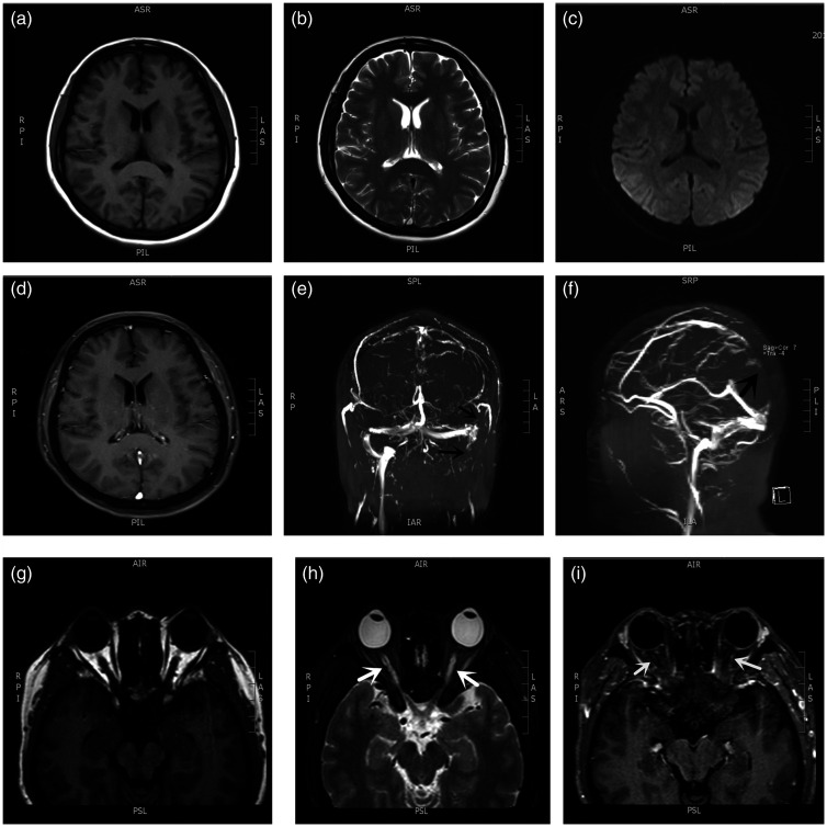
Cerebral venous sinus thrombosis (CVST) typically presents as headache, papilledema, and seizures. A dural arteriovenous fistula (DAVF) is a rare intracranial vascular malformation, and common symptoms include headache, pulsatile tinnitus, and stroke. The occurrence of CVST as a complication of DAVF is infrequent. Moreover, optic neuropathy presenting as the initial symptom of CVST and secondary DAVF is also unusual. We present a case of a patient with optic neuropathy and persistent intracranial hypertension who underwent head magnetic resonance imaging, which indicated CVST. She received normative anticoagulant and dehydration therapy; however, a repeated lumbar puncture showed dramatically increased intracranial pressure. Further digital subtraction angiography revealed an intracranial DAVF. The patient was finally diagnosed with a DAVF and secondary CVST. This case indicates that intractable optic neuropathy could be an uncommon indicator for CVST and secondary DAVF. Early diagnosis and early treatment are essential for visual rehabilitation and prognosis improvement.
Keywords: Optic neuropathy; cerebral venous sinus thrombosis; digital subtraction angiography, magnetic resonance imaging; dural arteriovenous fistula; intracranial hypertension; papilledema.
Conflict of interest statement
Figures


Similar articles
-
Dural arteriovenous fistula in the setting of cerebral venous sinus thrombosis and COVID-19 infection.Neurosurg Focus. 2024 Mar;56(3):E17. doi: 10.3171/2023.12.FOCUS23794. Neurosurg Focus. 2024. PMID: 38427997
-
A case report of oral contraceptive misuse induced cerebral venous sinus thrombosis and dural arteriovenous fistula.Medicine (Baltimore). 2019 Aug;98(33):e16440. doi: 10.1097/MD.0000000000016440. Medicine (Baltimore). 2019. PMID: 31415348 Free PMC article.
-
Clinical characteristics and outcome of dural arteriovenous fistulas secondary to cerebral venous sinus thrombosis: a primary or secondary event?BMC Neurol. 2023 Mar 30;23(1):131. doi: 10.1186/s12883-023-03141-6. BMC Neurol. 2023. PMID: 36997877 Free PMC article.
-
Dural Arteriovenous Fistulae: Imaging and Management.Neuroimaging Clin N Am. 2016 May;26(2):247-58. doi: 10.1016/j.nic.2015.12.003. Epub 2016 Feb 28. Neuroimaging Clin N Am. 2016. PMID: 27154607 Review.
-
Ruptured dural arteriovenous fistula and sinus venous thrombosis following surgical resection of a vestibular schwannoma: Case report and review of the literature.Neurochirurgie. 2022 Dec;68(6):688-692. doi: 10.1016/j.neuchi.2022.03.008. Epub 2022 May 20. Neurochirurgie. 2022. PMID: 35599062 Review.
References
-
- Micieli JA, Derkatch S, Pereira VM, et al.. Development of dural arteriovenous fistulas after cerebral venous sinus thrombosis. J Neuroophthalmol 2016; 36: 53–57. - PubMed
-
- Ahmed RM, Khoury B, Wilkinson M, et al.. Venous hypertension as the cause of intracranial hypertension in patients with transverse sinus dural arteriovenous fistula. J Neuroophthalmol 2013; 33: 102–105. - PubMed
-
- Gagnier JJ, Kienle G, Altman DG, et al.. The CARE guidelines: consensus-based clinical case reporting guideline development. Headache 2013; 53: 1541–1547. - PubMed
-
- Stam J. Thrombosis of the cerebral veins and sinuses. N Engl J Med 2005; 352: 1791–1798. - PubMed
-
- Kobayashi A, Al-Shahi Salman R. Prognosis and treatment of intracranial dural arteriovenous fistulae: a systematic review and meta-analysis. Int J Stroke 2014; 9: 670–677. - PubMed
Publication types
MeSH terms
LinkOut - more resources
Full Text Sources
Medical
